

Tested Applications
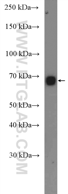
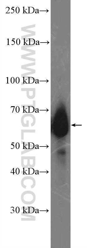
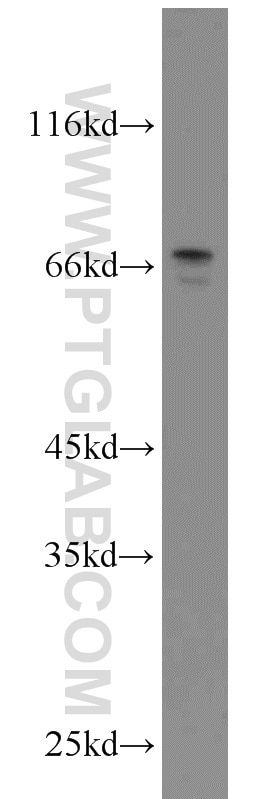
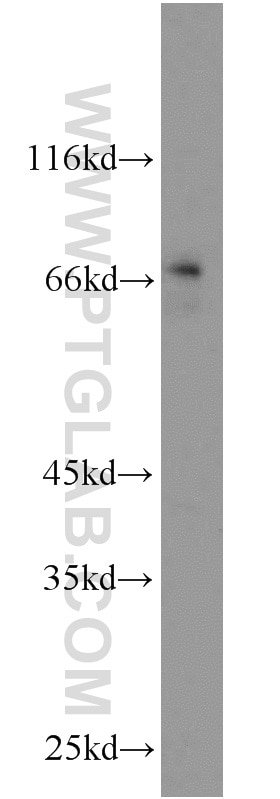
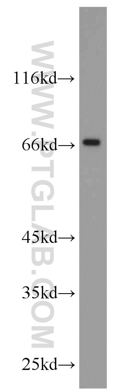
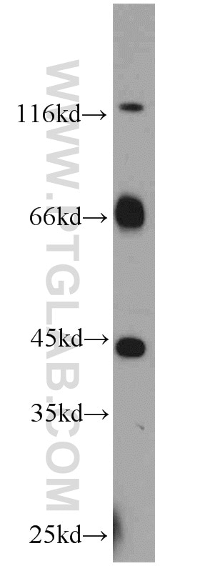
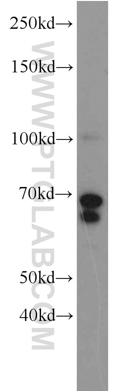
| Positive WB detected in | human placenta tissue, SKOV-3 cells, C6 cells, mouse brain tissue, SH-SY5Y cells, rat brain tissue |
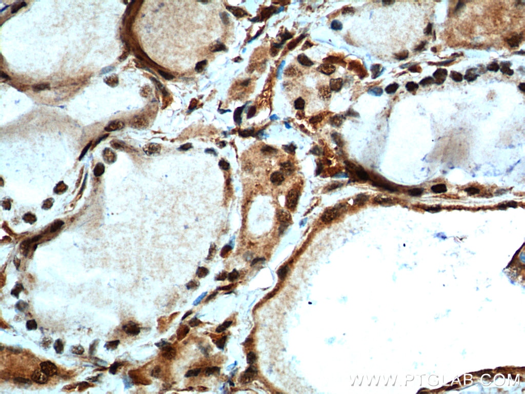
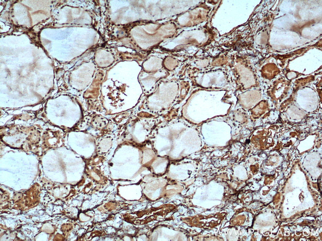
| Positive IHC detected in | human thyroid cancer tissue Note: suggested antigen retrieval with TE buffer pH 9.0; (*) Alternatively, antigen retrieval may be performed with citrate buffer pH 6.0 |
Recommended dilution
| Application | Dilution |
|---|---|
| Western Blot (WB) | WB : 1:500-1:3000 |
| Immunohistochemistry (IHC) | IHC : 1:50-1:500 |
| It is recommended that this reagent should be titrated in each testing system to obtain optimal results. | |
| Sample-dependent, Check data in validation data gallery. | |
Product Information
10159-2-AP targets MYCN in WB, IHC, IF, ELISA applications and shows reactivity with human, mouse, rat samples.
| Tested Reactivity | human, mouse, rat |
| Cited Reactivity | human, mouse |
| Host / Isotype | Rabbit / IgG |
| Class | Polyclonal |
| Type | Antibody |
| Immunogen |
CatNo: Ag0193 Product name: Recombinant human MYCN protein Source: e coli.-derived, PGEX-4T Tag: GST Domain: 289-464 aa of BC002712 Sequence: SNTKAVTTFTITVRPKNAALGPGRAQSSELILKRCLPIHQQHNYAAPSPYVESEDAPPQKKIKSEASPRPLKSVIPPKAKSLSPRNSDSEDSERRRNHNILERQRRNDLRSSFLTLRDHVPELVKNEKAAKVVILKKATEYVHSLQAEEHQLLLEKEKLQARQQQLLKKIEHARTC Predict reactive species |
| Full Name | v-myc myelocytomatosis viral related oncogene, neuroblastoma derived (avian) |
| Calculated Molecular Weight | 50 kDa |
| Observed Molecular Weight | 65-70 kDa |
| GenBank Accession Number | BC002712 |
| Gene Symbol | MYCN |
| Gene ID (NCBI) | 4613 |
| RRID | AB_2266881 |
| Conjugate | Unconjugated |
| Form | Liquid |
| Purification Method | Antigen affinity purification |
| UNIPROT ID | P04198 |
| Storage Buffer | PBS with 0.02% sodium azide and 50% glycerol, pH 7.3. |
| Storage Conditions | Store at -20°C. Stable for one year after shipment. Aliquoting is unnecessary for -20oC storage. 20ul sizes contain 0.1% BSA. |
Background Information
MYCN, also named as BHLHE37 and NMYC, is a transcription factor. It is primarily expressed in normal developing embryos and is thought to be critical in brain and other neural development. It is often amplified in human neuroblastomas. IR34A can suppress MYCN expression, it suggest MYCN has a role in cell growth. The expression of the MCYN is upregulated in a variety of human tumors, most frequently neuroblastoma, where the level of expression appears to increase as the tumor progresses. The calculated molecuar weight of MYCN is 50 kDa, but Phosphorylated MYCN is about 65-70 kDa. MYCN exsits two isoforms and MV of isoform is respectively 65 kDa and 45 kDa. (PMID: 19615087 )
Protocols
| Product Specific Protocols | |
|---|---|
| IHC protocol for MYCN antibody 10159-2-AP | Download protocol |
| WB protocol for MYCN antibody 10159-2-AP | Download protocol |
| Standard Protocols | |
|---|---|
| Click here to view our Standard Protocols |
Publications
What published studies show
Anti-MYCN (Cat# 10159-2-AP) has 29 citations published across 27 journals, including Nature, Molecular Cancer, Nature Communications, Science Advances, Clinical Cancer Research, Cancer, eLife, Frontiers in Oncology, and Scientific Reports. Research fields predominantly span neuroblastoma, oncology/cancer biology, signaling pathways (MYCN-driven), cell biology, and developmental biology. Applications include IHC, WB, and IF in human and mouse models.
| Species | Application | Title |
|---|---|---|
Mol Cancer N-Myc promotes therapeutic resistance development of neuroendocrine prostate cancer by differentially regulating miR-421/ATM pathway. | ||
Nat Commun The transcriptional co-repressor Runx1t1 is essential for MYCN-driven neuroblastoma tumorigenesis | ||
Sci Adv Clinically relevant treatment of PDX models reveals patterns of neuroblastoma chemoresistance | ||
Clin Cancer Res Amplification of extrachromosomal MYC paralogs shapes immunosuppressive tumor microenvironment in small cell lung cancer | ||
J Transl Med 7,7"-dimethoxyagastisflavone induced MYCN/MYBL2-dependent apoptosis and metabolism reprogramming in Sonic Hedgehog medulloblastoma. |
Reviews
What customers say
Users report this MYCN antibody performs well for Western blotting, producing a strong, specific band around 60 kDa in mouse heart tissue and neuroblastoma cell lines. One reviewer noted the band appears larger than expected molecular weight, likely due to post-translational modifications. For immunofluorescence, staining was strong but showed some cytoplasmic signal alongside nuclear localization. Specificity was confirmed as the antibody recognized MYCN-expressing BE2C cells but not c-myc-expressing Sy5Y cells. Overall, reviewers rate it favorably at 4–5 stars with working dilutions of 1:100 to 1:2000.
The reviews below have been submitted by verified Proteintech customers who received an incentive for providing their feedback.
CHAO (Verified Customer) (03-31-2022) | Strong bands.
|
Tom (Verified Customer) (08-22-2020) | I saw a specific ~60 kDa band when performing western blots using this MYCN antibody. The band is bigger than molecular weight of MYCN, probably due to post-translational modifications.
|
Tom (Verified Customer) (08-22-2020) | I performed immunofluorescence using this antibody. I observed very strong MYCN signal in mouse heart tissue sections. However, I saw cytoplasmic staining in addition to nuclear staining. It is probably due to MYCN's localization to both cytoplasm and nuclei or this antibody has some background staining.
|
Jessica (Verified Customer) (10-31-2018) | Strongest band observed was around 60 kDa with some much weaker bands much higher and much lower observable with higher exposures. Ab had affinity for BE2C cell lysate, but did not bind the c-myc expressed in Sy5Y cells on the same blot. I used 2 % skim milk PBST for incubation. As MYCN Abs go this one is pretty good!
|


