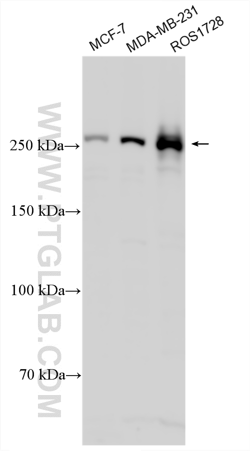
MCF7 cells were subjected to SDS PAGE followed by western blot with 20657-1-AP (MTOR antibody) at dilution of 1:500 incubated at room temperature for 1.5 hours.
WB analysis of HeLa using 20657-1-AP
HeLa cells were subjected to SDS PAGE followed by western blot with 20657-1-AP (MTOR antibody) at dilution of 1:500 incubated at room temperature for 1.5 hours.
WB analysis of MDA-MB-453s using 20657-1-AP
MDA-MB-453s cells were subjected to SDS PAGE followed by western blot with 20657-1-AP (MTOR Antibody) at dilution of 1:100 incubated at room temperature for 1.5 hours.
IHC staining of mouse testis using 20657-1-AP
Immunohistochemical analysis of paraffin-embedded mouse testis tissue slide using 20657-1-AP (mTOR antibody) at dilution of 1:200 (under 40x lens. Heat mediated antigen retrieval with Tris-EDTA buffer (pH 9.0).
Immunohistochemical analysis of paraffin-embedded mouse testis tissue slide using 20657-1-AP (mTOR antibody) at dilution of 1:200 (under 10x lens. Heat mediated antigen retrieval with Tris-EDTA buffer (pH 9.0).
IHC staining of human breast cancer using 20657-1-AP
Immunohistochemical analysis of paraffin-embedded human breast cancer tissue slide using 20657-1-AP (mTOR antibody) at dilution of 1:200 (under 10x lens. Heat mediated antigen retrieval with Tris-EDTA buffer (pH 9.0).
IHC staining of human breast cancer using 20657-1-AP
Immunohistochemical analysis of paraffin-embedded human breast cancer tissue slide using 20657-1-AP (mTOR antibody) at dilution of 1:200 (under 40x lens. Heat mediated antigen retrieval with Tris-EDTA buffer (pH 9.0).
IF Staining of HeLa using 20657-1-AP
Immunofluorescent analysis of HeLa cells using 20657-1-AP (MTOR antibody) at dilution of 1:50 and Alexa Fluor 488-conjugated Goat Anti-Rabbit IgG(H+L).
The Proteintech guarantee covers Proteintech antibodies in any species and any application, including those not listed on the datasheet. If the antibody doesn’t perform, you can receive a hassle-free refund or credit note.
mouse testis tissue, human breast cancer tissue Note: suggested antigen retrieval with TE buffer pH 9.0; (*) Alternatively, antigen retrieval may be performed with citrate buffer pH 6.0
Positive IF/ICC detected in
HeLa cells
Recommended dilution
Application
Dilution
Western Blot (WB)
WB : 1:500-1:1000
Immunohistochemistry (IHC)
IHC : 1:50-1:500
Immunofluorescence (IF)/ICC
IF/ICC : 1:50-1:500
It is recommended that this reagent should be titrated in each testing system to obtain optimal results.
Sample-dependent, Check data in validation data gallery.
PBS with 0.02% sodium azide and 50% glycerol pH 7.3.
Storage Conditions
Store at -20°C. Stable for one year after shipment. Aliquoting is unnecessary for -20oC storage. 20ul sizes contain 0.1% BSA.
Background Information
MTOR, also named as FRAP1, FRAP, FRAP2 and RAPT1, belongs to the PI3/PI4-kinase family. MTOR is a Ser/Thr protein kinase that functions as an ATP and amino acid sensor to balance nutrient availability and cell growth. MTOR is Kinase subunit of both mTORC1 and mTORC2, which regulate cell growth and survival in response to nutrient and hormonal signals. mTORC1 is activated in response to growth factors or amino-acids. mTORC2 is also activated by growth factors, but seems to be nutrient-insensitive. mTORC2 seems to function upstream of Rho GTPases to regulate the actin cytoskeleton, probably by activating one or more Rho-type guanine nucleotide exchange factors. mTORC2 promotes the serum-induced formation of stress-fibers or F-actin. MTOR has a calculated molecular mass of 289 kDa, and always can be detected at about 250 kDa due to some modifications (PMID: 14578359). The antibody is specific to MTOR.
MCF7 cells were subjected to SDS PAGE followed by western blot with 20657-1-AP (MTOR antibody) at dilution of 1:500 incubated at room temperature for 1.5 hours.
WB analysis of HeLa using 20657-1-AP
HeLa cells were subjected to SDS PAGE followed by western blot with 20657-1-AP (MTOR antibody) at dilution of 1:500 incubated at room temperature for 1.5 hours.
WB analysis of MDA-MB-453s using 20657-1-AP
MDA-MB-453s cells were subjected to SDS PAGE followed by western blot with 20657-1-AP (MTOR Antibody) at dilution of 1:100 incubated at room temperature for 1.5 hours.
IHC Figures
IHC staining of mouse testis using 20657-1-AP
Immunohistochemical analysis of paraffin-embedded mouse testis tissue slide using 20657-1-AP (mTOR antibody) at dilution of 1:200 (under 40x lens. Heat mediated antigen retrieval with Tris-EDTA buffer (pH 9.0).
IHC staining of mouse testis using 20657-1-AP
Immunohistochemical analysis of paraffin-embedded mouse testis tissue slide using 20657-1-AP (mTOR antibody) at dilution of 1:200 (under 10x lens. Heat mediated antigen retrieval with Tris-EDTA buffer (pH 9.0).
IHC staining of human breast cancer using 20657-1-AP
Immunohistochemical analysis of paraffin-embedded human breast cancer tissue slide using 20657-1-AP (mTOR antibody) at dilution of 1:200 (under 10x lens. Heat mediated antigen retrieval with Tris-EDTA buffer (pH 9.0).
IHC staining of human breast cancer using 20657-1-AP
Immunohistochemical analysis of paraffin-embedded human breast cancer tissue slide using 20657-1-AP (mTOR antibody) at dilution of 1:200 (under 40x lens. Heat mediated antigen retrieval with Tris-EDTA buffer (pH 9.0).
IF Figures
IF Staining of HeLa using 20657-1-AP
Immunofluorescent analysis of HeLa cells using 20657-1-AP (MTOR antibody) at dilution of 1:50 and Alexa Fluor 488-conjugated Goat Anti-Rabbit IgG(H+L).
The species listed in Tested Reactivity are in-house verified and applicable species. For unlisted species, please refer to the homology analysis of the immunogen sequence and related species. For rabbit polyclonal antibodies, homology >70% is recommended. For mouse monoclonal antibodies and rabbit recombinant antibodies, homology >90% is recommended. Generally, the higher the homology, the greater the applicability. However, there will be certain differences in protein expression in different species, tissues or cells. Therefore, the homology analysis results are for reference only and do not serve as a guarantee.
At Proteintech, we pride ourselves on our antibody quality, customer service and transparency. As such, we are comparing our antibodies with other vendors, enabling easy identification and comparisons of key data to help you choose the suitable antibody for your needs.
We have selected the top cited antibodies from these vendors for you to compare.