Tested Applications
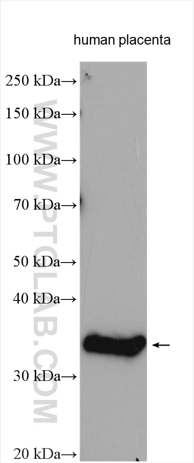
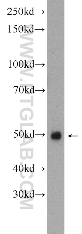
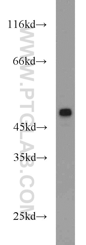
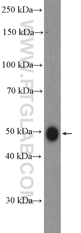
| Positive WB detected in | human placenta tissue, mouse liver tissue, PC-3 cells, mouse heart tissue |
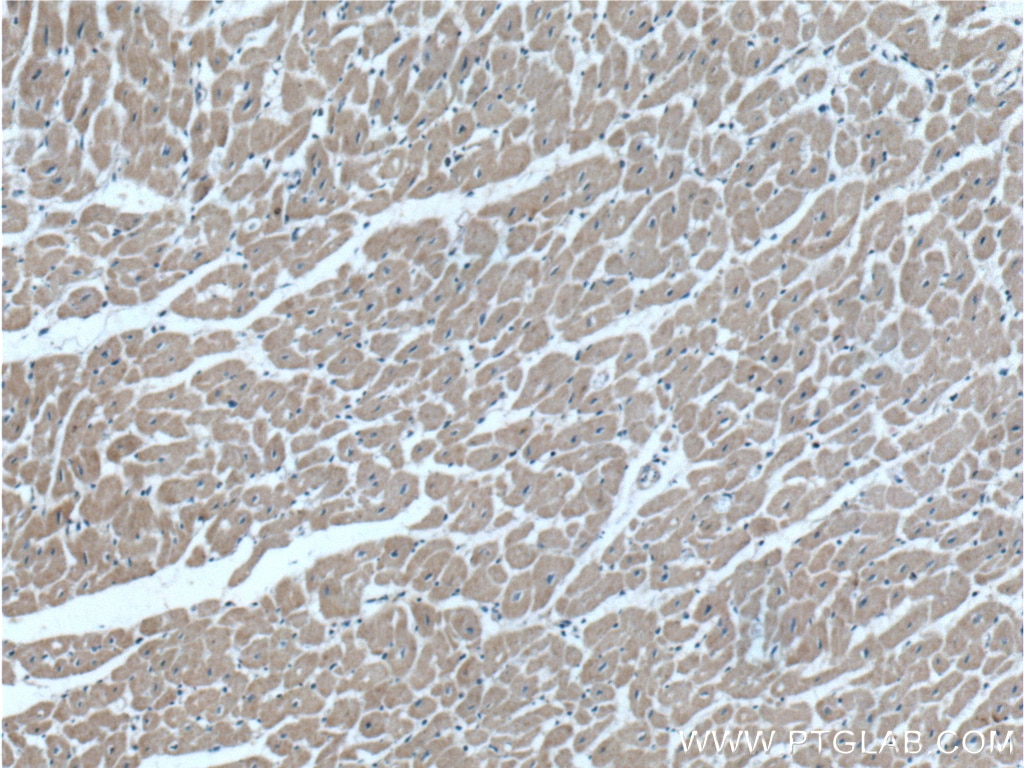
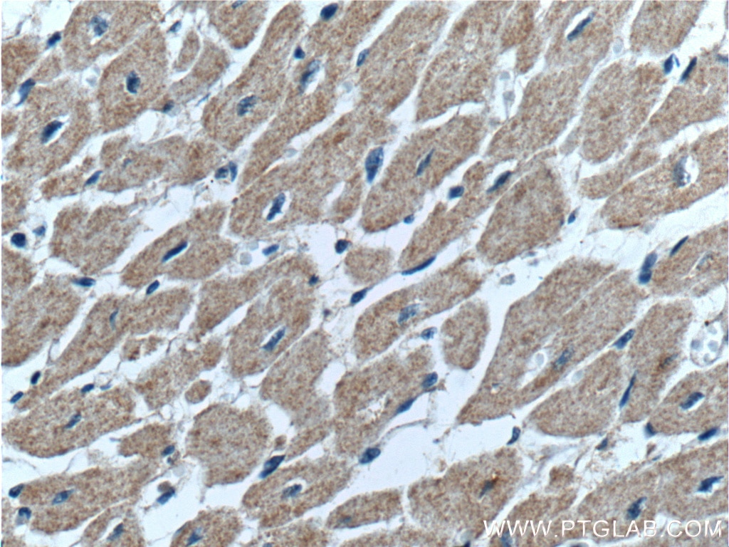
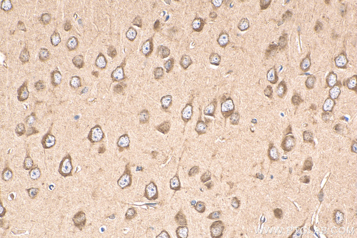
| Positive IHC detected in | human heart tissue, mouse brain tissue Note: suggested antigen retrieval with TE buffer pH 9.0; (*) Alternatively, antigen retrieval may be performed with citrate buffer pH 6.0 |
Recommended dilution
| Application | Dilution |
|---|---|
| Western Blot (WB) | WB : 1:500-1:2000 |
| Immunohistochemistry (IHC) | IHC : 1:50-1:500 |
| It is recommended that this reagent should be titrated in each testing system to obtain optimal results. | |
| Sample-dependent, Check data in validation data gallery. | |
Published Applications
| KD/KO | See 3 publications below |
| WB | See 32 publications below |
| IHC | See 7 publications below |
| IF | See 3 publications below |
Product Information
19142-1-AP targets GDF8/Myostatin in WB, IHC, IF, ELISA applications and shows reactivity with human, mouse samples.
| Tested Reactivity | human, mouse |
| Cited Reactivity | human, mouse, rat, pig, zebrafish, bovine, sheep |
| Host / Isotype | Rabbit / IgG |
| Class | Polyclonal |
| Type | Antibody |
| Immunogen | GDF8/Myostatin fusion protein Ag13605 Predict reactive species |
| Full Name | myostatin |
| Calculated Molecular Weight | 375 aa, 43 kDa |
| Observed Molecular Weight | 50 and 35 kDa |
| GenBank Accession Number | BC074757 |
| Gene Symbol | MSTN |
| Gene ID (NCBI) | 2660 |
| RRID | AB_10638615 |
| Conjugate | Unconjugated |
| Form | Liquid |
| Purification Method | Antigen affinity purification |
| UNIPROT ID | O14793 |
| Storage Buffer | PBS with 0.02% sodium azide and 50% glycerol , pH 7.3 |
| Storage Conditions | Store at -20°C. Stable for one year after shipment. Aliquoting is unnecessary for -20oC storage. 20ul sizes contain 0.1% BSA. |
Background Information
Myostatin, also known as GDF-8, is a member of the TGF-beta superfamily. Myostatin is expressed in the myotome and developing skeletal muscles. Myostatin negatively regulates skeletal muscle growth and development through the regulation of anabolic and catabolic pathways in skeletal muscles. Myostatin immunoreactive bands could be identified at ∼50, 32-35, and 16 kDa, which correspond to the precursor, glycosylated dimmer, and monomer, respectively (PMID: 17711997).
Protocols
| Product Specific Protocols | |
|---|---|
| WB protocol for GDF8/Myostatin antibody 19142-1-AP | Download protocol |
| IHC protocol for GDF8/Myostatin antibody 19142-1-AP | Download protocol |
| Standard Protocols | |
|---|---|
| Click here to view our Standard Protocols |
Publications
| Species | Application | Title |
|---|---|---|
J Cachexia Sarcopenia Muscle Potential therapeutic interventions for chronic kidney disease-associated sarcopenia via indoxyl sulfate-induced mitochondrial dysfunction. | ||
Br J Pharmacol Inhibition of heat shock protein (HSP) 90 reverses signal transducer and activator of transcription (STAT) 3-mediated muscle wasting in cancer cachexia mice. | ||
Int J Mol Sci Renal Ischemia/Reperfusion Early Induces Myostatin and PCSK9 Expression in Rat Kidneys and HK-2 Cells.
| ||
iScience Phosphate depletion in insulin-insensitive skeletal muscle drives AMPD activation and sarcopenia in chronic kidney disease | ||
Aging (Albany NY) Vitellogenin 2 promotes muscle development and stimulates the browning of white fat | ||
Sci Rep Indoxyl sulfate potentiates skeletal muscle atrophy by inducing the oxidative stress-mediated expression of myostatin and atrogin-1. |
