Tested Applications
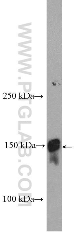
| Positive WB detected in | human plasma tissue |
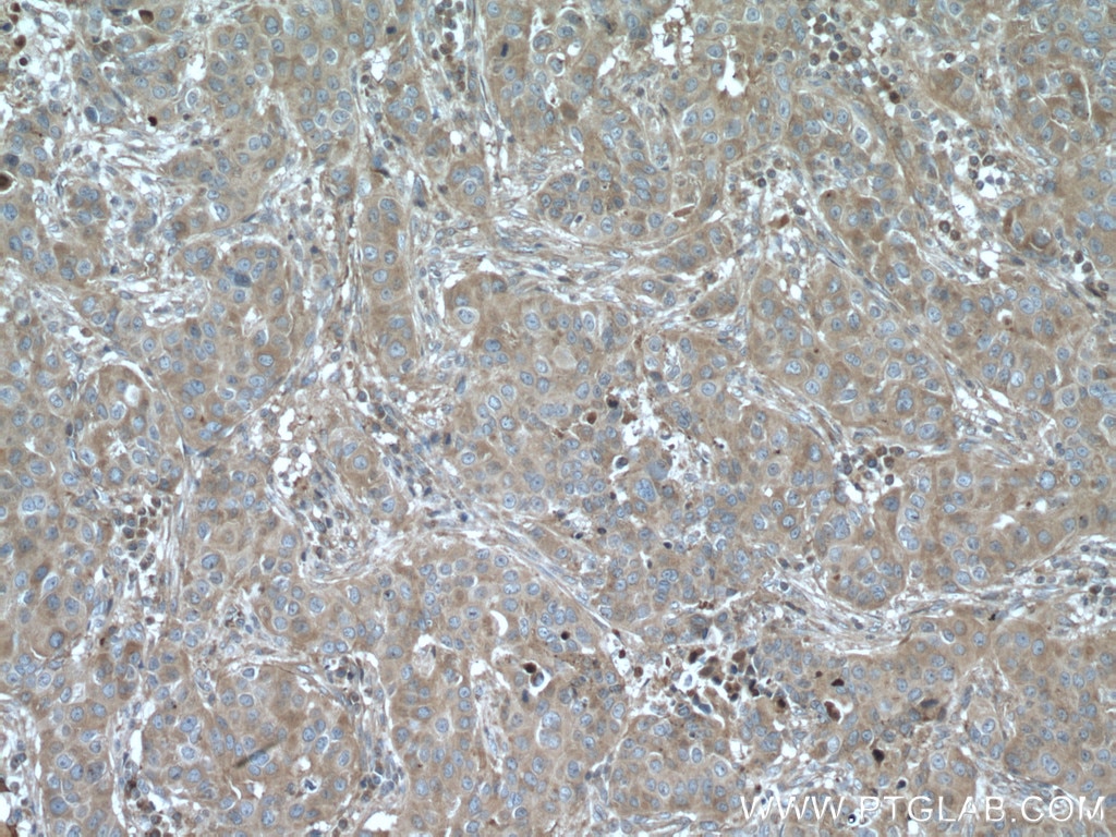
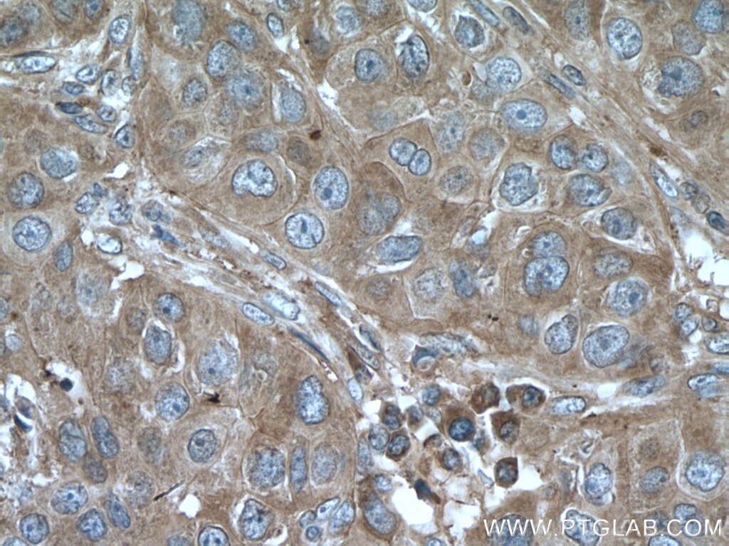
| Positive IHC detected in | human breast cancer tissue Note: suggested antigen retrieval with TE buffer pH 9.0; (*) Alternatively, antigen retrieval may be performed with citrate buffer pH 6.0 |
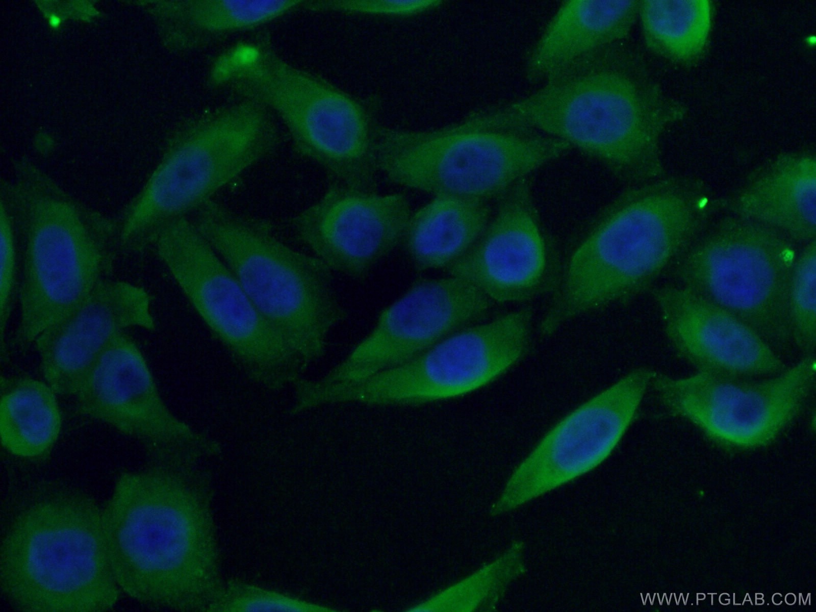
| Positive IF/ICC detected in | HeLa cells |
Recommended dilution
| Application | Dilution |
|---|---|
| Western Blot (WB) | WB : 1:500-1:3000 |
| Immunohistochemistry (IHC) | IHC : 1:50-1:500 |
| Immunofluorescence (IF)/ICC | IF/ICC : 1:50-1:500 |
| It is recommended that this reagent should be titrated in each testing system to obtain optimal results. | |
| Sample-dependent, Check data in validation data gallery. | |
Published Applications
| KD/KO | See 1 publications below |
| WB | See 4 publications below |
| IHC | See 2 publications below |
| IF | See 2 publications below |
Product Information
26855-1-AP targets LTBP1 in WB, IHC, IF/ICC, ELISA applications and shows reactivity with human samples.
| Tested Reactivity | human |
| Cited Reactivity | human, mouse |
| Host / Isotype | Rabbit / IgG |
| Class | Polyclonal |
| Type | Antibody |
| Immunogen | LTBP1 fusion protein Ag25392 Predict reactive species |
| Full Name | latent transforming growth factor beta binding protein 1 |
| Calculated Molecular Weight | 1721 aa, 187 kDa |
| Observed Molecular Weight | 150 kDa |
| GenBank Accession Number | BC130289 |
| Gene Symbol | LTBP1 |
| Gene ID (NCBI) | 4052 |
| RRID | AB_2880658 |
| Conjugate | Unconjugated |
| Form | Liquid |
| Purification Method | Antigen affinity purification |
| UNIPROT ID | Q14766 |
| Storage Buffer | PBS with 0.02% sodium azide and 50% glycerol , pH 7.3 |
| Storage Conditions | Store at -20°C. Stable for one year after shipment. Aliquoting is unnecessary for -20oC storage. 20ul sizes contain 0.1% BSA. |
Background Information
Latent TGFβ binding protein 1 (LTBP1) is a large extracellular protein which belongs to the family of latent TGF-beta binding proteins (LTBPs). LTBP1 is essential for TGF-beta folding, secretion, matrix localization and activation. LTBP1 has also been demonstrated to interact with a number of insoluble extracellular matrix components, such as fibrillin. We got 150-240 kDa in western blotting due to protein glycosylation of LTBP1 (PMID: 18086923, PMID: 9923648 ).
Protocols
| Product Specific Protocols | |
|---|---|
| WB protocol for LTBP1 antibody 26855-1-AP | Download protocol |
| IHC protocol for LTBP1 antibody 26855-1-AP | Download protocol |
| IF protocol for LTBP1 antibody 26855-1-AP | Download protocol |
| Standard Protocols | |
|---|---|
| Click here to view our Standard Protocols |
Publications
| Species | Application | Title |
|---|---|---|
Cell Rep Integrative single-cell meta-analysis reveals disease-relevant vascular cell states and markers in human atherosclerosis | ||
J Transl Med LTBP1 promotes esophageal squamous cell carcinoma progression through epithelial-mesenchymal transition and cancer-associated fibroblasts transformation.
| ||
Cell Death Discov ERRFI1 induces apoptosis of hepatocellular carcinoma cells in response to tryptophan deficiency. | ||
medRxiv Secreted protein profiling of human aortic smooth muscle cells identifies vascular disease associations | ||
Adv Mater Engineered Surfaces that Promote Capture of Latent Proteins to Facilitate Integrin-Mediated Mechanical Activation of Growth Factors | ||
Front Immunol Hypoxia reconstructed colorectal tumor microenvironment weakening anti-tumor immunity: construction of a new prognosis predicting model through transcriptome analysis |
Reviews
The reviews below have been submitted by verified Proteintech customers who received an incentive for providing their feedback.
FH Udesh (Verified Customer) (10-05-2022) | The Ab was used at 1:1000 dilution in NFDM overnight 4 degrees and detected a band close to 220 kD in 20ug cell lysate. Also tested for for positive IF staining at 1:250 dilution in 2% BSA overnight at 4 degrees.
 |


