Tested Applications
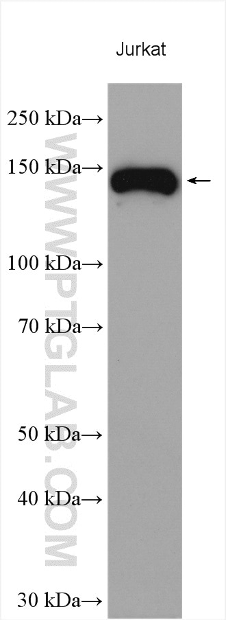
| Positive WB detected in | Jurkat cells |
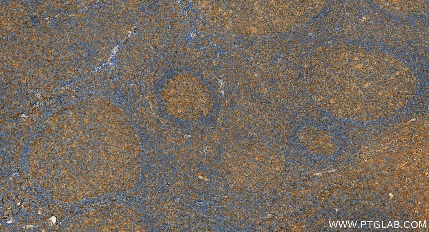
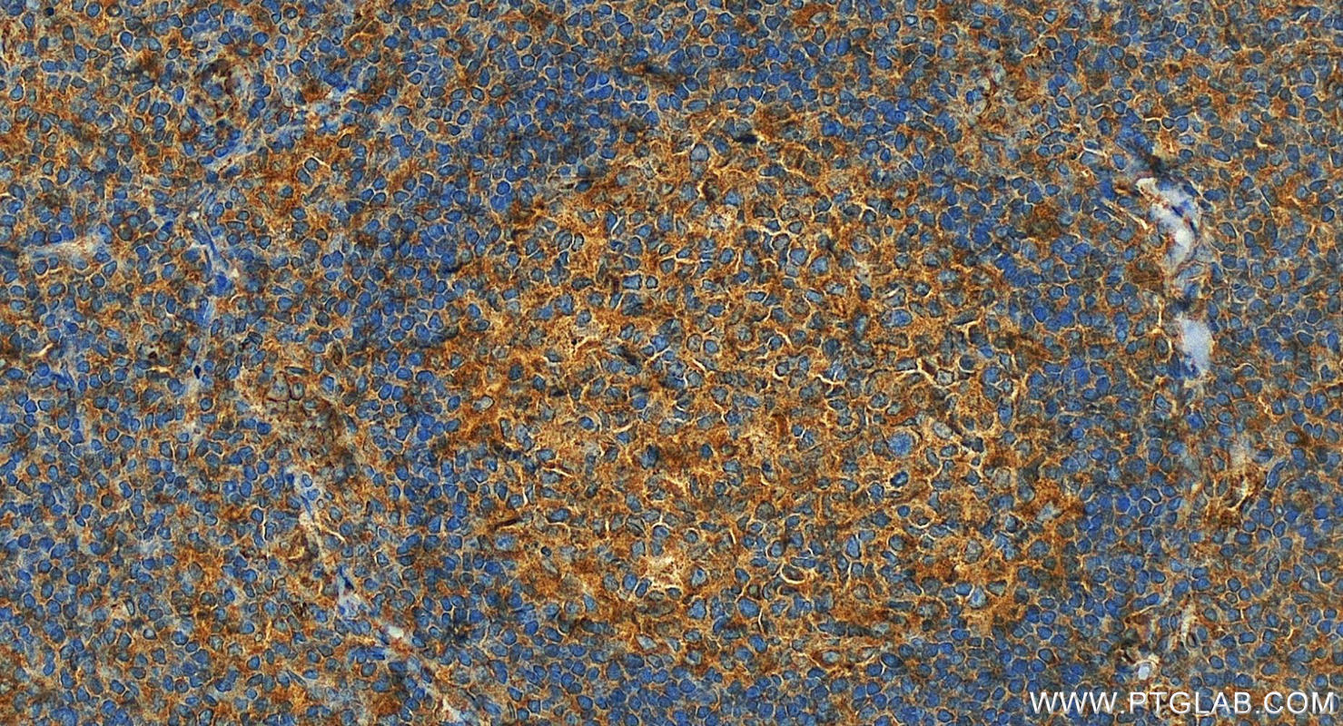
| Positive IHC detected in | human tonsillitis tissue Note: suggested antigen retrieval with TE buffer pH 9.0; (*) Alternatively, antigen retrieval may be performed with citrate buffer pH 6.0 |
Recommended dilution
| Application | Dilution |
|---|---|
| Western Blot (WB) | WB : 1:1000-1:6000 |
| Immunohistochemistry (IHC) | IHC : 1:200-1:800 |
| It is recommended that this reagent should be titrated in each testing system to obtain optimal results. | |
| Sample-dependent, Check data in validation data gallery. | |
Published Applications
| KD/KO | See 1 publications below |
| WB | See 20 publications below |
| IHC | See 4 publications below |
| IF | See 2 publications below |
Product Information
19676-1-AP targets Integrin alpha 4 in WB, IHC, IF, ELISA applications and shows reactivity with human samples.
| Tested Reactivity | human |
| Cited Reactivity | human, mouse, rat |
| Host / Isotype | Rabbit / IgG |
| Class | Polyclonal |
| Type | Antibody |
| Immunogen |
Peptide Predict reactive species |
| Full Name | integrin, alpha 4 (antigen CD49D, alpha 4 subunit of VLA-4 receptor) |
| Calculated Molecular Weight | 115 kDa |
| Observed Molecular Weight | 140-150 kDa |
| GenBank Accession Number | NM_000885 |
| Gene Symbol | Integrin alpha 4 |
| Gene ID (NCBI) | 3676 |
| RRID | AB_10640907 |
| Conjugate | Unconjugated |
| Form | Liquid |
| Purification Method | Antigen affinity purification |
| UNIPROT ID | P13612 |
| Storage Buffer | PBS with 0.02% sodium azide and 50% glycerol, pH 7.3. |
| Storage Conditions | Store at -20°C. Stable for one year after shipment. Aliquoting is unnecessary for -20oC storage. 20ul sizes contain 0.1% BSA. |
Background Information
Integrin alpha 4 (ITGA4), also named CD49D, belongs to the integrin alpha chain family. ITGA4 is a receptor for fibronectin, VCAM1, and MADCAM1. ITGA4 recognizes one or more domains within the alternatively spliced CS-1 and CS-5 regions of fibronectin. It recognizes the sequence Q-I-D-S in VCAM1. It recognizes the sequence L-D-T in MADCAM1. On activated endothelial cells ITGA4 triggers homotypic aggregation for most VLA-4-positive leukocyte cell lines. It may also participate in cytolytic T-cell interactions with target cells.
Protocols
| Product Specific Protocols | |
|---|---|
| IHC protocol for Integrin alpha 4 antibody 19676-1-AP | Download protocol |
| WB protocol for Integrin alpha 4 antibody 19676-1-AP | Download protocol |
| Standard Protocols | |
|---|---|
| Click here to view our Standard Protocols |
Publications
| Species | Application | Title |
|---|---|---|
Adv Sci (Weinh) Mesenchymal Stem Cell Membrane-Camouflaged Liposomes for Biomimetic Delivery of Cyclosporine A for Hepatic Ischemia-Reperfusion Injury Prevention | ||
Nat Commun Calreticulin and integrin alpha dissociation induces anti-inflammatory programming in animal models of inflammatory bowel disease. | ||
J Exp Med EP3 enhances adhesion and cytotoxicity of NK cells toward hepatic stellate cells in a murine liver fibrosis model. | ||
Int J Biol Macromol Diethylaminoethyl-dextran and monocyte cell membrane coated 1,8-cineole delivery system for intracellular delivery and synergistic treatment of atherosclerosis | ||
Int J Mol Sci Sesquiterpene Lactone Deoxyelephantopin Isolated from Elephantopus scaber and Its Derivative DETD-35 Suppress BRAFV600E Mutant Melanoma Lung Metastasis in Mice. | ||
Int J Mol Sci Induced Pluripotent Stem Cell-Derived Conditioned Medium Promotes Endogenous Leukemia Inhibitory Factor to Attenuate Endotoxin-Induced Acute Lung Injury. |
Reviews
The reviews below have been submitted by verified Proteintech customers who received an incentive for providing their feedback.
FH Boyan (Verified Customer) (03-11-2019) | For WB, the expected band was very weak, and was much weaker than those non-specific bands; for IF, it could label the cell cortex localisation of Itga4, partially co-localised with Actin.
|


