Tested Applications
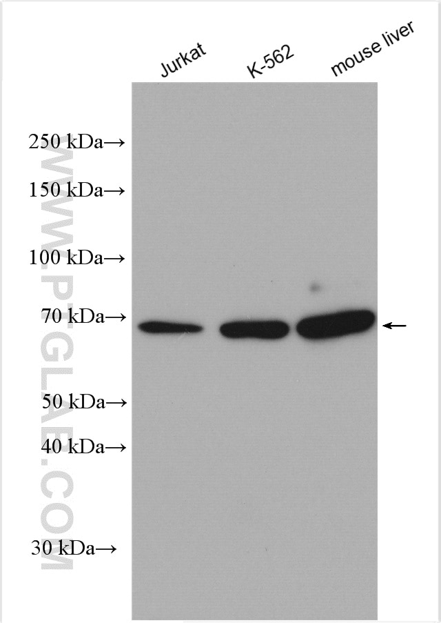
| Positive WB detected in | Jurkat cells, K-562 cell, mouse liver tissue |
Recommended dilution
| Application | Dilution |
|---|---|
| Western Blot (WB) | WB : 1:500-1:2000 |
| It is recommended that this reagent should be titrated in each testing system to obtain optimal results. | |
| Sample-dependent, Check data in validation data gallery. | |
Published Applications
| KD/KO | See 3 publications below |
| WB | See 14 publications below |
| IF | See 1 publications below |
| IP | See 1 publications below |
Product Information
13083-1-AP targets IFNAR1 in WB, IF, IP, ELISA applications and shows reactivity with human, mouse samples.
| Tested Reactivity | human, mouse |
| Cited Reactivity | human, mouse, pig, monkey |
| Host / Isotype | Rabbit / IgG |
| Class | Polyclonal |
| Type | Antibody |
| Immunogen |
CatNo: Ag3739 Product name: Recombinant human IFNAR1 protein Source: e coli.-derived, PGEX-4T Tag: GST Domain: 82-427 aa of BC021825 Sequence: ITSTKCNFSSLKLNVYEEIKLRIRAEKENTSSWYEVDSFTPFRKAQIGPPEVHLEAEDKAIVIHISPGTKDSVMWALDGLSFTYSLVIWKNSSGVEERIENIYSRHKIYKLSPETTYCLKVKAALLTSWKIGVYSPVHCIKTTVENELPPPENIEVSVQNQNYVLKWDYTYANMTFQVQWLHAFLKRNPGNHLYKWKQIPDCENVKTTQCVFPQNVFQKGIYLLRVQASDGNNTSFWSEEIKFDTEIQAFLLPPVFNIRSLSDSFHIYIGAPKQSGNTPVIQDYPLIYEIIFWENTSNAERKIIEKKTDVTVPNLKPLTVYCVKARAHTMDEKLNKSSVFSDAVCE Predict reactive species |
| Full Name | interferon (alpha, beta and omega) receptor 1 |
| Calculated Molecular Weight | 557 aa, 64 kDa |
| Observed Molecular Weight | 64 kDa |
| GenBank Accession Number | BC021825 |
| Gene Symbol | IFNAR1 |
| Gene ID (NCBI) | 3454 |
| RRID | AB_2122626 |
| Conjugate | Unconjugated |
| Form | Liquid |
| Purification Method | Antigen affinity purification |
| UNIPROT ID | P17181 |
| Storage Buffer | PBS with 0.02% sodium azide and 50% glycerol, pH 7.3. |
| Storage Conditions | Store at -20°C. Stable for one year after shipment. Aliquoting is unnecessary for -20oC storage. 20ul sizes contain 0.1% BSA. |
Background Information
Interferon alpha and beta receptor subunit 1 (IFNAR1) is a type I membrane protein that associates with IFNAR2 to form the receptor for type I interferons, including interferons-alpha, -beta, and -lambda. Binding and activation of the receptor stimulates Janus protein kinases, which in turn phosphorylate several proteins, including STAT1 and STAT2, and then increase transcription of the IFN-induced genes whose products exert antiviral, immunomodulatory, and antiproliferative effects.
Publications
| Species | Application | Title |
|---|---|---|
Oncogene Reciprocal regulation between ferroptosis and STING-type I interferon pathway suppresses head and neck squamous cell carcinoma growth through dendritic cell maturation
| ||
EMBO Rep Degradation of WTAP blocks antiviral responses by reducing the m 6 A levels of IRF3 and IFNAR1 mRNA | ||
Elife The oncoprotein BCL6 enables solid tumor cells to evade genotoxic stress.
| ||
J Virol African swine fever virus pH240R enhances viral replication via inhibition of the type I IFN signaling pathway | ||
Reviews
The reviews below have been submitted by verified Proteintech customers who received an incentive for providing their feedback.
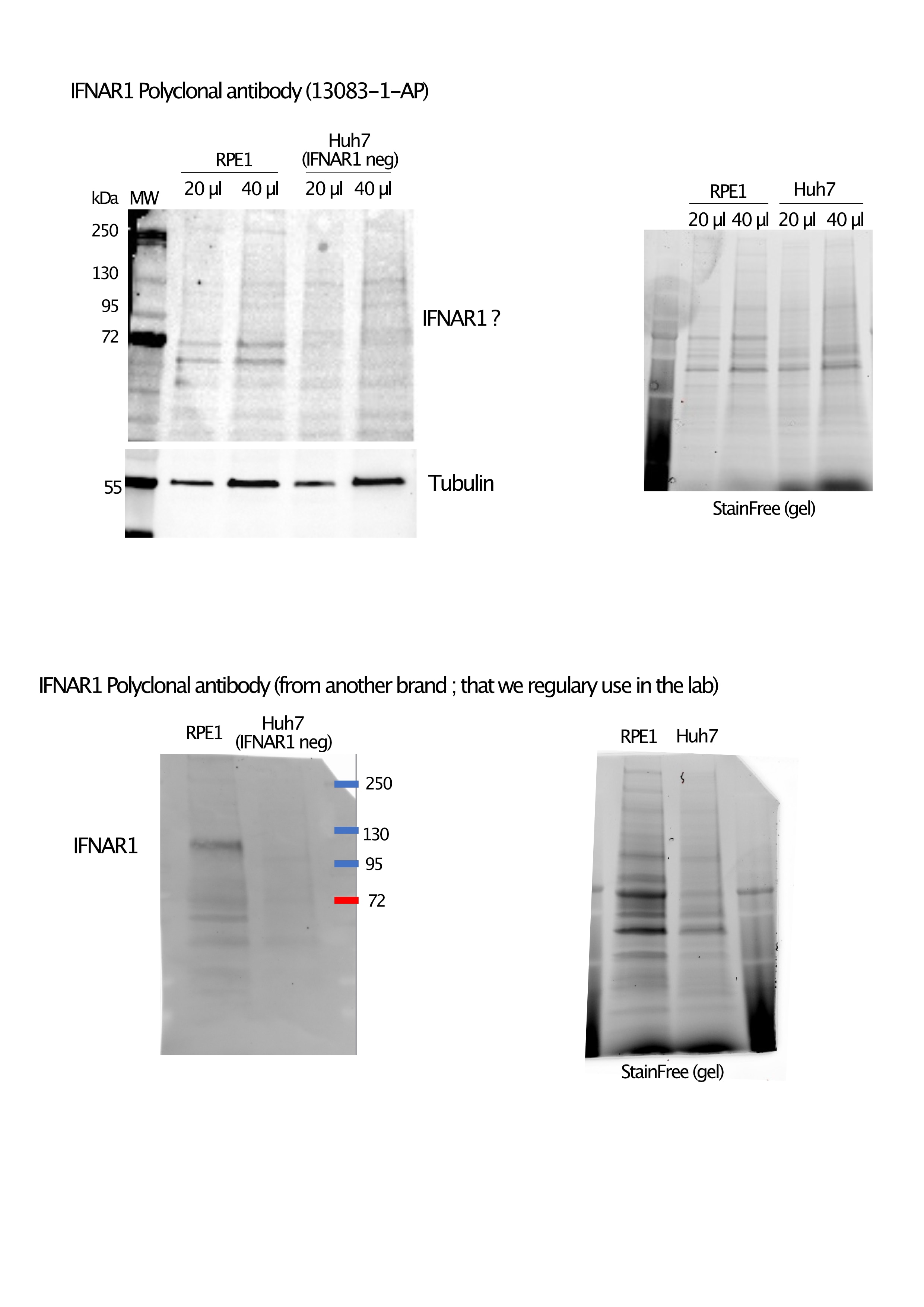
FH Christine (Verified Customer) (07-12-2022) | Both cell lines are human ones, only the RPE1 cells express the IFNAR1 receptor. IFNAR1 is described in the literature as heavily glycosylated and hence its molecular weight can vary from 64-135 kDa depending on its glycosylation level. We generally observed in RPE1 cells fully matured 110-130 kDa major band on western blot, which is not observed at all here with the 13083-1-AP antibody. The 72 kDa bands seems to be present only in RPE1 and not in the IFNAR1 null Huh7 cells, indicating it could indeed be immature non glycosylated forms of IFNAR1. (We also observed these minor bands on RPE1 cells with another IFNAR1 antibody , but at a much lower intensity than the 110 kDa one)
 |


