Various lysates were subjected to SDS PAGE followed by western blot with 80735-1-RR (HSP70 antibody) at dilution of 1:10000 incubated at room temperature for 1.5 hours.
Various lysates were subjected to SDS PAGE followed by western blot with 80735-1-RR (HSP70 antibody) at dilution of 1:10000 incubated at room temperature for 1.5 hours.
IHC staining of human breast cancer using 80735-1-RR
Immunohistochemical analysis of paraffin-embedded human breast cancer tissue slide using 80735-1-RR (HSP70 antibody) at dilution of 1:200 (under 10x lens). Heat mediated antigen retrieval with Tris-EDTA buffer (pH 9.0).
Immunohistochemical analysis of paraffin-embedded human breast cancer tissue slide using 80735-1-RR (HSP70 antibody) at dilution of 1:200 (under 10x lens). Heat mediated antigen retrieval with Tris-EDTA buffer (pH 9.0).
IHC staining of human lung squamous cell carcinoma using 80735-1-RR
Immunohistochemical analysis of paraffin-embedded human lung squamous cell carcinoma tissue slide using 80735-1-RR (HSP70 antibody) at dilution of 1:4000 (under 20x lens). Heat mediated antigen retrieval with Tris-EDTA buffer (pH 9.0).
Immunohistochemical analysis of paraffin-embedded human lung squamous cell carcinoma tissue slide using 80735-1-RR (HSP70 antibody) at dilution of 1:4000 (under 20x lens). Heat mediated antigen retrieval with Tris-EDTA buffer (pH 9.0).
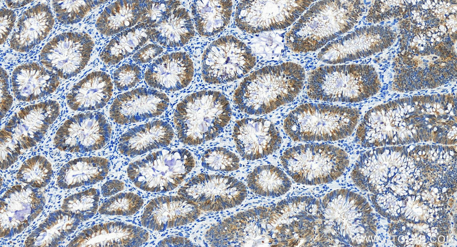
IHC staining of human colon cancer using 80735-1-RR
Immunohistochemical analysis of paraffin-embedded human colon cancer tissue slide using 80735-1-RR (HSP70 antibody) at dilution of 1:6000 (under 20x lens). Heat mediated antigen retrieval with Tris-EDTA buffer (pH 9.0).
Immunohistochemical analysis of paraffin-embedded human colon cancer tissue slide using 80735-1-RR (HSP70 antibody) at dilution of 1:6000 (under 20x lens). Heat mediated antigen retrieval with Tris-EDTA buffer (pH 9.0).
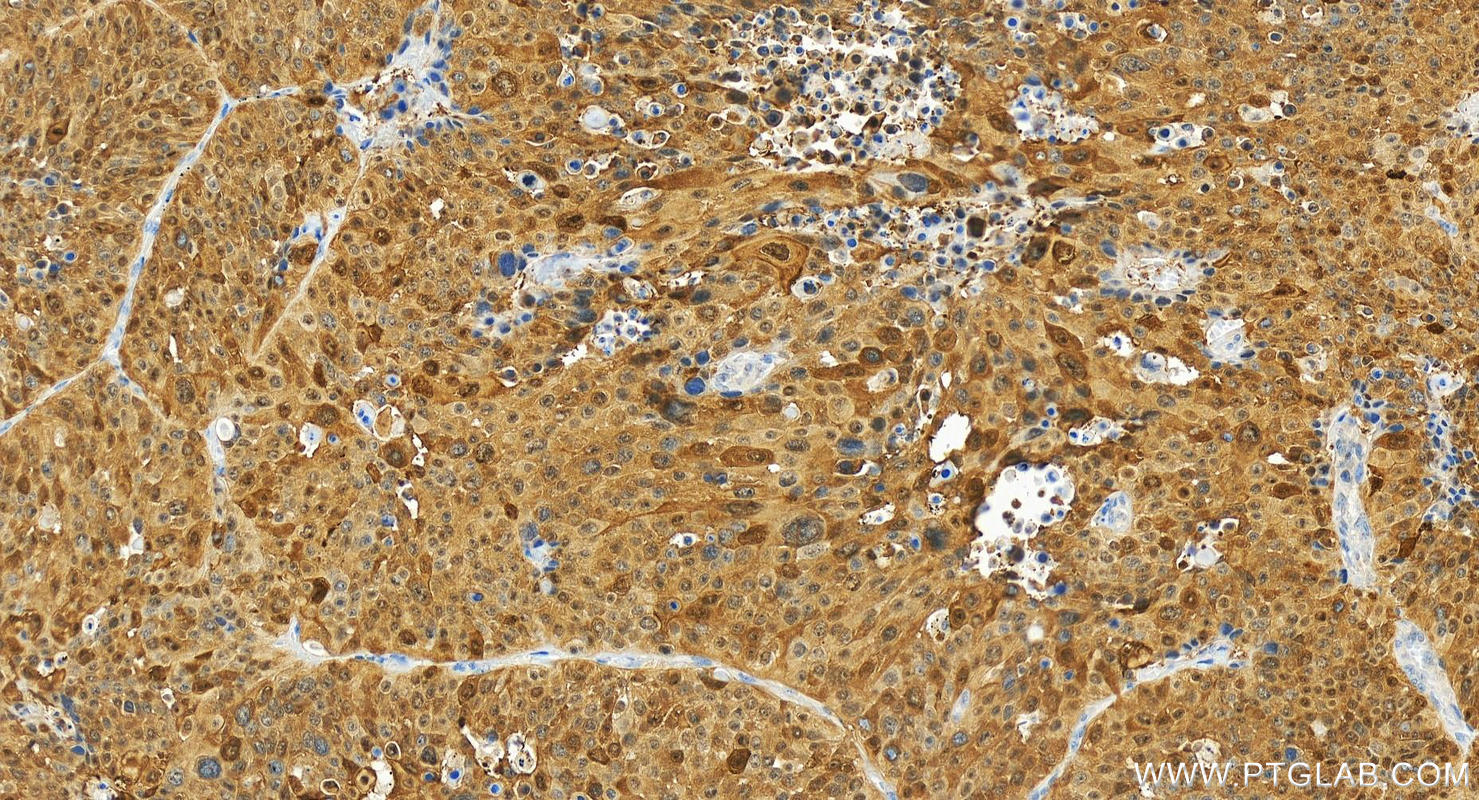
IHC staining of human liver cancer using 80735-1-RR
Immunohistochemical analysis of paraffin-embedded human liver cancer tissue slide using 80735-1-RR (HSP70 antibody) at dilution of 1:200 (under 10x lens). Heat mediated antigen retrieval with Tris-EDTA buffer (pH 9.0).
Immunohistochemical analysis of paraffin-embedded human liver cancer tissue slide using 80735-1-RR (HSP70 antibody) at dilution of 1:200 (under 10x lens). Heat mediated antigen retrieval with Tris-EDTA buffer (pH 9.0).
IF Staining of HeLa using 80735-1-RR
Immunofluorescent analysis of (-20°C Methanol) fixed HeLa cells using HSP70 antibody (80735-1-RR, Clone: 4E10 ) at dilution of 1:400 and CoraLite®488-Conjugated AffiniPure Goat Anti-Rabbit IgG(H+L).
Immunofluorescent analysis of (-20°C Methanol) fixed HeLa cells using HSP70 antibody (80735-1-RR, Clone: 4E10 ) at dilution of 1:400 and CoraLite®488-Conjugated AffiniPure Goat Anti-Rabbit IgG(H+L).
The Proteintech guarantee covers Proteintech antibodies in any species and any application, including those not listed on the datasheet. If the antibody doesn’t perform, you can receive a hassle-free refund or credit note.
HEK-293 cells, HeLa cells, K-562 cells, NIH/3T3 cells, C6 cells, the whole yeast
Positive IHC detected in
human breast cancer tissue, human colon cancer tissue, human liver cancer tissue, human lung squamous cell carcinoma tissue Note: suggested antigen retrieval with TE buffer pH 9.0; (*) Alternatively, antigen retrieval may be performed with citrate buffer pH 6.0
Positive IF/ICC detected in
HeLa cells
Recommended dilution
Application
Dilution
Western Blot (WB)
WB : 1:5000-1:20000
Immunohistochemistry (IHC)
IHC : 1:200-1:6000
Immunofluorescence (IF)/ICC
IF/ICC : 1:200-1:800
It is recommended that this reagent should be titrated in each testing system to obtain optimal results.
Sample-dependent, Check data in validation data gallery.
PBS with 0.02% sodium azide and 50% glycerol, pH 7.3.
Storage Conditions
Store at -20°C. Stable for one year after shipment. Aliquoting is unnecessary for -20oC storage. 20ul sizes contain 0.1% BSA.
Background Information
HSPA1A , collectively known as HSP70 (also referred to HSP72), is a stress-inducible member of heat-shock protein 70 (HSP70) proteins which are highly conserved chaperons implicated in protein folding, protein refolding, protein transport, and protein targeting. Encoded by two closely linked, intronless and stress-inducible genes, HSPA1A and HSPA1B differ by only two amino acids and are believed to be fully interchangeable proteins. HSPA1A is a cytosol nuclear protein able to translocate between cytoplasm and nucleus. Generally, HSPA1A is thought to be expressed in unstressed normal cells at low or undetectable levels. Expression of HSPA1A protein can be highly activated by various stressful stimuli. Significant up-regulation of HSPA1A has been found in various tumors. Recently it has been reported that HSPA1A can be constitutively expressed in selected cell types. HSP70 is also used as exosomal marker
Various lysates were subjected to SDS PAGE followed by western blot with 80735-1-RR (HSP70 antibody) at dilution of 1:10000 incubated at room temperature for 1.5 hours.
IHC Figures
IHC staining of human breast cancer using 80735-1-RR
Immunohistochemical analysis of paraffin-embedded human breast cancer tissue slide using 80735-1-RR (HSP70 antibody) at dilution of 1:200 (under 10x lens). Heat mediated antigen retrieval with Tris-EDTA buffer (pH 9.0).
IHC staining of human lung squamous cell carcinoma using 80735-1-RR
Immunohistochemical analysis of paraffin-embedded human lung squamous cell carcinoma tissue slide using 80735-1-RR (HSP70 antibody) at dilution of 1:4000 (under 20x lens). Heat mediated antigen retrieval with Tris-EDTA buffer (pH 9.0).
IHC staining of human colon cancer using 80735-1-RR
Immunohistochemical analysis of paraffin-embedded human colon cancer tissue slide using 80735-1-RR (HSP70 antibody) at dilution of 1:6000 (under 20x lens). Heat mediated antigen retrieval with Tris-EDTA buffer (pH 9.0).
IHC staining of human liver cancer using 80735-1-RR
Immunohistochemical analysis of paraffin-embedded human liver cancer tissue slide using 80735-1-RR (HSP70 antibody) at dilution of 1:200 (under 10x lens). Heat mediated antigen retrieval with Tris-EDTA buffer (pH 9.0).
IF/ICC Figures
IF Staining of HeLa using 80735-1-RR
Immunofluorescent analysis of (-20°C Methanol) fixed HeLa cells using HSP70 antibody (80735-1-RR, Clone: 4E10 ) at dilution of 1:400 and CoraLite®488-Conjugated AffiniPure Goat Anti-Rabbit IgG(H+L).
The species listed in Tested Reactivity are in-house verified and applicable species. For unlisted species, please refer to the homology analysis of the immunogen sequence and related species. For rabbit polyclonal antibodies, homology >70% is recommended. For mouse monoclonal antibodies and rabbit recombinant antibodies, homology >90% is recommended. Generally, the higher the homology, the greater the applicability. However, there will be certain differences in protein expression in different species, tissues or cells. Therefore, the homology analysis results are for reference only and do not serve as a guarantee.
At Proteintech, we pride ourselves on our antibody quality, customer service and transparency. As such, we are comparing our antibodies with other vendors, enabling easy identification and comparisons of key data to help you choose the suitable antibody for your needs.
We have selected the top cited antibodies from these vendors for you to compare.
 at dilution of 1:10000 incubated at room temperature for 1.5 hours.")
 at dilution of 1:200 (under 10x lens). Heat mediated antigen retrieval with Tris-EDTA buffer (pH 9.0).")
 at dilution of 1:4000 (under 20x lens). Heat mediated antigen retrieval with Tris-EDTA buffer (pH 9.0).")
 at dilution of 1:6000 (under 20x lens). Heat mediated antigen retrieval with Tris-EDTA buffer (pH 9.0).")
 at dilution of 1:200 (under 10x lens). Heat mediated antigen retrieval with Tris-EDTA buffer (pH 9.0).")
 fixed HeLa cells using HSP70 antibody (80735-1-RR, Clone: 4E10 ) at dilution of 1:400 and CoraLite®488-Conjugated AffiniPure Goat Anti-Rabbit IgG(H+L).")