WB result of HRPT2, CDC73 antibody (66490-1-Ig; 1:50000; incubated at room temperature for 1.5 hours) with sh-Control and sh-HRPT2, CDC73 transfected HeLa cells.
WB result of HRPT2, CDC73 antibody (66490-1-Ig; 1:50000; incubated at room temperature for 1.5 hours) with sh-Control and sh-HRPT2, CDC73 transfected HeLa cells.
WB analysis using 66490-1-Ig
Various lysates were subjected to SDS PAGE followed by western blot with 66490-1-Ig (HRPT2, CDC73 antibody) at dilution of 1:10000 incubated at room temperature for 1.5 hours. The membrane was stripped and reblotted with HRP-conjugated Beta Actin Monoclonal antibody (HRP-66009) as loading control.
Various lysates were subjected to SDS PAGE followed by western blot with 66490-1-Ig (HRPT2, CDC73 antibody) at dilution of 1:10000 incubated at room temperature for 1.5 hours. The membrane was stripped and reblotted with HRP-conjugated Beta Actin Monoclonal antibody (HRP-66009) as loading control.
WB analysis using 66490-1-Ig
Various lysates were subjected to SDS PAGE followed by western blot with 66490-1-Ig (HRPT2, CDC73 antibody) at dilution of 1:10000 incubated at room temperature for 1.5 hours.
Various lysates were subjected to SDS PAGE followed by western blot with 66490-1-Ig (HRPT2, CDC73 antibody) at dilution of 1:10000 incubated at room temperature for 1.5 hours.
WB analysis of pig brain using 66490-1-Ig
pig brain tissue were subjected to SDS PAGE followed by western blot with 66490-1-Ig (HRPT2, CDC73 antibody) at dilution of 1:10000 incubated at room temperature for 1.5 hours.
pig brain tissue were subjected to SDS PAGE followed by western blot with 66490-1-Ig (HRPT2, CDC73 antibody) at dilution of 1:10000 incubated at room temperature for 1.5 hours.
WB analysis of mouse brain using 66490-1-Ig
mouse brain tissue were subjected to SDS PAGE followed by western blot with 66490-1-Ig (HRPT2, CDC73 antibody) at dilution of 1:10000 incubated at room temperature for 1.5 hours.
mouse brain tissue were subjected to SDS PAGE followed by western blot with 66490-1-Ig (HRPT2, CDC73 antibody) at dilution of 1:10000 incubated at room temperature for 1.5 hours.
IHC staining of human breast cancer using 66490-1-Ig
Immunohistochemical analysis of paraffin-embedded human breast cancer tissue slide using 66490-1-Ig (HRPT2, CDC73 antibody) at dilution of 1:400 (under 10x lens). Heat mediated antigen retrieval with Tris-EDTA buffer (pH 9.0).
Immunohistochemical analysis of paraffin-embedded human breast cancer tissue slide using 66490-1-Ig (HRPT2, CDC73 antibody) at dilution of 1:400 (under 10x lens). Heat mediated antigen retrieval with Tris-EDTA buffer (pH 9.0).
IHC staining of human breast cancer using 66490-1-Ig
Immunohistochemical analysis of paraffin-embedded human breast cancer tissue slide using 66490-1-Ig (HRPT2; CDC73 antibody) at dilution of 1:400 (under 40x lens. Heat mediated antigen retrieval with Tris-EDTA buffer (pH 9.0).
Immunohistochemical analysis of paraffin-embedded human breast cancer tissue slide using 66490-1-Ig (HRPT2; CDC73 antibody) at dilution of 1:400 (under 40x lens. Heat mediated antigen retrieval with Tris-EDTA buffer (pH 9.0).
IF Staining of human breast cancer using 66490-1-Ig
Immunofluorescent analysis of (4% PFA) fixed human breast cancer tissue using HRPT2, CDC73 antibody (66490-1-Ig, Clone: 4B4H2 ) at dilution of 1:100 and CoraLite488-Conjugated AffiniPure Goat Anti-Mouse IgG(H+L).
Immunofluorescent analysis of (4% PFA) fixed human breast cancer tissue using HRPT2, CDC73 antibody (66490-1-Ig, Clone: 4B4H2 ) at dilution of 1:100 and CoraLite488-Conjugated AffiniPure Goat Anti-Mouse IgG(H+L).
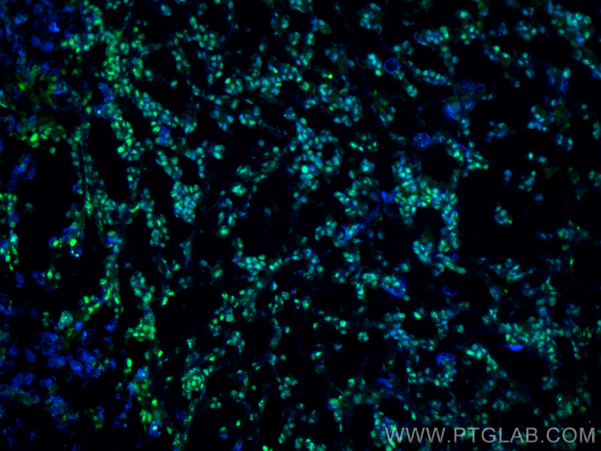
IF Staining of mouse breast cancer using 66490-1-Ig
Immunofluorescent analysis of (4% PFA) fixed frozen OCT-embedded mouse breast cancer using HRPT2, CDC73 antibody (66490-1-Ig, Clone: 4B4H2 ) at dilution of 1:400 and CoraLite®488-Conjugated Goat Anti-Mouse IgG(H+L) (SA00013-1).
Immunofluorescent analysis of (4% PFA) fixed frozen OCT-embedded mouse breast cancer using HRPT2, CDC73 antibody (66490-1-Ig, Clone: 4B4H2 ) at dilution of 1:400 and CoraLite®488-Conjugated Goat Anti-Mouse IgG(H+L) (SA00013-1).
The Proteintech guarantee covers Proteintech antibodies in any species and any application, including those not listed on the datasheet. If the antibody doesn’t perform, you can receive a hassle-free refund or credit note.
pig brain tissue, LNCaP cells, HeLa cells, mouse brain tissue, HEK-293 cells, Caco-2 cells, RAW 264.7 cells
Positive IHC detected in
human breast cancer tissue Note: suggested antigen retrieval with TE buffer pH 9.0; (*) Alternatively, antigen retrieval may be performed with citrate buffer pH 6.0
Positive IF-P detected in
human breast cancer tissue
Positive IF-Fro detected in
mouse breast cancer
Recommended dilution
Application
Dilution
Western Blot (WB)
WB : 1:5000-1:50000
Immunohistochemistry (IHC)
IHC : 1:200-1:800
Immunofluorescence (IF)-P
IF-P : 1:50-1:500
Immunofluorescence (IF)-FRO
IF-FRO : 1:200-1:800
It is recommended that this reagent should be titrated in each testing system to obtain optimal results.
Sample-dependent, Check data in validation data gallery.
PBS with 0.02% sodium azide and 50% glycerol, pH 7.3.
Storage Conditions
Store at -20°C. Stable for one year after shipment. Aliquoting is unnecessary for -20oC storage. 20ul sizes contain 0.1% BSA.
Background Information
Parafibromin is a product of the hyperparathyroidism-jaw tumor syndrome gene HRPT2/CDC73, a putative tumor suppressor gene recently implicated in the autosomal dominant hyperparathyroidism-jaw tumor familial cancer syndrome, sporadic parathyroid cancer, and a minority of families with isolated hyperparathyroidism (PMID: 15580289). Defects in CDC73 are causes of hyperparathyroidism type 1 (HRPT1) and hyperparathyroidism type 2 (HRPT2) as well as parathyroid carcinoma (PRTC) (PMID: 12434154). Tumor suppressor parafibromin to the transcription elongation and RNA processing pathway as a PAF1 complex- and RNA polymerase II-bound protein (PMID: 15923622). Besides, parafibromin is also involved in post-transcriptional control pathways (PMID: 16116486). Recent report has revealed that pathogenic mutation, such as CDC73 gene mutation, is able to affect histone monoubiquitination (PMID: 22021426).
WB result of HRPT2, CDC73 antibody (66490-1-Ig; 1:50000; incubated at room temperature for 1.5 hours) with sh-Control and sh-HRPT2, CDC73 transfected HeLa cells.
WB analysis using 66490-1-Ig
Various lysates were subjected to SDS PAGE followed by western blot with 66490-1-Ig (HRPT2, CDC73 antibody) at dilution of 1:10000 incubated at room temperature for 1.5 hours. The membrane was stripped and reblotted with HRP-conjugated Beta Actin Monoclonal antibody (HRP-66009) as loading control.
WB analysis using 66490-1-Ig
Various lysates were subjected to SDS PAGE followed by western blot with 66490-1-Ig (HRPT2, CDC73 antibody) at dilution of 1:10000 incubated at room temperature for 1.5 hours.
WB analysis of pig brain using 66490-1-Ig
pig brain tissue were subjected to SDS PAGE followed by western blot with 66490-1-Ig (HRPT2, CDC73 antibody) at dilution of 1:10000 incubated at room temperature for 1.5 hours.
WB analysis of mouse brain using 66490-1-Ig
mouse brain tissue were subjected to SDS PAGE followed by western blot with 66490-1-Ig (HRPT2, CDC73 antibody) at dilution of 1:10000 incubated at room temperature for 1.5 hours.
IHC Figures
IHC staining of human breast cancer using 66490-1-Ig
Immunohistochemical analysis of paraffin-embedded human breast cancer tissue slide using 66490-1-Ig (HRPT2, CDC73 antibody) at dilution of 1:400 (under 10x lens). Heat mediated antigen retrieval with Tris-EDTA buffer (pH 9.0).
IHC staining of human breast cancer using 66490-1-Ig
Immunohistochemical analysis of paraffin-embedded human breast cancer tissue slide using 66490-1-Ig (HRPT2; CDC73 antibody) at dilution of 1:400 (under 40x lens. Heat mediated antigen retrieval with Tris-EDTA buffer (pH 9.0).
IF-P Figures
IF Staining of human breast cancer using 66490-1-Ig
Immunofluorescent analysis of (4% PFA) fixed human breast cancer tissue using HRPT2, CDC73 antibody (66490-1-Ig, Clone: 4B4H2 ) at dilution of 1:100 and CoraLite488-Conjugated AffiniPure Goat Anti-Mouse IgG(H+L).
IF-FRO Figures
IF Staining of mouse breast cancer using 66490-1-Ig
Immunofluorescent analysis of (4% PFA) fixed frozen OCT-embedded mouse breast cancer using HRPT2, CDC73 antibody (66490-1-Ig, Clone: 4B4H2 ) at dilution of 1:400 and CoraLite®488-Conjugated Goat Anti-Mouse IgG(H+L) (SA00013-1).
The species listed in Tested Reactivity are in-house verified and applicable species. For unlisted species, please refer to the homology analysis of the immunogen sequence and related species. For rabbit polyclonal antibodies, homology >70% is recommended. For mouse monoclonal antibodies and rabbit recombinant antibodies, homology >90% is recommended. Generally, the higher the homology, the greater the applicability. However, there will be certain differences in protein expression in different species, tissues or cells. Therefore, the homology analysis results are for reference only and do not serve as a guarantee.
At Proteintech, we pride ourselves on our antibody quality, customer service and transparency. As such, we are comparing our antibodies with other vendors, enabling easy identification and comparisons of key data to help you choose the suitable antibody for your needs.
We have selected the top cited antibodies from these vendors for you to compare.
 with sh-Control and sh-HRPT2, CDC73 transfected HeLa cells.")
 at dilution of 1:10000 incubated at room temperature for 1.5 hours. The membrane was stripped and reblotted with HRP-conjugated Beta Actin Monoclonal antibody (HRP-66009) as loading control.")
 at dilution of 1:10000 incubated at room temperature for 1.5 hours. The membrane was stripped and reblotted with HRP-conjugated Beta Actin Monoclonal antibody (HRP-66009) as loading control.")
 at dilution of 1:10000 incubated at room temperature for 1.5 hours.")
 at dilution of 1:10000 incubated at room temperature for 1.5 hours.")
 at dilution of 1:10000 incubated at room temperature for 1.5 hours.")
 at dilution of 1:400 (under 10x lens). Heat mediated antigen retrieval with Tris-EDTA buffer (pH 9.0).")
 at dilution of 1:400 (under 40x lens. Heat mediated antigen retrieval with Tris-EDTA buffer (pH 9.0).")
 fixed human breast cancer tissue using HRPT2, CDC73 antibody (66490-1-Ig, Clone: 4B4H2 ) at dilution of 1:100 and CoraLite488-Conjugated AffiniPure Goat Anti-Mouse IgG(H+L).")
 fixed frozen OCT-embedded mouse breast cancer using HRPT2, CDC73 antibody (66490-1-Ig, Clone: 4B4H2 ) at dilution of 1:400 and CoraLite®488-Conjugated Goat Anti-Mouse IgG(H+L) (SA00013-1).")