at dilution of 1:1500 incubated at room temperature for 1.5 hours.")
 at dilution of 1:300 incubated at room temperature for 1.5 hours.")
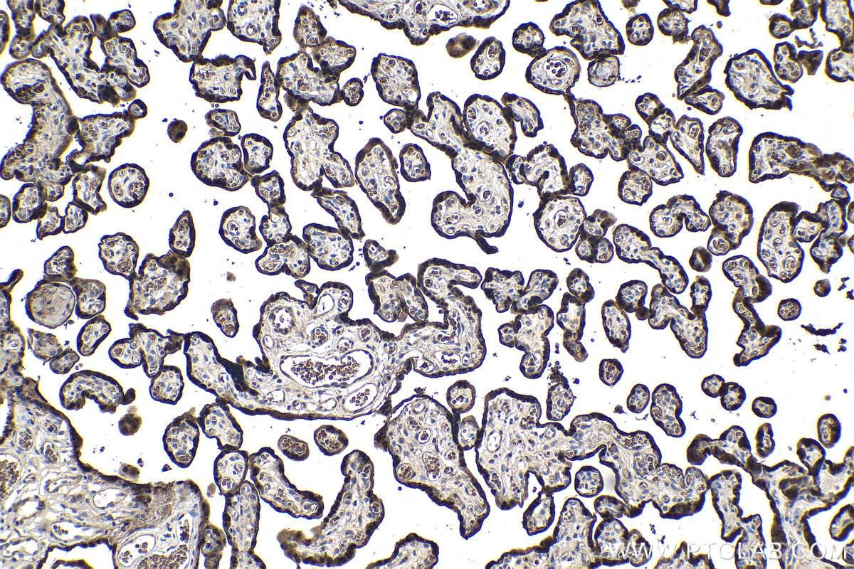
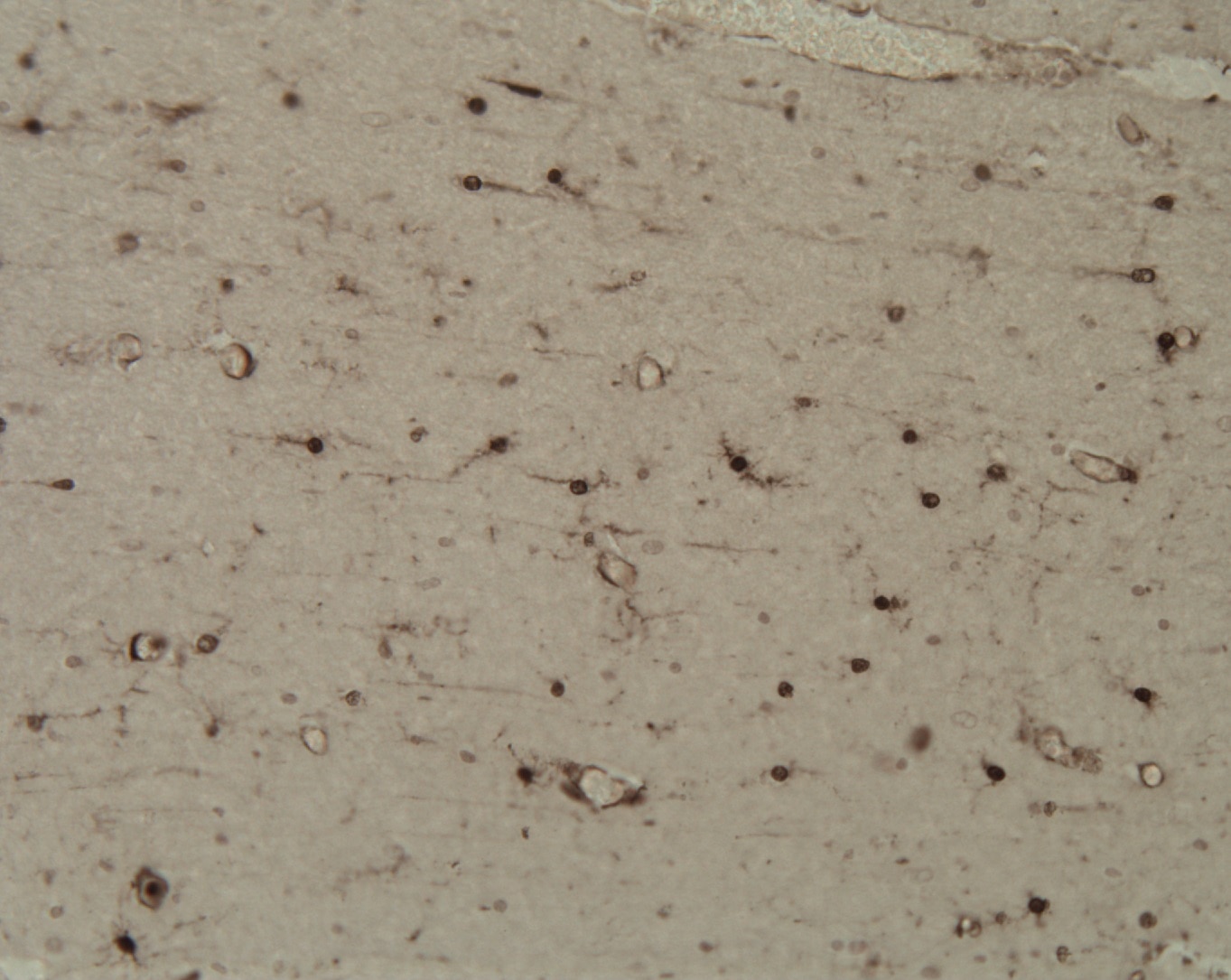
 at dilution of 1:1000 (under 10x lens). Heat mediated antigen retrieval with Tris-EDTA buffer (pH 9.0).")
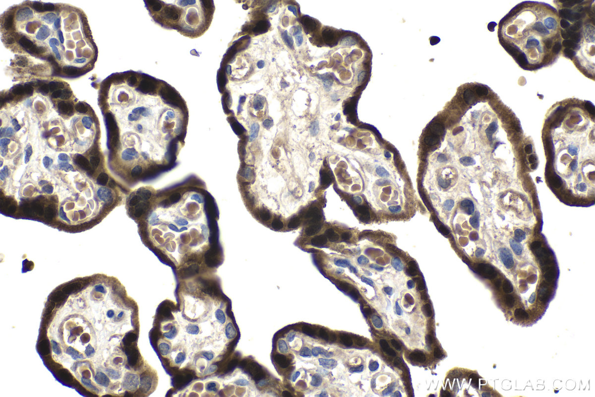
 at dilution of 1:1000 (under 40x lens). Heat mediated antigen retrieval with Tris-EDTA buffer (pH 9.0).")
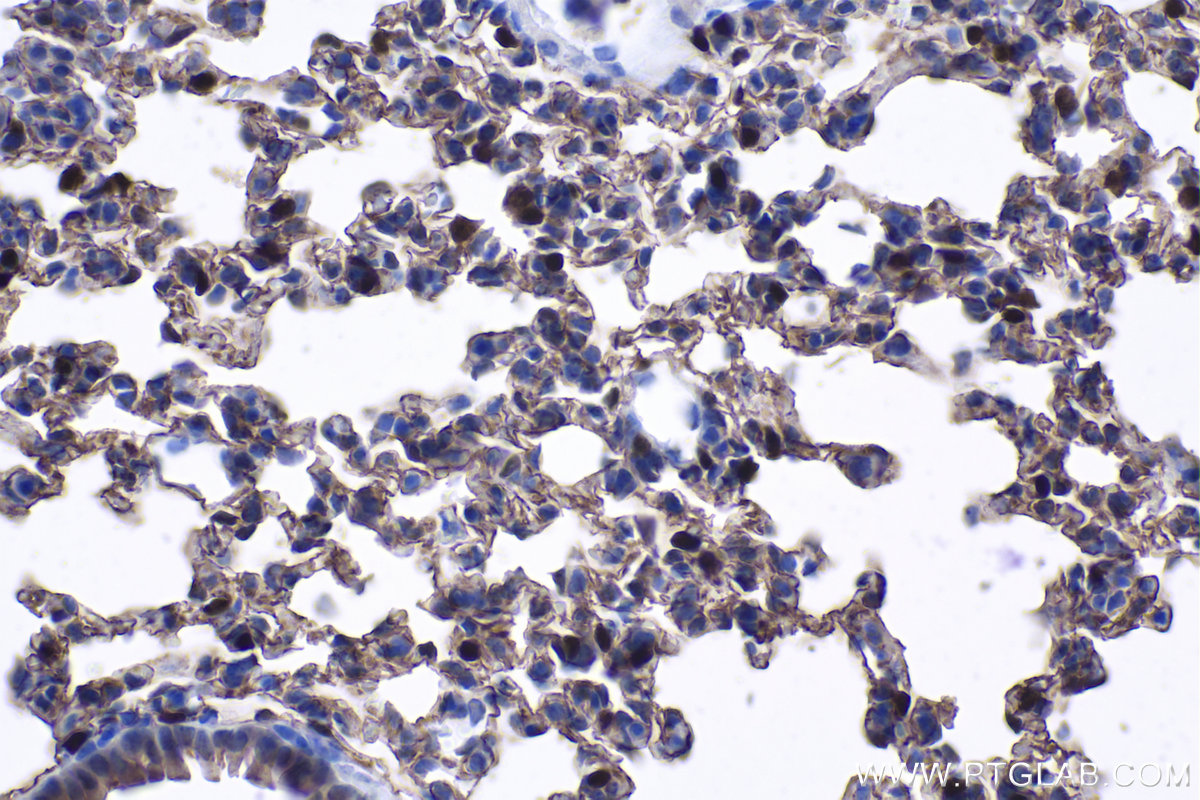
 at dilution of 1:4000 (under 10x lens). Heat mediated antigen retrieval with Tris-EDTA buffer (pH 9.0).")
 at dilution of 1:4000 (under 40x lens). Heat mediated antigen retrieval with Tris-EDTA buffer (pH 9.0).")
Tested Applications
| Positive WB detected in | mouse lung tissue, mouse small intestine tissue, rat lung tissue |
| Positive IHC detected in | human placenta tissue, mouse lung tissue Note: suggested antigen retrieval with TE buffer pH 9.0; (*) Alternatively, antigen retrieval may be performed with citrate buffer pH 6.0 |
Recommended dilution
| Application | Dilution |
|---|---|
| Western Blot (WB) | WB : 1:500-1:3000 |
| Immunohistochemistry (IHC) | IHC : 1:500-1:2000 |
| It is recommended that this reagent should be titrated in each testing system to obtain optimal results. | |
| Sample-dependent, Check data in validation data gallery. | |
Published Applications
| KD/KO | See 1 publications below |
| WB | See 10 publications below |
| IHC | See 11 publications below |
| IF | See 29 publications below |
Product Information
11419-1-AP targets HOPX in WB, IHC, IF, ELISA applications and shows reactivity with human, mouse, rat samples.
| Tested Reactivity | human, mouse, rat |
| Cited Reactivity | human, mouse, rat |
| Host / Isotype | Rabbit / IgG |
| Class | Polyclonal |
| Type | Antibody |
| Immunogen |
CatNo: Ag1979 Product name: Recombinant human HOPX protein Source: e coli.-derived, PGEX-4T Tag: GST Domain: 1-73 aa of BC014225 Sequence: MSAETASGPTEDQVEILEYNFNKVDKHPDSTTLCLIAAEAGLSEEETQKWFKQRLAKWRRSEGLPSECRSVID Predict reactive species |
| Full Name | HOP homeobox |
| Calculated Molecular Weight | 73 aa, 8 kDa |
| Observed Molecular Weight | 8-12 kDa |
| GenBank Accession Number | BC014225 |
| Gene Symbol | HOPX |
| Gene ID (NCBI) | 84525 |
| RRID | AB_10693525 |
| Conjugate | Unconjugated |
| Form | Liquid |
| Purification Method | Antigen affinity purification |
| UNIPROT ID | Q9BPY8 |
| Storage Buffer | PBS with 0.02% sodium azide and 50% glycerol, pH 7.3. |
| Storage Conditions | Store at -20°C. Stable for one year after shipment. Aliquoting is unnecessary for -20oC storage. 20ul sizes contain 0.1% BSA. |
Background Information
HOPX (Homeodomain-only protein) gene has various synonyms including HOD, HOP, OB1, LAGY and NECC1. The protein encoded by this gene is an unusual homeodomain protein that lacks certain conserved residues required for DNA binding. HOPX has diverse effects on cardiac growth. Manipulation of Hopx function in murine models is associated with cardiac hypertrophy, dilation and fibrosis. HOPX protein acts as an antagonist to the serum response factor (SRF), which regulates the opposing processes of cell proliferation and differentiation. Overexpression of HOPX causes cardiac hypertrophy. HOPX protein can inhibit SRF-dependent transcriptional activation by recruiting histone deacetylase (HDAC) activity.
Protocols
| Product Specific Protocols | |
|---|---|
| IHC protocol for HOPX antibody 11419-1-AP | Download protocol |
| WB protocol for HOPX antibody 11419-1-AP | Download protocol |
| Standard Protocols | |
|---|---|
| Click here to view our Standard Protocols |
Publications
| Species | Application | Title |
|---|---|---|
Cancer Cell Epigenomic State Transitions Characterize Tumor Progression in Mouse Lung Adenocarcinoma. | ||
Cancer Cell Control of alveolar differentiation by the lineage transcription factors GATA6 and HOPX inhibits lung adenocarcinoma metastasis. | ||
Cell Stem Cell LIF signaling regulates outer radial glial to interneuron fate during human cortical development | ||
Protein Cell BMP7 expression in mammalian cortical radial glial cells increases the length of the neurogenic period |
Reviews
The reviews below have been submitted by verified Proteintech customers who received an incentive for providing their feedback.
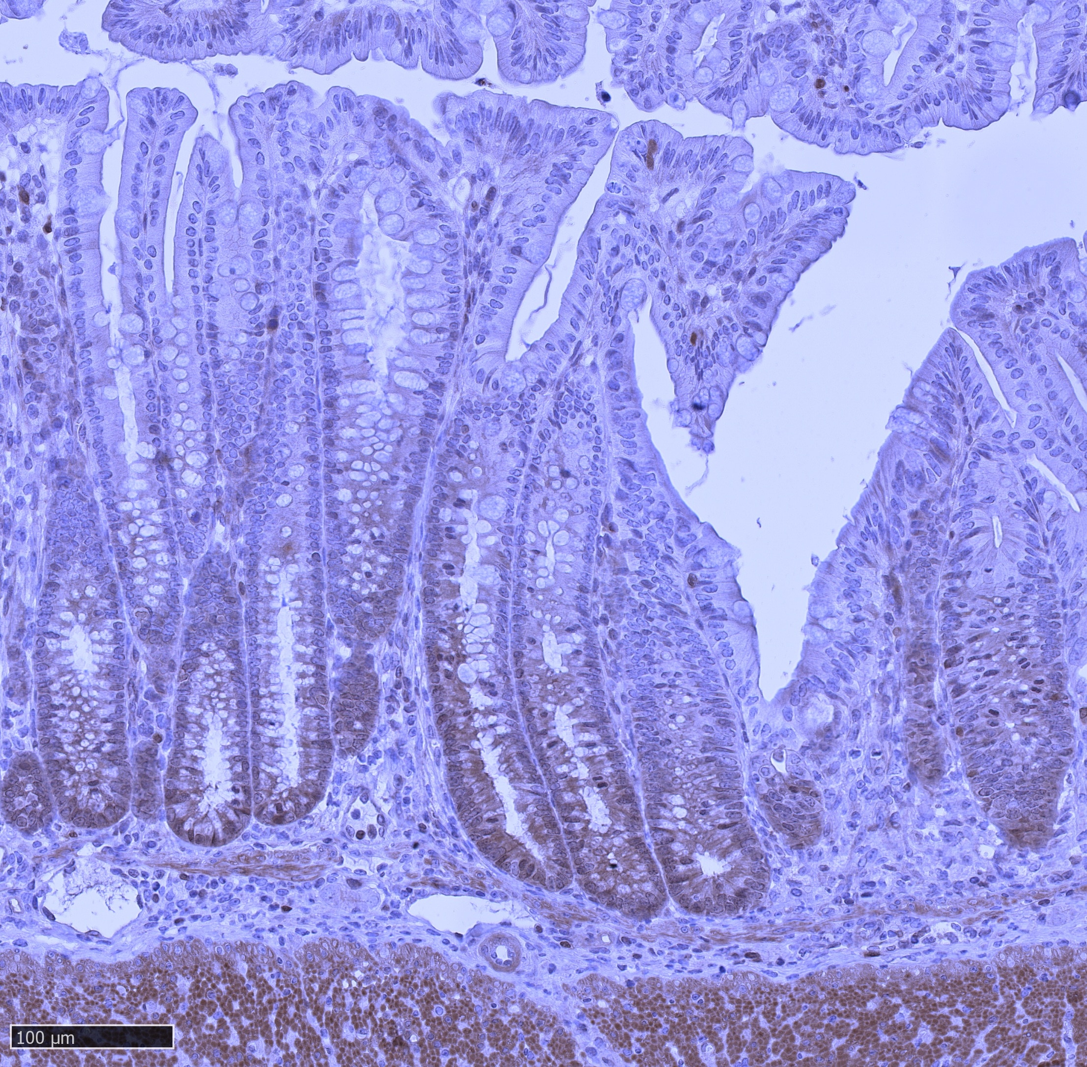
FH Bénédicte (Verified Customer) (02-13-2026) | IHC staining of mouse colon after induction of colitis using 11419-1-AP Immunohistochemical analysis of paraffin-embedded mouse colon tissue (mouse subjected to colitis) using 11419-1-AP (HOPX antibody) at dilution of 1:200 (30x on NDPview). Heat mediated antigen retrieval with Tris-EDTA buffer (pH 9.0).
 |
FH Nada (Verified Customer) (12-13-2024) | Performs well for immunoblotting and flow cytometry (performed internal validation prior to using)
|
FH Ana (Verified Customer) (11-10-2020) | We recently published with this paper: PubMed ID: 32819430 Images are within the manuscript.Protocol information: We used Citirc Acid Buffer Antigen Retrieval
 |
FH Nicole (Verified Customer) (07-23-2019) | Used to mark stem cell cultures for differentiation into neural like cells
|