Filter:
 at dilution of 1:300 incubated at room temperature for 1.5 hours.")
 at dilution of 1:300 incubated at room temperature for 1.5 hours.")
 at dilution of 1:300 incubated at room temperature for 1.5 hours.")
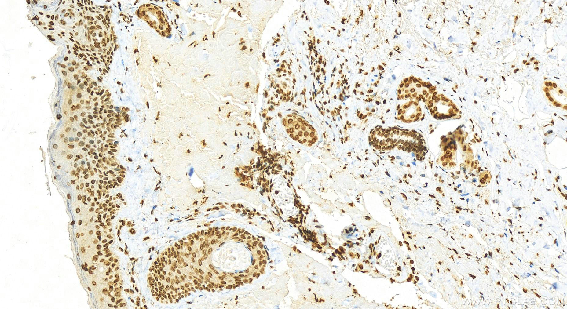
 at dilution of 1:200 (under 20x lens). Heat mediated antigen retrieval with Tris-EDTA buffer (pH 9.0).")
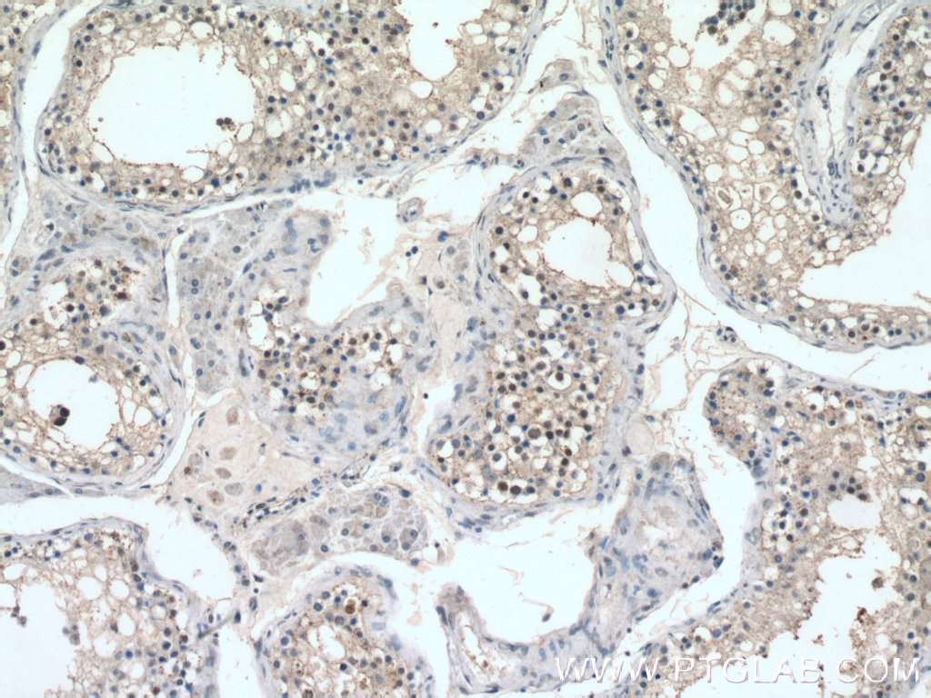
 at dilution of 1:200 (under 10x lens).")
 fixed HeLa cells using 16441-1-AP (Histone H2A.z antibody) at dilution of 1:50 and Alexa Fluor 488-conjugated AffiniPure Goat Anti-Rabbit IgG(H+L).")
Tested Applications
| Positive WB detected in | human brain tissue, mouse kidney tissue |
| Positive IHC detected in | human skin cancer tissue, human testis tissue Note: suggested antigen retrieval with TE buffer pH 9.0; (*) Alternatively, antigen retrieval may be performed with citrate buffer pH 6.0 |
| Positive IF/ICC detected in | HeLa cells |
Recommended dilution
| Application | Dilution |
|---|---|
| Western Blot (WB) | WB : 1:500-1:2000 |
| Immunohistochemistry (IHC) | IHC : 1:50-1:500 |
| Immunofluorescence (IF)/ICC | IF/ICC : 1:20-1:200 |
| It is recommended that this reagent should be titrated in each testing system to obtain optimal results. | |
| Sample-dependent, Check data in validation data gallery. | |
Published Applications
| KD/KO | See 1 publications below |
| WB | See 5 publications below |
| IF | See 3 publications below |
Product Information
16441-1-AP targets Histone H2A.z in WB, IHC, IF/ICC, ELISA applications and shows reactivity with human, mouse, rat samples.
| Tested Reactivity | human, mouse, rat |
| Cited Reactivity | human, mouse, fish, oratosquilla oratoria |
| Host / Isotype | Rabbit / IgG |
| Class | Polyclonal |
| Type | Antibody |
| Immunogen |
CatNo: Ag9762 Product name: Recombinant human Histone H2A.z protein Source: e coli.-derived, PGEX-4T Tag: GST Domain: 1-128 aa of BC018002 Sequence: MAGGKAGKDSGKAKTKAVSRSQRAGLQFPVGRIHRHLKSRTTSHGRVGATAAVYSAAILEYLTAEVLELAGNASKDLKVKRITPRHLQLAIRGDEELDSLIKATIAGGGVIPHIHKSLIGKKGQQKTV Predict reactive species |
| Full Name | H2A histone family, member Z |
| Calculated Molecular Weight | 128 aa, 14 kDa |
| Observed Molecular Weight | 14 kDa |
| GenBank Accession Number | BC018002 |
| Gene Symbol | Histone H2A.z |
| Gene ID (NCBI) | 3015 |
| RRID | AB_2115113 |
| Conjugate | Unconjugated |
| Form | Liquid |
| Purification Method | Antigen affinity purification |
| UNIPROT ID | P0C0S5 |
| Storage Buffer | PBS with 0.02% sodium azide and 50% glycerol, pH 7.3. |
| Storage Conditions | Store at -20°C. Stable for one year after shipment. Aliquoting is unnecessary for -20oC storage. 20ul sizes contain 0.1% BSA. |
Protocols
| Product Specific Protocols | |
|---|---|
| IF protocol for Histone H2A.z antibody 16441-1-AP | Download protocol |
| IHC protocol for Histone H2A.z antibody 16441-1-AP | Download protocol |
| WB protocol for Histone H2A.z antibody 16441-1-AP | Download protocol |
| Standard Protocols | |
|---|---|
| Click here to view our Standard Protocols |
Publications
| Species | Application | Title |
|---|---|---|
J Biol Chem The chromatin remodeler protein Chd4 maintains embryonic stem cell identity by controlling pluripotency- and differentiation-associated genes.
| ||
Biomed Pharmacother Limonin enhances the radiosensitivity of nasopharyngeal carcinoma cells via attenuating Stat3-induced cell stemness. | ||
J Morphol Ultrastructure of spermiogenesis and the distribution of spermatozoal nuclear histones in the Japanese mantis shrimp, Oratosquilla oratoria (Crustacea: Stomatopoda). | ||
J Biol Chem Role of noncanonical histone H2A variant, H2A.Z, to maintain proper centromeric transcription and chromosome segregation |