

Tested Applications
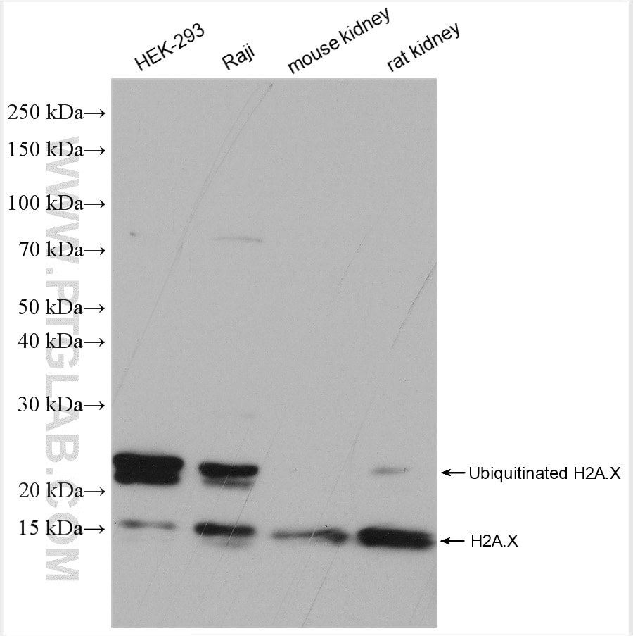
| Positive WB detected in | HEK-293 cells, HL-60 cells, HEK-293 ells, mouse heart, mouse kidney, rat kidney |
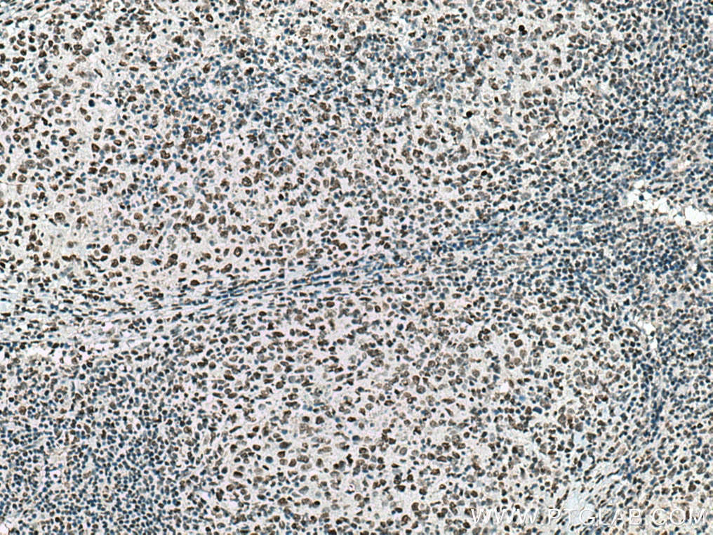
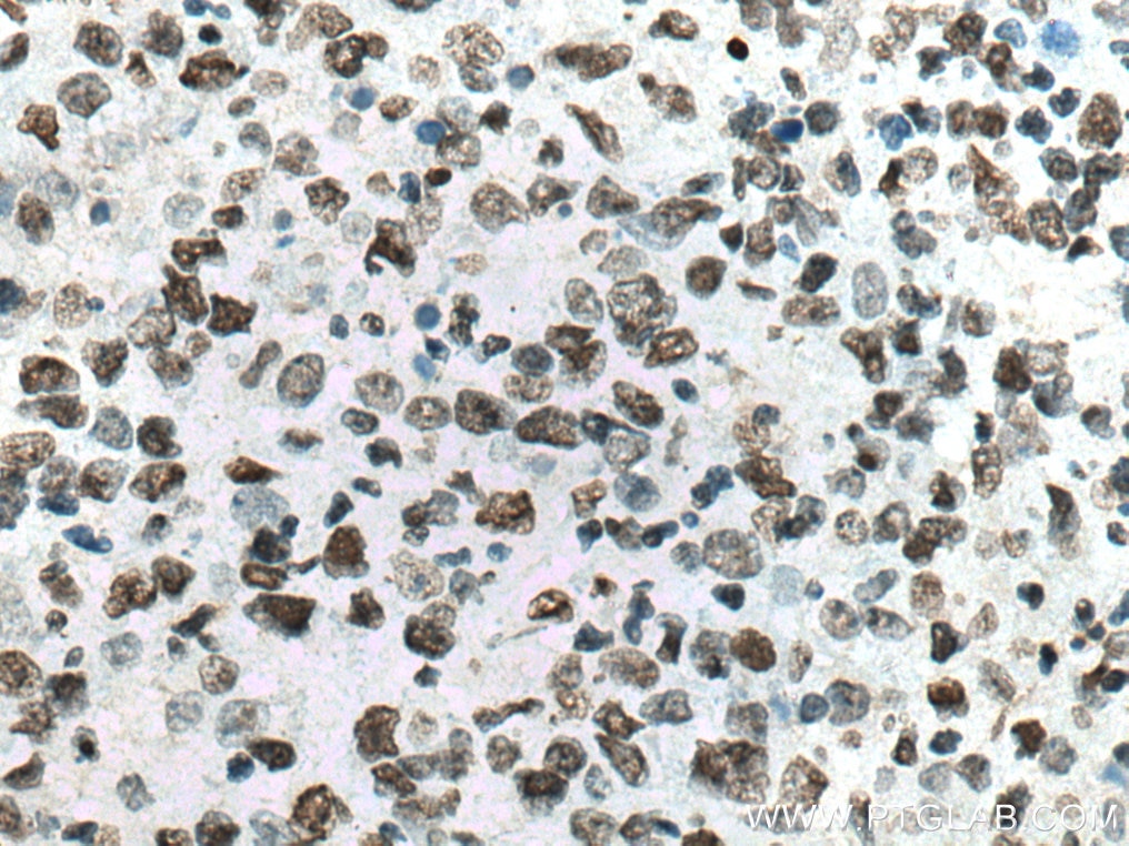
| Positive IHC detected in | human lymphoma tissue Note: suggested antigen retrieval with TE buffer pH 9.0; (*) Alternatively, antigen retrieval may be performed with citrate buffer pH 6.0 |
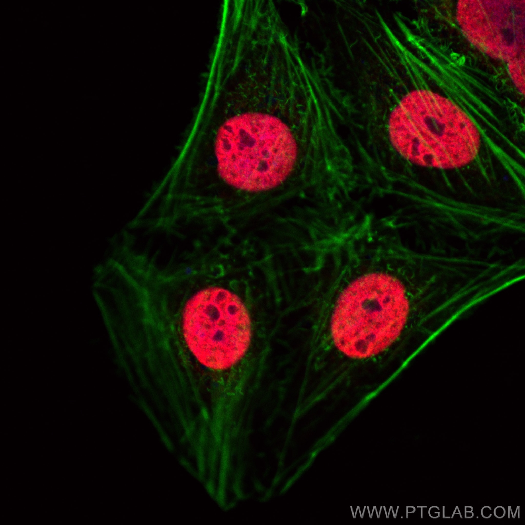
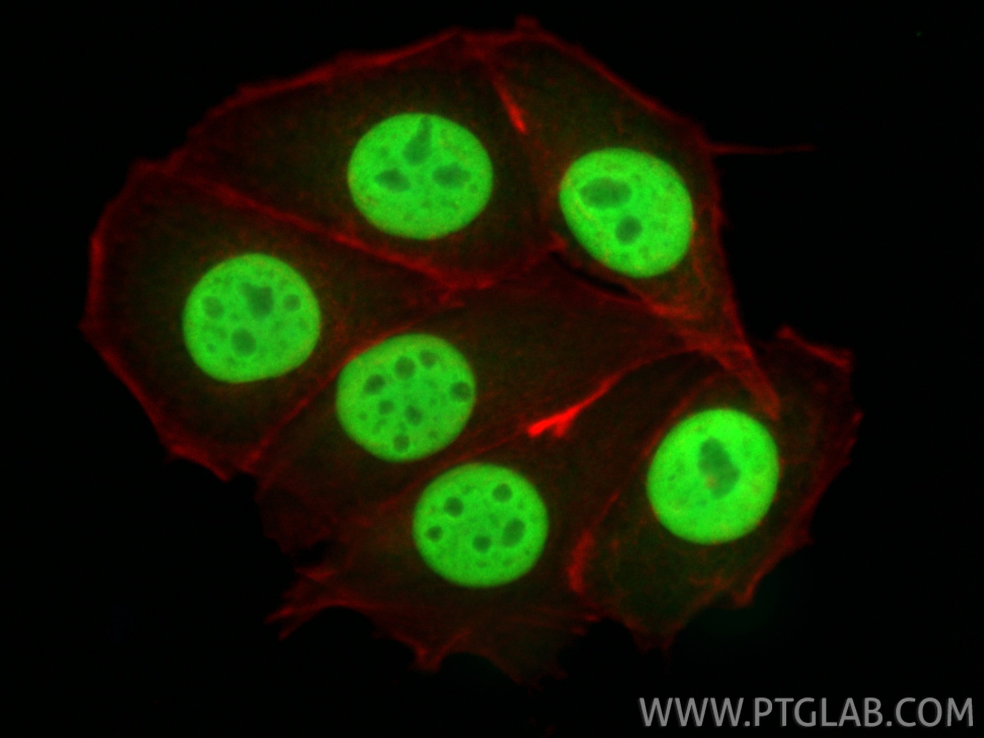
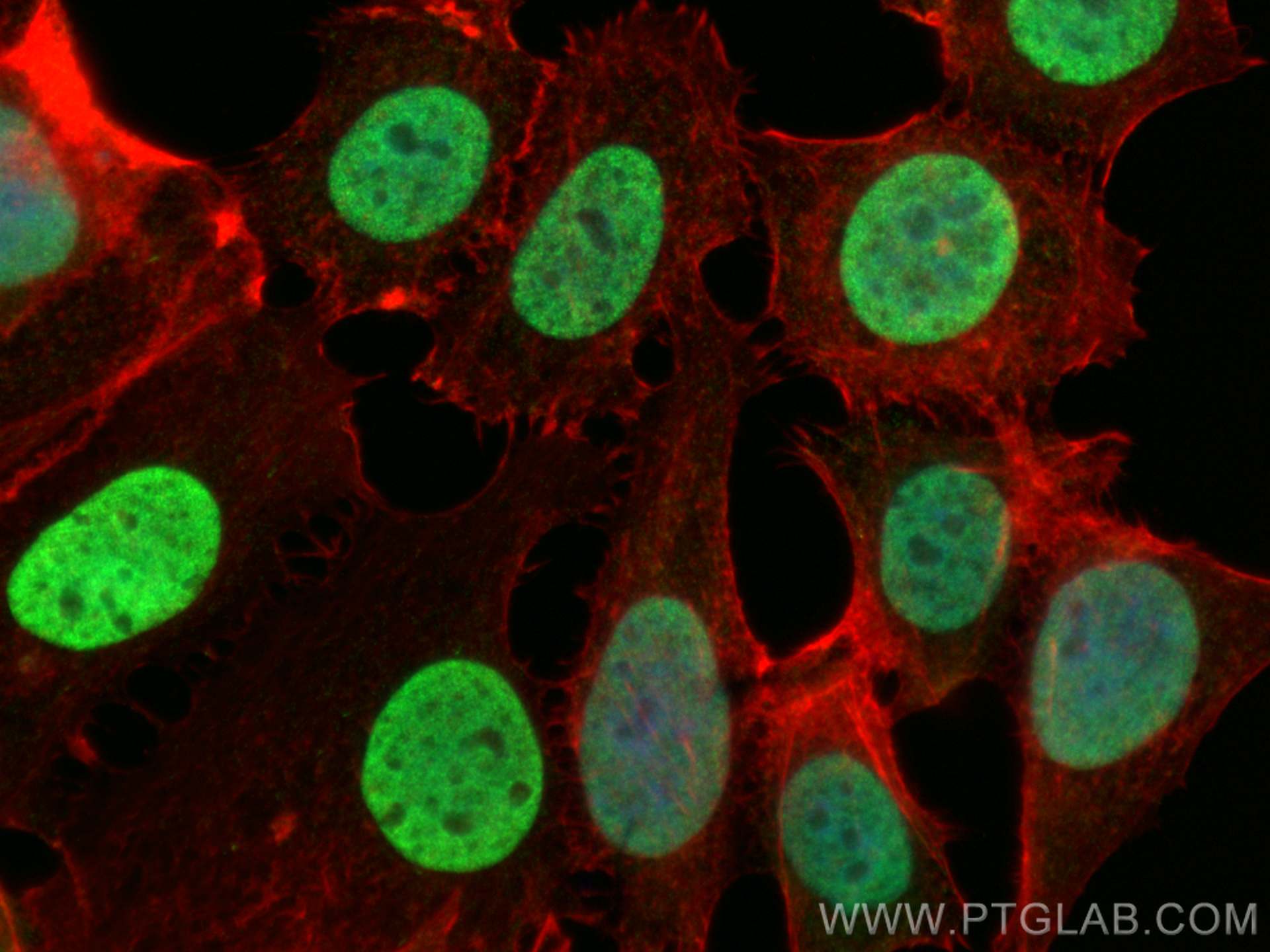
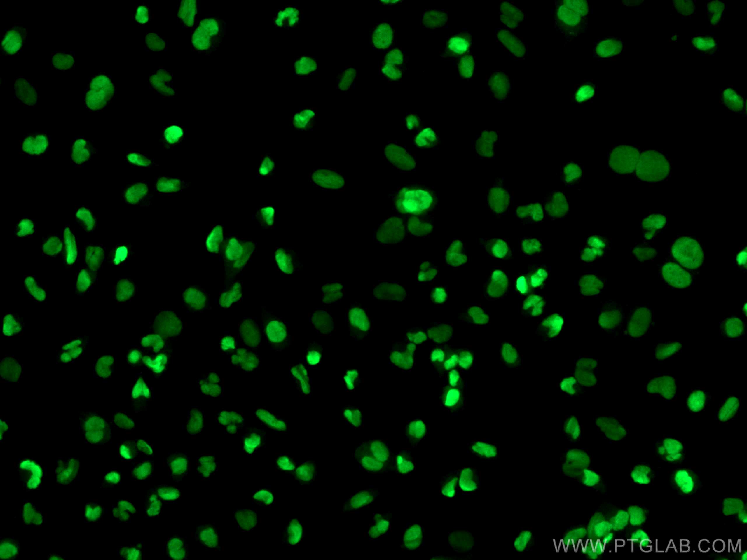
| Positive IF/ICC detected in | MCF-7 cells, U-251 cells, U2OS cells |
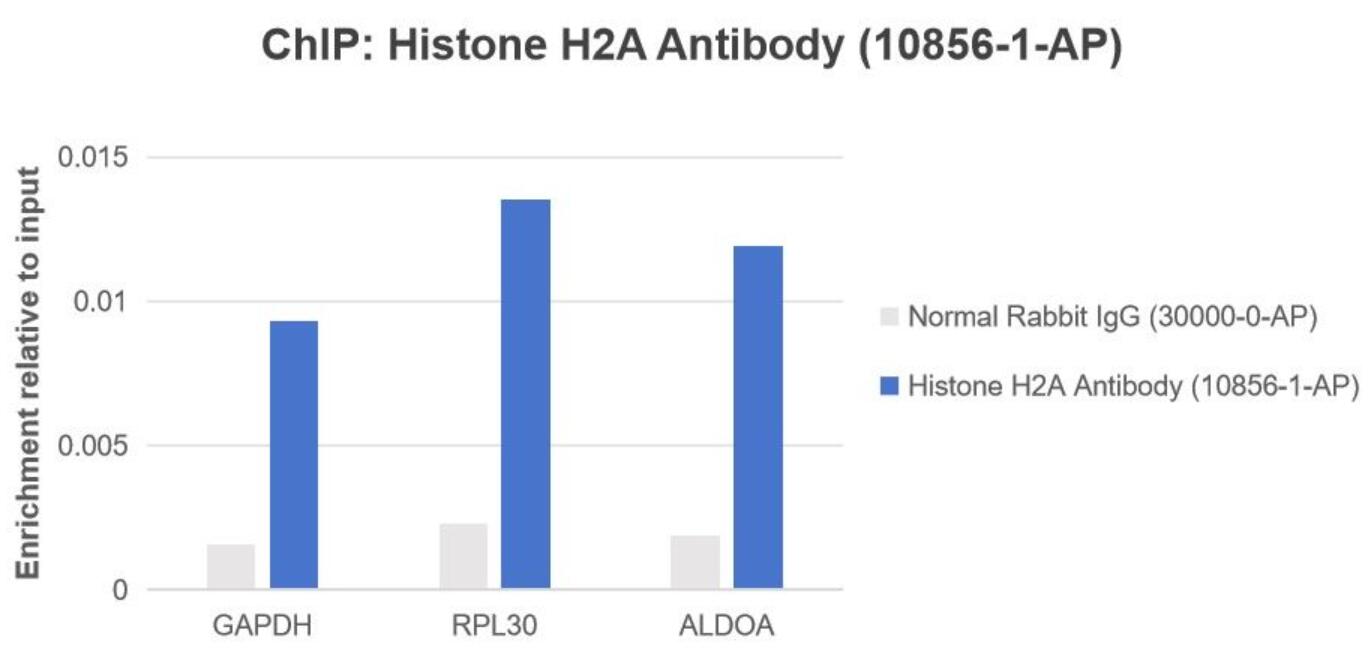
| Positive ChIP detected in | HEK-293 cells |
Recommended dilution
| Application | Dilution |
|---|---|
| Western Blot (WB) | WB : 1:1000-1:4000 |
| Immunohistochemistry (IHC) | IHC : 1:50-1:500 |
| Immunofluorescence (IF)/ICC | IF/ICC : 1:1000-1:4000 |
| Chromatin immunoprecipitation (ChIP) | CHIP : 1:10-1:100 |
| It is recommended that this reagent should be titrated in each testing system to obtain optimal results. | |
| Sample-dependent, Check data in validation data gallery. | |
Product Information
10856-1-AP targets Histone H2A.X in WB, IHC, IF/ICC, CoIP, ChIP, ELISA applications and shows reactivity with human, mouse, rat samples.
| Tested Reactivity | human, mouse, rat |
| Cited Reactivity | human, mouse, rat, pig, chicken, zebrafish |
| Host / Isotype | Rabbit / IgG |
| Class | Polyclonal |
| Type | Antibody |
| Immunogen |
CatNo: Ag1305 Product name: Recombinant human Histone H2A.X protein Source: e coli.-derived, PGEX-4T Tag: GST Domain: 1-143 aa of BC013416 Sequence: MSGRGKTGGKARAKAKSRSSRAGLQFPVGRVHRLLRKGHYAERVGAGAPVYLAAVLEYLTAEILELAGNAARDNKKTRIIPRHLQLAIRNDEELNKLLGGVTIAQGGVLPNIQAVLLPKKTSATVGPKAPSGGKKATQASQEY Predict reactive species |
| Full Name | H2A histone family, member X |
| Calculated Molecular Weight | 15 kDa |
| Observed Molecular Weight | 15-18 kDa |
| GenBank Accession Number | BC013416 |
| Gene Symbol | Histone H2A.X |
| Gene ID (NCBI) | 3014 |
| RRID | AB_2114985 |
| Conjugate | Unconjugated |
| Form | Liquid |
| Purification Method | Antigen affinity purification |
| UNIPROT ID | P16104 |
| Storage Buffer | PBS with 0.02% sodium azide and 50% glycerol, pH 7.3. |
| Storage Conditions | Store at -20°C. Stable for one year after shipment. Aliquoting is unnecessary for -20oC storage. 20ul sizes contain 0.1% BSA. |
Background Information
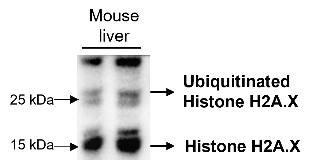
Histone H2A.X belongs to the histone H2A family, which is synthesized in G1 and S phase. It is involved in nucleosomal organization of chromatin together with other histone proteins, and is specially important for recombination between immunoglobulin switch regions. H2A.X becomes phosphorylated on serine 139 (to form gamma-H2AFX or H2AX139ph) in response to DNA double strand breaks (DSBs) generated by exogenous genotoxic agents and by stalled replication forks, which promotes DNA repair and maintains genomic stability. The calculated molecular weight of H2AX is 15 kDa, but the ubiquitinated H2A.X is about 22 kDa.
Protocols
| Product Specific Protocols | |
|---|---|
| IF protocol for Histone H2A.X antibody 10856-1-AP | Download protocol |
| IHC protocol for Histone H2A.X antibody 10856-1-AP | Download protocol |
| WB protocol for Histone H2A.X antibody 10856-1-AP | Download protocol |
| Standard Protocols | |
|---|---|
| Click here to view our Standard Protocols |
Publications
What published studies show
The anti-phospho-H2AX antibody (Cat# 10856-1-AP) has accumulated 186 citations across diverse journals including Nature Communications, Nature Neuroscience, Cell Metabolism, Nucleic Acids Research, Advanced Science, Cancer Research, Theranostics, Biomaterials, and EMBO Journal. Research fields span DNA damage and repair, oncology, oxidative stress, ferroptosis, radiation therapy, cellular senescence and aging, neurodegeneration, immunology, stem cell biology, cardiovascular disease, renal fibrosis, reproductive biology, toxicology, and nanomedicine-based drug delivery systems.
| Species | Application | Title |
|---|---|---|
Cell Metab Sensing steroid hormone 17α-hydroxypregnenolone by GPR56 enables protection from ferroptosis-induced liver injury | ||
Adv Mater Selective Organ-Targeting Hafnium Oxide Nanoparticles with Multienzyme-Mimetic Activities Attenuate Radiation-Induced Tissue Damage | ||
Nat Aging Single-cell and spatial RNA sequencing identify divergent microenvironments and progression signatures in early- versus late-onset prostate cancer | ||
Cell Mol Immunol Mapping the holonomic signaling network that drives pathological changes in endotoxic shock. | ||
Cancer Res ZMYND8 Expression in Breast Cancer Cells Blocks T-Lymphocyte Surveillance to Promote Tumor Growth. | ||
Cancer Res Combined inactivation of CTPS1 and ATR is synthetically lethal to MYC-overexpressing cancer cells. |
Reviews
What customers say
All three reviewers used this antibody for Western Blot and reported positive results. One user achieved clear 15 kDa bands in mouse liver tissue at 1:2000 dilution. Another obtained strong signal in HEK293FT cells at 1:3000 and recommended adding benzonase to the lysis buffer to recover chromatin-bound material. A third user confirmed good performance at 1:1000 in liver cell lines using milk/TBST blocking buffer. Overall, users rate it highly for sensitivity and specificity across different sample types and dilutions.
The reviews below have been submitted by verified Proteintech customers who received an incentive for providing their feedback.
Kamal (Verified Customer) (02-27-2025) | Mouse liver lysates were subjected to SDS-PAGE and immunoblotted with Histone H2A.X Polyclonal antibody (1:2000 in 1XTBST). Bands appeared at 15 kDa.
 |
Andreas (Verified Customer) (03-23-2022) | Antibody gives very good signal, hence the dilution factor. One important note is that I include 1:2000 dilution of benzonase in my lysis buffer to recover most of the chromatin-bound material, which big part of it is otherwise lost during the clarification step.
|
kes (Verified Customer) (01-17-2022) | used for WB (1:1000) in milk+TBST buffer and got good results
|


