at dilution of 1:5000 incubated at room temperature for 1.5 hours.")
 at dilution of 1:5000 incubated at room temperature for 1.5 hours.")
 at dilution of 1:5000 incubated at room temperature for 1.5 hours.")
 at dilution of 1:5000 incubated at room temperature for 1.5 hours.")
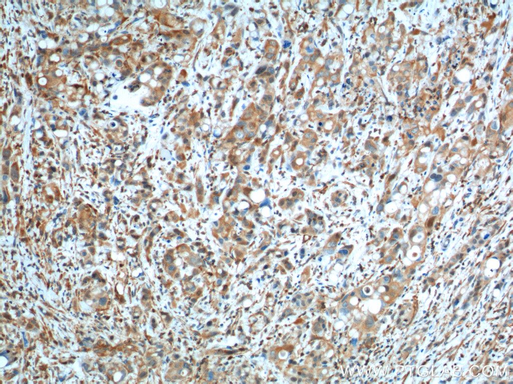
 at dilution of 1:200 (under 10x lens). Heat mediated antigen retrieval with Tris-EDTA buffer (pH 9.0).")
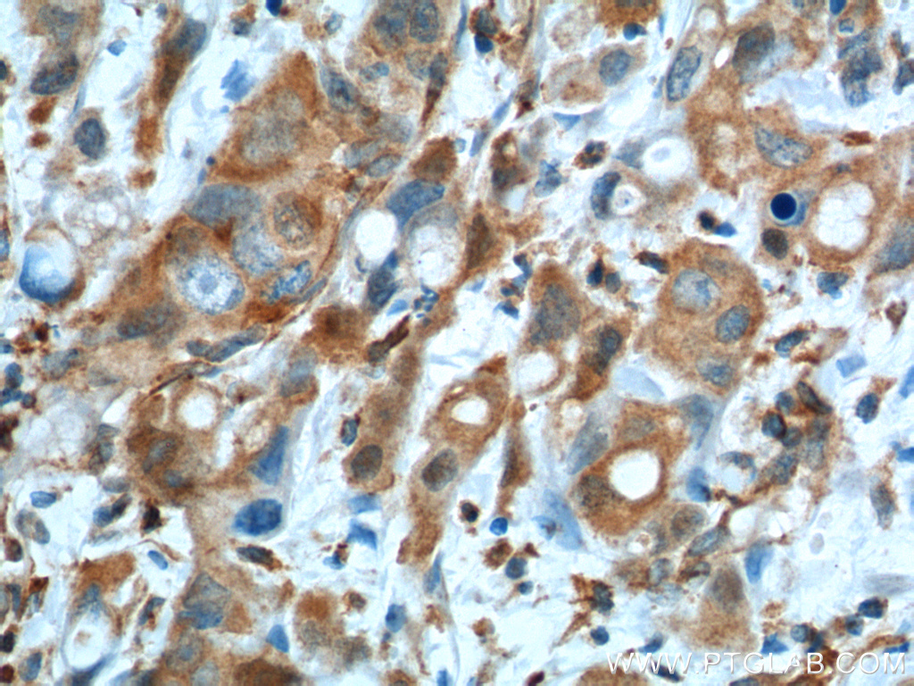
 at dilution of 1:200 (under 40x lens). Heat mediated antigen retrieval with Tris-EDTA buffer (pH 9.0).")
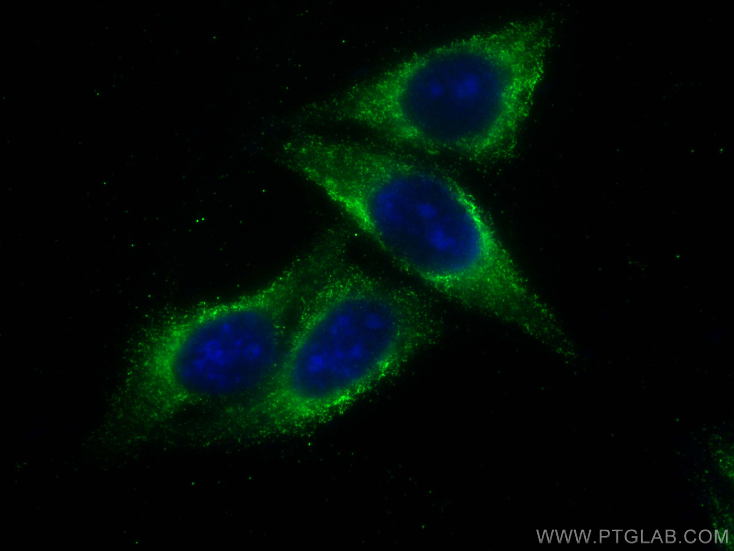
 fixed HepG2 cells using GOT1 antibody (60317-1-Ig, Clone: 5A12E10 ) at dilution of 1:400 and CoraLite®488-Conjugated Goat Anti-Mouse IgG(H+L).")
Tested Applications
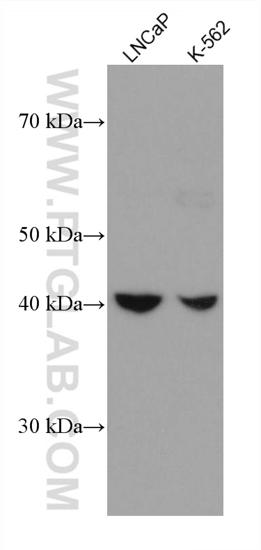
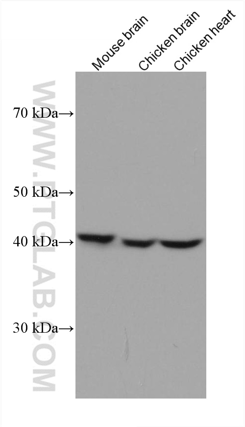
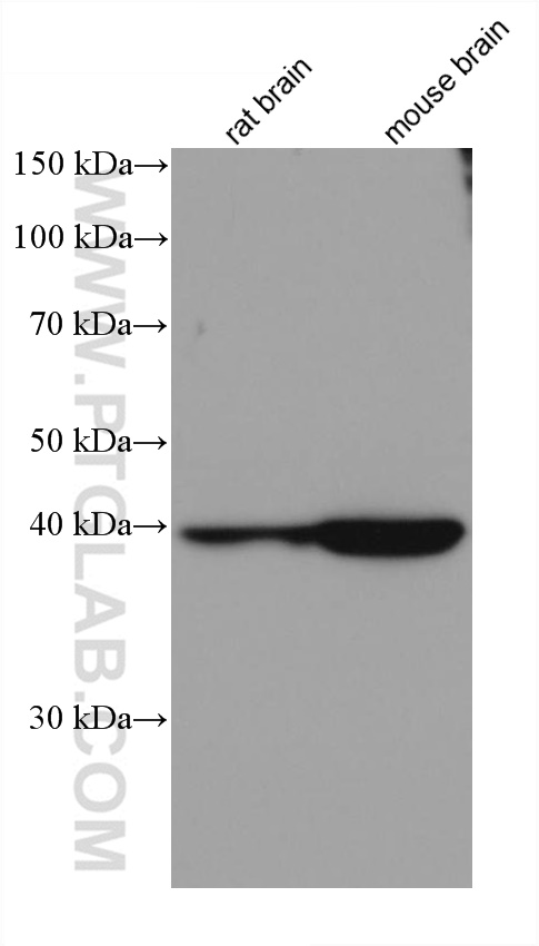
| Positive WB detected in | LNCaP cells, rat brain tissue, rat heart tissue, mouse brain tissue, K-562 cells, chicken brain, chicken heart |
| Positive IHC detected in | human liver cancer tissue Note: suggested antigen retrieval with TE buffer pH 9.0; (*) Alternatively, antigen retrieval may be performed with citrate buffer pH 6.0 |
| Positive IF/ICC detected in | HepG2 cells |
Recommended dilution
| Application | Dilution |
|---|---|
| Western Blot (WB) | WB : 1:2000-1:10000 |
| Immunohistochemistry (IHC) | IHC : 1:20-1:200 |
| Immunofluorescence (IF)/ICC | IF/ICC : 1:200-1:800 |
| It is recommended that this reagent should be titrated in each testing system to obtain optimal results. | |
| Sample-dependent, Check data in validation data gallery. | |
Published Applications
| KD/KO | See 2 publications below |
| WB | See 4 publications below |
| IF | See 1 publications below |
| IP | See 1 publications below |
Product Information
60317-1-Ig targets GOT1 in WB, IHC, IF/ICC, IP, ELISA applications and shows reactivity with human, mouse, rat, chicken samples.
| Tested Reactivity | human, mouse, rat, chicken |
| Cited Reactivity | human, mouse |
| Host / Isotype | Mouse / IgG1 |
| Class | Monoclonal |
| Type | Antibody |
| Immunogen |
CatNo: Ag7012 Product name: Recombinant human GOT1 protein Source: e coli.-derived, PET28a Tag: 6*His Domain: 1-413 aa of BC000498 Sequence: MAPPSVFAEVPQAQPVLVFKLTADFREDPDPRKVNLGVGAYRTDDCHPWVLPVVKKVEQKIANDNSLNHEYLPILGLAEFRSCASRLALGDDSPALKEKRVGGVQSLGGTGALRIGADFLARWYNGTNNKNTPVYVSSPTWENHNAVFSAAGFKDIRSYRYWDAEKRGLDLQGFLNDLENAPEFSIVVLHACAHNPTGIDPTPEQWKQIASVMKHRFLFPFFDSAYQGFASGNLERDAWAIRYFVSEGFEFFCAQSFSKNFGLYNERVGNLTVVGKEPESILQVLSQMEKIVRITWSNPPAQGARIVASTLSNPELFEEWTGNVKTMADRILTMRSELRARLEALKTPGTWNHITDQIGMFSFTGLNPKQVEYLVNEKHIYLLPSGRINVSGLTTKNLDYVATSIHEAVTKIQ Predict reactive species |
| Full Name | glutamic-oxaloacetic transaminase 1, soluble (aspartate aminotransferase 1) |
| Calculated Molecular Weight | 46 kDa |
| Observed Molecular Weight | 40-46 kDa |
| GenBank Accession Number | BC000498 |
| Gene Symbol | GOT1 |
| Gene ID (NCBI) | 2805 |
| RRID | AB_2881428 |
| Conjugate | Unconjugated |
| Form | Liquid |
| Purification Method | Protein G purification |
| UNIPROT ID | P17174 |
| Storage Buffer | PBS with 0.02% sodium azide and 50% glycerol, pH 7.3. |
| Storage Conditions | Store at -20°C. Stable for one year after shipment. Aliquoting is unnecessary for -20oC storage. 20ul sizes contain 0.1% BSA. |
Background Information
Glutamate oxaloacetate transaminase 1 (GOT1) catalyzes the reversible reaction of L-aspartate and alpha-ketoglutarate into oxaloacetate and L-glutamate and plays a key role in carbon and nitrogen metabolism. GOT1 can potentially control the intracellular levels of reactive oxygen species (ROS) through NADPH synthesis and enhances tumor growth. GOT1 expression correlates with the growth of several tumors.
Protocols
| Product Specific Protocols | |
|---|---|
| IF protocol for GOT1 antibody 60317-1-Ig | Download protocol |
| IHC protocol for GOT1 antibody 60317-1-Ig | Download protocol |
| WB protocol for GOT1 antibody 60317-1-Ig | Download protocol |
| Standard Protocols | |
|---|---|
| Click here to view our Standard Protocols |
Publications
| Species | Application | Title |
|---|---|---|
Free Radic Biol Med Sulfur dioxide controls M1 macrophage polarization by sulphenylation of prolyl hydroxylase 2 at cysteine 260
| ||
J Cell Mol Med HIF-2α regulates non-canonical glutamine metabolism via activation of PI3K/mTORC2 pathway in human pancreatic ductal adenocarcinoma. | ||
Antioxid Redox Signal Sulfur Dioxide Alleviates Aortic Dissection Through Inhibiting Vascular Smooth Muscle Cell Phenotype Switch, Migration, and Proliferation via miR-184-3p/Cyp26b1 Axis | ||
Nat Cell Biol De novo pyrimidine biosynthetic complexes support cancer cell proliferation and ferroptosis defence
| ||
Mol Cell Functional nutrient-genetic profiling reveals biotin and FBXW7 are essential to bypass glutamine addiction |