
 at dilution of 1:600 incubated at room temperature for 1.5 hours.")
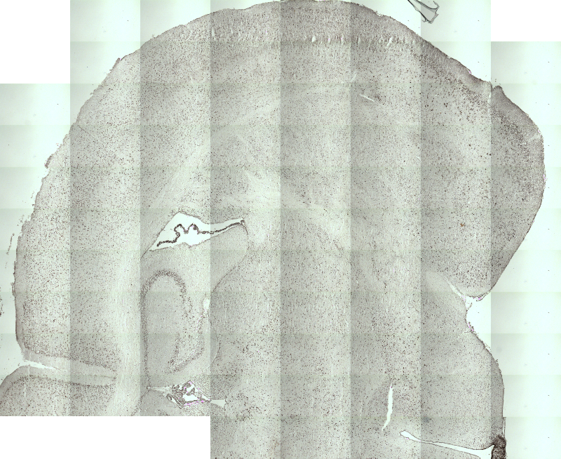
 at dilution of 1:200 (under 10x lens). Heat mediated antigen retrieval with Tris-EDTA buffer (pH 9.0).")
 at dilution of 1:200 (under 40x lens). Heat mediated antigen retrieval with Tris-EDTA buffer (pH 9.0).")
 at dilution of 1:200 (under 10x lens). Heat mediated antigen retrieval with Tris-EDTA buffer (pH 9.0).")
 at dilution of 1:200 (under 40x lens). Heat mediated antigen retrieval with Tris-EDTA buffer (pH 9.0).")
Tested Applications
| Positive WB detected in | RAW 264.7 cells |
| Positive IHC detected in | mouse brain tissue, rat brain tissue Note: suggested antigen retrieval with TE buffer pH 9.0; (*) Alternatively, antigen retrieval may be performed with citrate buffer pH 6.0 |
Recommended dilution
| Application | Dilution |
|---|---|
| Western Blot (WB) | WB : 1:500-1:1000 |
| Immunohistochemistry (IHC) | IHC : 1:50-1:500 |
| It is recommended that this reagent should be titrated in each testing system to obtain optimal results. | |
| Sample-dependent, Check data in validation data gallery. | |
Published Applications
| WB | See 13 publications below |
| IHC | See 11 publications below |
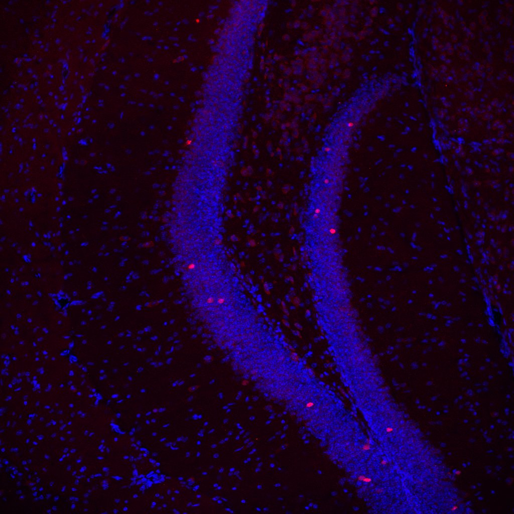
| IF | See 10 publications below |
Product Information
26192-1-AP targets c-Fos in WB, IHC, IF-P, ELISA applications and shows reactivity with human, mouse, rat samples.
| Tested Reactivity | human, mouse, rat |
| Cited Reactivity | human, mouse, rat, canine |
| Host / Isotype | Rabbit / IgG |
| Class | Polyclonal |
| Type | Antibody |
| Immunogen |
CatNo: Ag24340 Product name: Recombinant human FOS protein Source: e coli.-derived, PGEX-4T Tag: GST Domain: 196-341 aa of BC004490 Sequence: ILAAHRPACKIPDDLGFPEEMSVASLDLTGGLPEVATPESEEAFTLPLLNDPEPKPSVEPVKSISSMELKTEPFDDFLFPASSRPSGSETARSVPDMDLSGSFYAADWEPLHSGSLGMGPMATELEPLCTPVVTCTPSCTAYTSSF Predict reactive species |
| Full Name | FOS |
| Calculated Molecular Weight | 41 kDa |
| Observed Molecular Weight | 65 kDa |
| GenBank Accession Number | BC004490 |
| Gene Symbol | c-Fos |
| Gene ID (NCBI) | 2353 |
| Conjugate | Unconjugated |
| Form | Liquid |
| Purification Method | Antigen affinity purification |
| UNIPROT ID | P01100 |
| Storage Buffer | PBS with 0.02% sodium azide and 50% glycerol, pH 7.3. |
| Storage Conditions | Store at -20°C. Stable for one year after shipment. Aliquoting is unnecessary for -20oC storage. 20ul sizes contain 0.1% BSA. |
Background Information
c-Fos, also named as FOS and G0/G1 switch regulatory protein 7, is a 380 amino acid protein, which contains 1 bZIP (basic-leucine zipper) domain and belongs to the bZIP family. c-Fos is expressed at very low levels in quiescent cells. When cells are stimulated to reenter growth, c-Fos undergo 2 waves of expression, the first one peaks 7.5 minutes following FBS induction. At this stage, the c-Fos protein is localized endoplasmic reticulum. The second wave of expression occurs at about 20 minutes after induction and peaks at 1 hour. At this stage, the c-FOS protein becomes nuclear. c-Fos is a very short-lived intracellular protein, which is very easy to degrade. The calculated molecular weight of c-Fos is 40 kDa, but Phosphorylated c-Fos protein is about 60-65 kDa. It is involved in important cellular events, including cell proliferation, differentiation and survival; genes associated with hypoxia; and angiogenesis; which makes its dysregulation an important factor for cancer development. It can also induce a loss of cell polarity and epithelial-mesenchymal transition, leading to invasive and metastatic growth in mammary epithelial cells. Expression of c-Fos is an indirect marker of neuronal activity because c-Fos is often expressed when neurons fire action potentials. Upregulation of c-Fos mRNA in a neuron indicates recent activity.
Publications
| Species | Application | Title |
|---|---|---|
Neuron Astrocytic ApoE reprograms neuronal cholesterol metabolism and histone-acetylation-mediated memory. | ||
Nat Commun Ketamine activates adult-born immature granule neurons to rapidly alleviate depression-like behaviors in mice. | ||
Biol Psychiatry Fighting Females: Neural and Behavioral Consequences of Social Defeat Stress in Female Mice. | ||
Reviews
The reviews below have been submitted by verified Proteintech customers who received an incentive for providing their feedback.
FH Matthias (Verified Customer) (09-10-2019) | Good Antibody, very specific.
 |
FH Elif (Verified Customer) (06-21-2019) | It works better with 2 overnight incubation in 4 degrees.
 |


