PC-3 cells and LNCap cells were subjected to SDS PAGE followed by western blot with 16864-1-AP (FMO5-specific antibody) at dilution of 1:800 incubated at room temperature for 1.5 hours.
PC-3 cells and LNCap cells were subjected to SDS PAGE followed by western blot with 16864-1-AP (FMO5-specific antibody) at dilution of 1:800 incubated at room temperature for 1.5 hours.
WB analysis using 16864-1-AP
Various lysates were subjected to SDS PAGE followed by western blot with 16864-1-AP (FMO5-specific antibody) at dilution of 1:3000 incubated at room temperature for 1.5 hours.
Various lysates were subjected to SDS PAGE followed by western blot with 16864-1-AP (FMO5-specific antibody) at dilution of 1:3000 incubated at room temperature for 1.5 hours.
Immunohistochemical analysis of paraffin-embedded human liver tissue slide using 16864-1-AP (FMO5-specific antibody) at dilution of 1:400 (under 10x lens). Heat mediated antigen retrieval with Tris-EDTA buffer (pH 9.0).
Immunohistochemical analysis of paraffin-embedded human liver tissue slide using 16864-1-AP (FMO5-specific antibody) at dilution of 1:400 (under 10x lens). Heat mediated antigen retrieval with Tris-EDTA buffer (pH 9.0).
IHC staining of human liver using 16864-1-AP
Immunohistochemical analysis of paraffin-embedded human liver tissue slide using 16864-1-AP (FMO5-specific antibody) at dilution of 1:400 (under 40x lens). Heat mediated antigen retrieval with Tris-EDTA buffer (pH 9.0).
Immunohistochemical analysis of paraffin-embedded human liver tissue slide using 16864-1-AP (FMO5-specific antibody) at dilution of 1:400 (under 40x lens). Heat mediated antigen retrieval with Tris-EDTA buffer (pH 9.0).
IHC staining of mouse liver using 16864-1-AP
Immunohistochemical analysis of paraffin-embedded mouse liver tissue slide using 16864-1-AP (FMO5-specific antibody) at dilution of 1:400 (under 10x lens). Heat mediated antigen retrieval with Tris-EDTA buffer (pH 9.0).
Immunohistochemical analysis of paraffin-embedded human breast cancer using 16864-1-AP (FMO5-specific antibody) at dilution of 1:50 (under 40x lens).
IF Staining of MCF-7 using 16864-1-AP
Immunofluorescent analysis of (-20℃ Ethanol) fixed MCF-7 cells using 16864-1-AP (FMO5-specific antibody) at dilution of 1:50 and Alexa Fluor 488-conjugated Goat Anti-Rabbit IgG(H+L).
Immunofluorescent analysis of (-20℃ Ethanol) fixed MCF-7 cells using 16864-1-AP (FMO5-specific antibody) at dilution of 1:50 and Alexa Fluor 488-conjugated Goat Anti-Rabbit IgG(H+L).
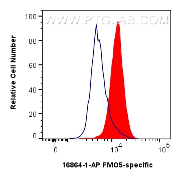
FC experiment of MCF-7 using 16864-1-AP
1x10^6 MCF-7 cells were intracellularly stained with 0.4 ug FMO5-specific Polyclonal antibody (16864-1-AP) and CoraLite®488-Conjugated Goat Anti-Rabbit IgG(H+L) (SA00013-2)(red), or 0.4 ug Rabbit IgG control Rabbit PolyAb (30000-0-AP) (blue). Cells were fixed with 4% PFA and permeabilized with Flow Cytometry Perm Buffer (PF00011-C).
1x10^6 MCF-7 cells were intracellularly stained with 0.4 ug FMO5-specific Polyclonal antibody (16864-1-AP) and CoraLite®488-Conjugated Goat Anti-Rabbit IgG(H+L) (SA00013-2)(red), or 0.4 ug Rabbit IgG control Rabbit PolyAb (30000-0-AP) (blue). Cells were fixed with 4% PFA and permeabilized with Flow Cytometry Perm Buffer (PF00011-C).
The Proteintech guarantee covers Proteintech antibodies in any species and any application, including those not listed on the datasheet. If the antibody doesn’t perform, you can receive a hassle-free refund or credit note.
PC-3 cells, mouse liver tissue, DU 145 cells, LNCaP cells, VCaP cells, RAW 264.7 cells, MCF-7 cells, rat liver tissue
Positive IHC detected in
human liver tissue, mouse liver tissue, human breast cancer tissue Note: suggested antigen retrieval with TE buffer pH 9.0; (*) Alternatively, antigen retrieval may be performed with citrate buffer pH 6.0
Positive IF/ICC detected in
MCF-7 cells
Positive FC (Intra) detected in
MCF-7 cells
Recommended dilution
Application
Dilution
Western Blot (WB)
WB : 1:500-1:1000
Immunohistochemistry (IHC)
IHC : 1:200-1:800
Immunofluorescence (IF)/ICC
IF/ICC : 1:50-1:500
Flow Cytometry (FC) (INTRA)
FC (INTRA) : 0.40 ug per 10^6 cells in a 100 µl suspension
It is recommended that this reagent should be titrated in each testing system to obtain optimal results.
Sample-dependent, Check data in validation data gallery.
PBS with 0.02% sodium azide and 50% glycerol , pH 7.3
Storage Conditions
Store at -20°C. Stable for one year after shipment. Aliquoting is unnecessary for -20oC storage. 20ul sizes contain 0.1% BSA.
Background Information
Microsomal flavin-containing monooxygenases (FMOs) [dimethylaniline monooxygenase (N-oxide forming) catalyze the FAD-, NADPH- and O2-dependent oxidation of a large number of structurally diverse compounds, including drugs, pesticides, and industrial chemicals containing a soft nucleophile(PMID:12488558 ). FMO5, which belongs to the FMO family, is a lesser component of human liver microsomes and is present at about one-third the level of FMO3. FMO5 protein is also present at very low levels in kidney, however, FMO5 exhibits a severely restricted substrate specificity for most drugs and other xenobiotics examined to date(PMID:10950857). It has 3 isoforms produced by alternative splicing.
PC-3 cells and LNCap cells were subjected to SDS PAGE followed by western blot with 16864-1-AP (FMO5-specific antibody) at dilution of 1:800 incubated at room temperature for 1.5 hours.
WB analysis using 16864-1-AP
Various lysates were subjected to SDS PAGE followed by western blot with 16864-1-AP (FMO5-specific antibody) at dilution of 1:3000 incubated at room temperature for 1.5 hours.
WB analysis of multi-cells using 16864-1-AP
WB result of 16864-1-AP from Dr.Soory.
IHC Figures
IHC staining of human liver using 16864-1-AP
Immunohistochemical analysis of paraffin-embedded human liver tissue slide using 16864-1-AP (FMO5-specific antibody) at dilution of 1:400 (under 10x lens). Heat mediated antigen retrieval with Tris-EDTA buffer (pH 9.0).
IHC staining of human liver using 16864-1-AP
Immunohistochemical analysis of paraffin-embedded human liver tissue slide using 16864-1-AP (FMO5-specific antibody) at dilution of 1:400 (under 40x lens). Heat mediated antigen retrieval with Tris-EDTA buffer (pH 9.0).
IHC staining of mouse liver using 16864-1-AP
Immunohistochemical analysis of paraffin-embedded mouse liver tissue slide using 16864-1-AP (FMO5-specific antibody) at dilution of 1:400 (under 10x lens). Heat mediated antigen retrieval with Tris-EDTA buffer (pH 9.0).
IHC staining of mouse liver using 16864-1-AP
Immunohistochemical analysis of paraffin-embedded mouse liver tissue slide using 16864-1-AP (FMO5-specific antibody) at dilution of 1:400 (under 40x lens). Heat mediated antigen retrieval with Tris-EDTA buffer (pH 9.0).
IHC staining of human breast cancer using 16864-1-AP
Immunohistochemical analysis of paraffin-embedded human breast cancer using 16864-1-AP (FMO5-specific antibody) at dilution of 1:50 (under 10x lens).
IHC staining of human breast cancer using 16864-1-AP
Immunohistochemical analysis of paraffin-embedded human breast cancer using 16864-1-AP (FMO5-specific antibody) at dilution of 1:50 (under 40x lens).
IF/ICC Figures
IF Staining of MCF-7 using 16864-1-AP
Immunofluorescent analysis of (-20℃ Ethanol) fixed MCF-7 cells using 16864-1-AP (FMO5-specific antibody) at dilution of 1:50 and Alexa Fluor 488-conjugated Goat Anti-Rabbit IgG(H+L).
FC (INTRA) Figures
FC experiment of MCF-7 using 16864-1-AP
1x10^6 MCF-7 cells were intracellularly stained with 0.4 ug FMO5-specific Polyclonal antibody (16864-1-AP) and CoraLite®488-Conjugated Goat Anti-Rabbit IgG(H+L) (SA00013-2)(red), or 0.4 ug Rabbit IgG control Rabbit PolyAb (30000-0-AP) (blue). Cells were fixed with 4% PFA and permeabilized with Flow Cytometry Perm Buffer (PF00011-C).
The species listed in Tested Reactivity are in-house verified and applicable species. For unlisted species, please refer to the homology analysis of the immunogen sequence and related species. For rabbit polyclonal antibodies, homology >70% is recommended. For mouse monoclonal antibodies and rabbit recombinant antibodies, homology >90% is recommended. Generally, the higher the homology, the greater the applicability. However, there will be certain differences in protein expression in different species, tissues or cells. Therefore, the homology analysis results are for reference only and do not serve as a guarantee.
At Proteintech, we pride ourselves on our antibody quality, customer service and transparency. As such, we are comparing our antibodies with other vendors, enabling easy identification and comparisons of key data to help you choose the suitable antibody for your needs.
We have selected the top cited antibodies from these vendors for you to compare.
Proteintech
KD/KO VALIDATED
FMO5-specific Polyclonal antibody
Catalog Number
16864-1-AP
Citations
2
Dilutions
WB : 1:500-1:1000 IHC : 1:200-1:800 IF/ICC : 1:50-1:500 FC (INTRA) : 0.40 ug per 10^6 cells in a 100 µl suspension
Applications
WB, IHC, IF/ICC, FC (Intra), ELISA
Reactivity
human, mouse, rat
Product Guarantee
Covers any species including not listed on datasheet
Covers any applications including not listed on datasheet
 at dilution of 1:800 incubated at room temperature for 1.5 hours.")
 at dilution of 1:3000 incubated at room temperature for 1.5 hours.")

 at dilution of 1:400 (under 10x lens). Heat mediated antigen retrieval with Tris-EDTA buffer (pH 9.0).")
 at dilution of 1:400 (under 40x lens). Heat mediated antigen retrieval with Tris-EDTA buffer (pH 9.0).")
 at dilution of 1:400 (under 10x lens). Heat mediated antigen retrieval with Tris-EDTA buffer (pH 9.0).")
 at dilution of 1:400 (under 40x lens). Heat mediated antigen retrieval with Tris-EDTA buffer (pH 9.0).")
 at dilution of 1:50 (under 10x lens).")
 at dilution of 1:50 (under 40x lens).")
 fixed MCF-7 cells using 16864-1-AP (FMO5-specific antibody) at dilution of 1:50 and Alexa Fluor 488-conjugated Goat Anti-Rabbit IgG(H+L).")
 and CoraLite®488-Conjugated Goat Anti-Rabbit IgG(H+L) (SA00013-2)(red), or 0.4 ug Rabbit IgG control Rabbit PolyAb (30000-0-AP) (blue). Cells were fixed with 4% PFA and permeabilized with Flow Cytometry Perm Buffer (PF00011-C).")