Various lysates were subjected to SDS PAGE followed by western blot with 13141-1-AP (FGD1 antibody) at dilution of 1:500 incubated at room temperature for 1.5 hours.
Various lysates were subjected to SDS PAGE followed by western blot with 13141-1-AP (FGD1 antibody) at dilution of 1:500 incubated at room temperature for 1.5 hours.
WB analysis using 13141-1-AP
Various lysates were subjected to SDS PAGE followed by western blot with 13141-1-AP (FGD1 antibody) at dilution of 1:300 incubated at room temperature for 1.5 hours.
Various lysates were subjected to SDS PAGE followed by western blot with 13141-1-AP (FGD1 antibody) at dilution of 1:300 incubated at room temperature for 1.5 hours.
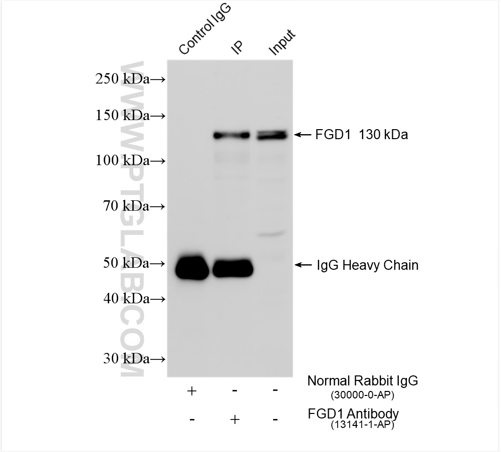
IP experiment of SK-N-SH using 13141-1-AP
IP result of anti-FGD1 (IP:13141-1-AP, 4ug; Detection:13141-1-AP 1:600) with SK-N-SH cells lysate 1160 ug.
Immunohistochemical analysis of paraffin-embedded mouse kidney tissue slide using 13141-1-AP (FGD1 antibody) at dilution of 1:200 (under 40x lens). Heat mediated antigen retrieval with Tris-EDTA buffer (pH 9.0).
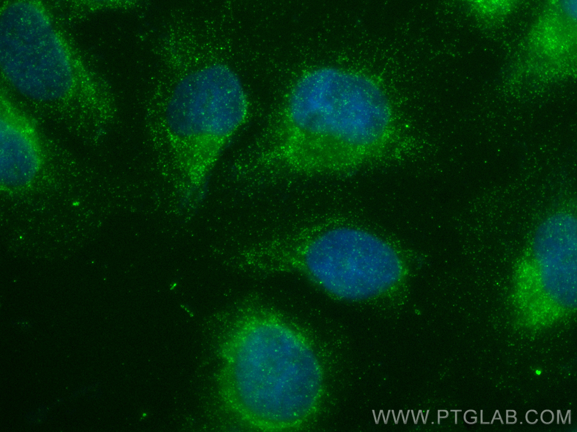
IF Staining of U2OS using 13141-1-AP
Immunofluorescent analysis of (-20°C Ethanol) fixed U2OS cells using FGD1 antibody (13141-1-AP) at dilution of 1:200 and Multi-rAb CoraLite ® Plus 488-Goat Anti-Rabbit Recombinant Secondary Antibody (H+L) (RGAR002).
Immunofluorescent analysis of (-20°C Ethanol) fixed U2OS cells using FGD1 antibody (13141-1-AP) at dilution of 1:200 and Multi-rAb CoraLite ® Plus 488-Goat Anti-Rabbit Recombinant Secondary Antibody (H+L) (RGAR002).
The Proteintech guarantee covers Proteintech antibodies in any species and any application, including those not listed on the datasheet. If the antibody doesn’t perform, you can receive a hassle-free refund or credit note.
mouse kidney tissue Note: suggested antigen retrieval with TE buffer pH 9.0; (*) Alternatively, antigen retrieval may be performed with citrate buffer pH 6.0
Positive IF/ICC detected in
U2OS cells
Recommended dilution
Application
Dilution
Western Blot (WB)
WB : 1:500-1:1000
Immunoprecipitation (IP)
IP : 0.5-4.0 ug for 1.0-3.0 mg of total protein lysate
Immunohistochemistry (IHC)
IHC : 1:50-1:500
Immunofluorescence (IF)/ICC
IF/ICC : 1:50-1:500
It is recommended that this reagent should be titrated in each testing system to obtain optimal results.
Sample-dependent, Check data in validation data gallery.
Product Information
13141-1-AP targets FGD1 in WB, IHC, IF/ICC, IP, ELISA applications and shows reactivity with human, mouse, rat samples.
PBS with 0.02% sodium azide and 50% glycerol , pH 7.3
Storage Conditions
Store at -20°C. Stable for one year after shipment. Aliquoting is unnecessary for -20oC storage. 20ul sizes contain 0.1% BSA.
Background Information
FGD1 is also named as FGDY (Faciogenital dysplasia 1 protein), ZFYVE3 (Zinc finger FYVE domain-containing protein 3), Rho/Rac guanine nucleotide exchange factor FGD1 (Rho/Rac GEF), FYVE, RhoGEF and PH domain-containing protein 1. FGD1 activates CDC42, a member of the Ras-like family of Rho- and Rac proteins, by exchanging bound GDP for free GTP. And FGD1 plays a role in regulating the actin cytoskeleton and cell shape. (PMID: 8969170). FGD1 was markedly overexpressed and might be a prognostic biomarker in osteosarcoma patient specimens (PMID: 32194840). FGD1 is specific for the Rho GTPase cell division cycle 42 (CDC42) (PMID: 22854039).
Various lysates were subjected to SDS PAGE followed by western blot with 13141-1-AP (FGD1 antibody) at dilution of 1:500 incubated at room temperature for 1.5 hours.
WB analysis using 13141-1-AP
Various lysates were subjected to SDS PAGE followed by western blot with 13141-1-AP (FGD1 antibody) at dilution of 1:300 incubated at room temperature for 1.5 hours.
IHC Figures
IHC staining of mouse kidney using 13141-1-AP
Immunohistochemical analysis of paraffin-embedded mouse kidney tissue slide using 13141-1-AP (FGD1 antibody) at dilution of 1:200 (under 40x lens). Heat mediated antigen retrieval with Tris-EDTA buffer (pH 9.0).
IP Figures
IP experiment of SK-N-SH using 13141-1-AP
IP result of anti-FGD1 (IP:13141-1-AP, 4ug; Detection:13141-1-AP 1:600) with SK-N-SH cells lysate 1160 ug.
IF/ICC Figures
IF Staining of U2OS using 13141-1-AP
Immunofluorescent analysis of (-20°C Ethanol) fixed U2OS cells using FGD1 antibody (13141-1-AP) at dilution of 1:200 and Multi-rAb CoraLite ® Plus 488-Goat Anti-Rabbit Recombinant Secondary Antibody (H+L) (RGAR002).
The species listed in Tested Reactivity are in-house verified and applicable species. For unlisted species, please refer to the homology analysis of the immunogen sequence and related species. For rabbit polyclonal antibodies, homology >70% is recommended. For mouse monoclonal antibodies and rabbit recombinant antibodies, homology >90% is recommended. Generally, the higher the homology, the greater the applicability. However, there will be certain differences in protein expression in different species, tissues or cells. Therefore, the homology analysis results are for reference only and do not serve as a guarantee.
At Proteintech, we pride ourselves on our antibody quality, customer service and transparency. As such, we are comparing our antibodies with other vendors, enabling easy identification and comparisons of key data to help you choose the suitable antibody for your needs.
We have selected the top cited antibodies from these vendors for you to compare.
Proteintech
FGD1 Polyclonal antibody
Catalog Number
13141-1-AP
Citations
-
Dilutions
WB : 1:500-1:1000 IP : 0.5-4.0 ug for IP and 0.5-4.0 ug for 1.0-3.0 mg of total protein lysate for WB IHC : 1:50-1:500 IF/ICC : 1:50-1:500
Applications
WB, IHC, IF/ICC, IP, ELISA
Reactivity
human, mouse, rat
Product Guarantee
Covers any species including not listed on datasheet
Covers any applications including not listed on datasheet
 at dilution of 1:500 incubated at room temperature for 1.5 hours.")
 at dilution of 1:300 incubated at room temperature for 1.5 hours.")
 with SK-N-SH cells lysate 1160 ug.")
 at dilution of 1:200 (under 40x lens). Heat mediated antigen retrieval with Tris-EDTA buffer (pH 9.0).")
 fixed U2OS cells using FGD1 antibody (13141-1-AP) at dilution of 1:200 and Multi-rAb CoraLite ® Plus 488-Goat Anti-Rabbit Recombinant Secondary Antibody (H+L) (RGAR002).")