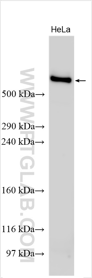
at dilution of 1:2500 incubated at room temperature for 1.5 hours.")
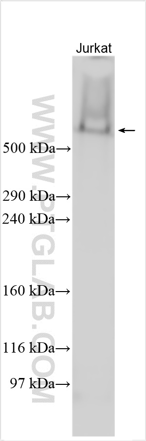
 at dilution of 1:2500 incubated at room temperature for 1.5 hours.")
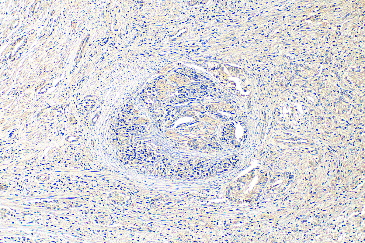
 at dilution of 1:200 (under 10x lens). Heat mediated antigen retrieval with Tris-EDTA buffer (pH 9.0).")
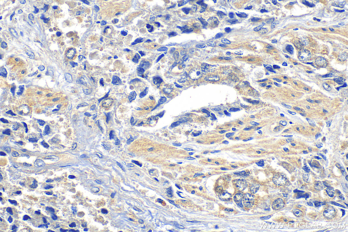
 at dilution of 1:200 (under 40x lens). Heat mediated antigen retrieval with Tris-EDTA buffer (pH 9.0).")
Tested Applications
| Positive WB detected in | HeLa cells, Jurkat cells |
| Positive IHC detected in | human prostate hyperplasia tissue Note: suggested antigen retrieval with TE buffer pH 9.0; (*) Alternatively, antigen retrieval may be performed with citrate buffer pH 6.0 |
Recommended dilution
| Application | Dilution |
|---|---|
| Western Blot (WB) | WB : 1:1000-1:5000 |
| Immunohistochemistry (IHC) | IHC : 1:50-1:500 |
| It is recommended that this reagent should be titrated in each testing system to obtain optimal results. | |
| Sample-dependent, Check data in validation data gallery. | |
Product Information
30733-1-AP targets FAT1 in WB, IHC, ELISA applications and shows reactivity with Human samples.
| Tested Reactivity | Human |
| Host / Isotype | Rabbit / IgG |
| Class | Polyclonal |
| Type | Antibody |
| Immunogen | FAT1 fusion protein Ag33957 Predict reactive species |
| Full Name | FAT tumor suppressor homolog 1 (Drosophila) |
| Observed Molecular Weight | 506-600 kDa |
| Gene Symbol | FAT1 |
| Gene ID (NCBI) | 2195 |
| RRID | AB_3086406 |
| Conjugate | Unconjugated |
| Form | Liquid |
| Purification Method | Antigen affinity purification |
| UNIPROT ID | Q14517 |
| Storage Buffer | PBS with 0.02% sodium azide and 50% glycerol , pH 7.3 |
| Storage Conditions | Store at -20°C. Stable for one year after shipment. Aliquoting is unnecessary for -20oC storage. 20ul sizes contain 0.1% BSA. |
Background Information
FAT1, also known as hFat1, belongs to a member of the cadherin superfamily, has been proposed to play roles in cerebral development, glomerular slit formation, and also to act as a tumor suppressor, but its mechanisms of action have not been elucidated. It is expected to be located in cell membrane and nucleus, which is expressed in many epithelial and some endothelial and smooth muscle cells. The calculated molecular weight of FAT1 is 506 kDa and there is glycosylation modification of the protein. To examine functions of the transmembrane and cytoplasmic domains, they were expressed in HEK-293 and HeLa cells as chimeric proteins in fusion with EGFP and extracellular domains derived from E-cadherin. Proteins comprising the transmembrane domain localized to the membrane fraction (PMID: 15922730, 26373379).
Protocols
| Product Specific Protocols | |
|---|---|
| WB protocol for FAT1 antibody 30733-1-AP | Download protocol |
| IHC protocol for FAT1 antibody 30733-1-AP | Download protocol |
| Standard Protocols | |
|---|---|
| Click here to view our Standard Protocols |