at dilution of 1:5000 incubated at room temperature for 1.5 hours.")
 with sh-Control and sh-EEF1B2 transfected HeLa cells.")
 at dilution of 1:5000 incubated at room temperature for 1.5 hours.")
 at dilution of 1:5000 incubated at room temperature for 1.5 hours.")
 at dilution of 1:5000 incubated at room temperature for 1.5 hours.")
 at dilution of 1:800 incubated at room temperature for 1.5 hours.")
 at dilution of 1:1000 incubated at room temperature for 1.5 hours.")
 with Jurkat cells lysate 1400 ug.")
 at dilution of 1:1000 (under 40x lens). Heat mediated antigen retrieval with Tris-EDTA buffer (pH 9.0).")
 at dilution of 1:1000 (under 10x lens). Heat mediated antigen retrieval with Tris-EDTA buffer (pH 9.0).")
 at dilution of 1:1000 (under 40x lens). Heat mediated antigen retrieval with Tris-EDTA buffer (pH 9.0).")
 at dilution of 1:1000 (under 10x lens). Heat mediated antigen retrieval with Tris-EDTA buffer (pH 9.0).")
 at dilution of 1:500 (under 10x lens). Heat mediated antigen retrieval with Tris-EDTA buffer (pH 9.0).")
 at dilution of 1:500 (under 40x lens). Heat mediated antigen retrieval with Tris-EDTA buffer (pH 9.0).")
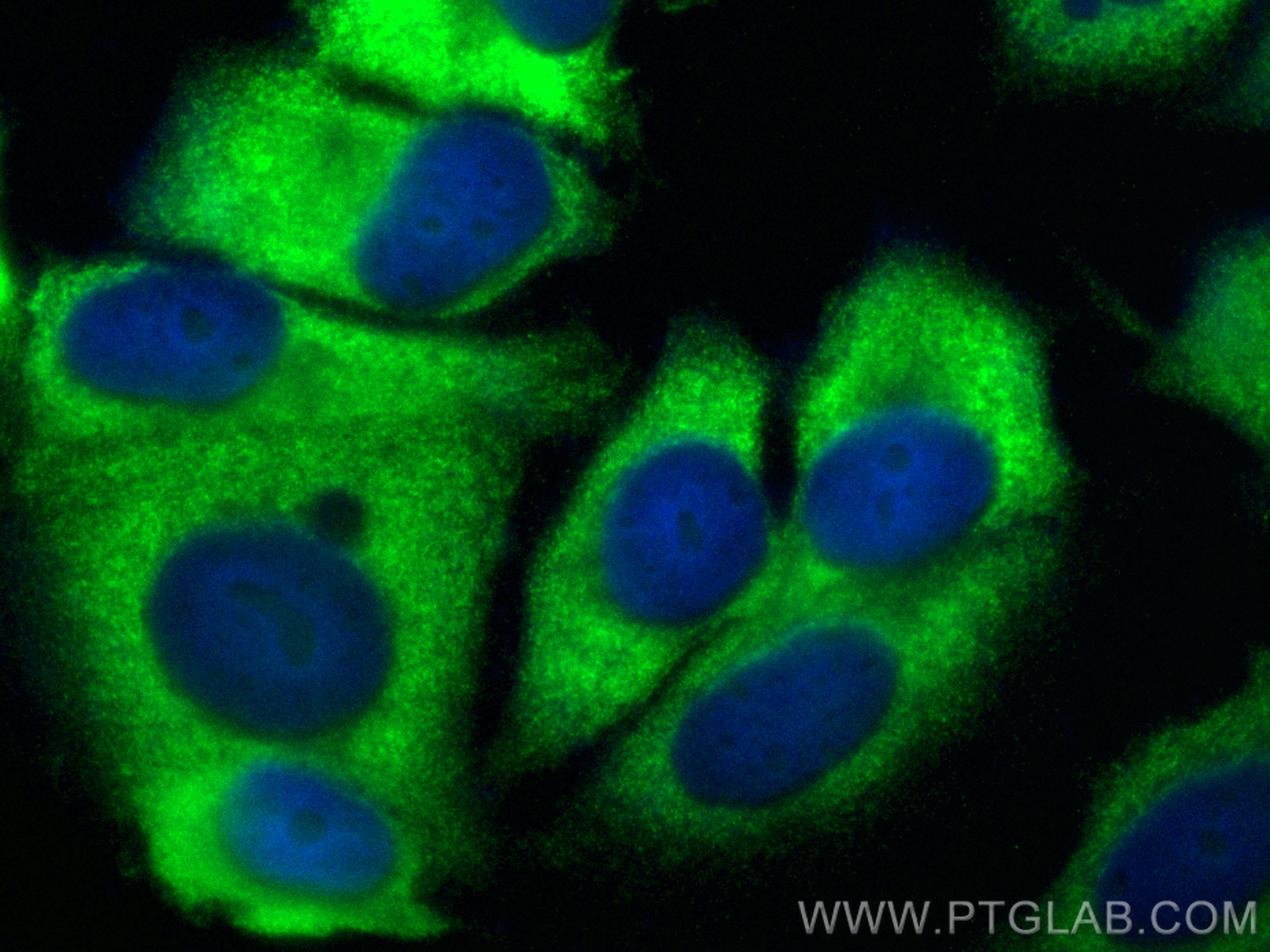
 fixed MCF-7 cells using EEF1B2 antibody (10095-2-AP) at dilution of 1:200 and CoraLite®488-Conjugated Goat Anti-Rabbit IgG(H+L) (SA00013-2).")
Tested Applications
| Positive WB detected in | PC-3 cells, RAW264.7, Jurkat cells, HEK-293 cells, HeLa cells, SKOV-3 cells |
| Positive IP detected in | Jurkat cells |
| Positive IHC detected in | human colon tissue, human breast cancer tissue, human pancreas cancer tissue Note: suggested antigen retrieval with TE buffer pH 9.0; (*) Alternatively, antigen retrieval may be performed with citrate buffer pH 6.0 |
| Positive IF/ICC detected in | MCF-7 cells |
Recommended dilution
| Application | Dilution |
|---|---|
| Western Blot (WB) | WB : 1:2000-1:10000 |
| Immunoprecipitation (IP) | IP : 0.5-4.0 ug for 1.0-3.0 mg of total protein lysate |
| Immunohistochemistry (IHC) | IHC : 1:250-1:1000 |
| Immunofluorescence (IF)/ICC | IF/ICC : 1:50-1:500 |
| It is recommended that this reagent should be titrated in each testing system to obtain optimal results. | |
| Sample-dependent, Check data in validation data gallery. | |
Published Applications
| KD/KO | See 1 publications below |
| WB | See 4 publications below |
| IHC | See 1 publications below |
| IF | See 2 publications below |
| IP | See 1 publications below |
| RIP | See 1 publications below |
Product Information
10095-2-AP targets EEF1B2 in WB, IHC, IF/ICC, IP, RIP, ELISA applications and shows reactivity with human, mouse, rat samples.
| Tested Reactivity | human, mouse, rat |
| Cited Reactivity | human, mouse |
| Host / Isotype | Rabbit / IgG |
| Class | Polyclonal |
| Type | Antibody |
| Immunogen |
CatNo: Ag0135 Product name: Recombinant human EEF1B2 protein Source: e coli.-derived, PGEX-4T Tag: GST Domain: 1-223 aa of BC000211 Sequence: MGFGDLKSPAGLQVLNDYLADKSYIEGYVPSQADVAVFEAVSSPPPADLCHALRWYNHIKSYEKEKASLPGVKKALGKYGPADVEDTTGSGATDSKDDDDIDLFGSDDEEESEEAKRLREERLAQYESKKAKKPALVAKSSILLDVKPWDDETDMAKLEECVRSIQADGLVWGSSKLVPVGYGIKKLQIQCVVEDDKVGTDMLEEQITAFEDYVQSMDVAAFN Predict reactive species |
| Full Name | eukaryotic translation elongation factor 1 beta 2 |
| Calculated Molecular Weight | 25 kDa |
| Observed Molecular Weight | 30-34 kDa |
| GenBank Accession Number | BC000211 |
| Gene Symbol | EEF1B2 |
| Gene ID (NCBI) | 1933 |
| RRID | AB_2096987 |
| Conjugate | Unconjugated |
| Form | Liquid |
| Purification Method | Antigen affinity purification |
| UNIPROT ID | P24534 |
| Storage Buffer | PBS with 0.02% sodium azide and 50% glycerol, pH 7.3. |
| Storage Conditions | Store at -20°C. Stable for one year after shipment. Aliquoting is unnecessary for -20oC storage. 20ul sizes contain 0.1% BSA. |
Background Information
In eukaryotes, the translation elongation factor eEF1A responsible for transporting amino-acylated tRNA to the ribosome forms a higher-order complex, eEF1H, with its guanine-nucleotide-exchange factor eEF1B. eEF1B consists of three subunits: eEF1B alpha, eEF1B beta and eEF1B gamma. The eEF1B2 possess the nucleotide-exchange activity. Although several models on the basis of in vitro experiments have been proposed for the macromolecular organization of the eEF1H complex, these models differ in various aspects. The human eukaryote elongation factor 1 beta 2 (eEF1B2) migrated as a 30-34 kDa protein in SDS-PAGE. This antibody is a rabbit polyclonal antibody raised against residues near the N terminus of human EEF1B2.
Protocols
| Product Specific Protocols | |
|---|---|
| IF protocol for EEF1B2 antibody 10095-2-AP | Download protocol |
| IHC protocol for EEF1B2 antibody 10095-2-AP | Download protocol |
| IP protocol for EEF1B2 antibody 10095-2-AP | Download protocol |
| WB protocol for EEF1B2 antibody 10095-2-AP | Download protocol |
| Standard Protocols | |
|---|---|
| Click here to view our Standard Protocols |
Publications
| Species | Application | Title |
|---|---|---|
Nat Cell Biol The MBNL3 splicing factor promotes hepatocellular carcinoma by increasing PXN expression through the alternative splicing of lncRNA-PXN-AS1. | ||
Front Microbiol Strain-Specific Contribution of Eukaryotic Elongation Factor 1 Gamma to the Translation of Influenza A Virus Proteins. | ||
PLoS One Characterisation of Translation Elongation Factor eEF1B Subunit Expression in Mammalian Cells and Tissues and Co-Localisation with eEF1A2.
| ||