SH-SY5Y cells were subjected to SDS PAGE followed by western blot with 18969-1-AP (CRMP4 antibody) at dilution of 1:800 incubated at room temperature for 1.5 hours.
SH-SY5Y cells were subjected to SDS PAGE followed by western blot with 18969-1-AP (CRMP4 antibody) at dilution of 1:800 incubated at room temperature for 1.5 hours.
WB analysis of SH-SY5Y using 18969-1-AP
SH-SY5Y cells were subjected to SDS PAGE followed by western blot with 18969-1-AP (CRMP4 antibody) at dilution of 1:800 incubated at room temperature for 1.5 hours.
SH-SY5Y cells were subjected to SDS PAGE followed by western blot with 18969-1-AP (CRMP4 antibody) at dilution of 1:800 incubated at room temperature for 1.5 hours.
WB analysis of mouse thymus using 18969-1-AP
mouse thymus tissue were subjected to SDS PAGE followed by western blot with 18969-1-AP (CRMP4 antibody) at dilution of 1:800 incubated at room temperature for 1.5 hours.
mouse thymus tissue were subjected to SDS PAGE followed by western blot with 18969-1-AP (CRMP4 antibody) at dilution of 1:800 incubated at room temperature for 1.5 hours.
WB analysis of Jurkat using 18969-1-AP
Jurkat cells were subjected to SDS PAGE followed by western blot with 18969-1-AP (CRMP4 antibody) at dilution of 1:800 incubated at room temperature for 1.5 hours.
Jurkat cells were subjected to SDS PAGE followed by western blot with 18969-1-AP (CRMP4 antibody) at dilution of 1:800 incubated at room temperature for 1.5 hours.
WB analysis of mouse cerebellum using 18969-1-AP
mouse cerebellum tissue were subjected to SDS PAGE followed by western blot with 18969-1-AP (CRMP4 antibody) at dilution of 1:800 incubated at room temperature for 1.5 hours.
mouse cerebellum tissue were subjected to SDS PAGE followed by western blot with 18969-1-AP (CRMP4 antibody) at dilution of 1:800 incubated at room temperature for 1.5 hours.
IP experiment of SH-SY5Y using 18969-1-AP
IP result of anti-CRMP4 (IP:18969-1-AP, 3ug; Detection:18969-1-AP 1:700) with SH-SY5Y cells lysate 3000ug.
IP result of anti-CRMP4 (IP:18969-1-AP, 3ug; Detection:18969-1-AP 1:700) with SH-SY5Y cells lysate 3000ug.
IHC staining of human liver cancer using 18969-1-AP
Immunohistochemical analysis of paraffin-embedded human liver cancer tissue slide using 18969-1-AP (CRMP4 antibody at dilution of 1:200 (under 10x lens). Heat mediated antigen retrieval with Tris-EDTA buffer (pH 9.0).
Immunohistochemical analysis of paraffin-embedded human liver cancer tissue slide using 18969-1-AP (CRMP4 antibody at dilution of 1:200 (under 10x lens). Heat mediated antigen retrieval with Tris-EDTA buffer (pH 9.0).
IHC staining of human liver cancer using 18969-1-AP
Immunohistochemical analysis of paraffin-embedded human liver cancer tissue slide using 18969-1-AP (CRMP4 antibody at dilution of 1:200 (under 40x lens). Heat mediated antigen retrieval with Tris-EDTA buffer (pH 9.0).
Immunohistochemical analysis of paraffin-embedded human liver cancer tissue slide using 18969-1-AP (CRMP4 antibody at dilution of 1:200 (under 40x lens). Heat mediated antigen retrieval with Tris-EDTA buffer (pH 9.0).
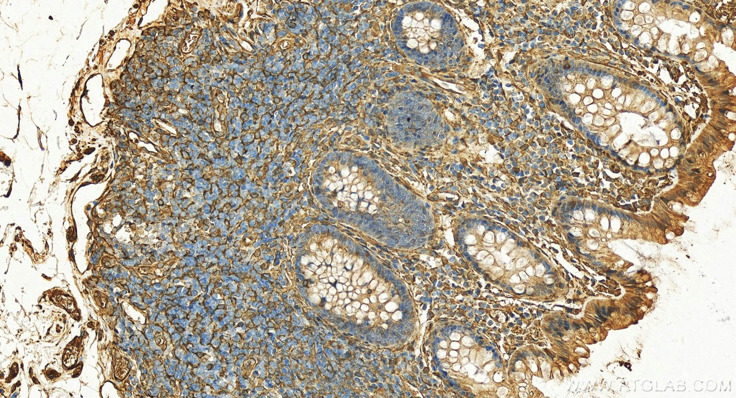
IHC staining of human colon using 18969-1-AP
Immunohistochemical analysis of paraffin-embedded human normal colon slide using 18969-1-AP (CRMP4 antibody) at dilution of 1:300 (under 20x lens). Heat mediated antigen retrieval with Tris-EDTA buffer (pH 9.0).
Immunohistochemical analysis of paraffin-embedded human normal colon slide using 18969-1-AP (CRMP4 antibody) at dilution of 1:300 (under 20x lens). Heat mediated antigen retrieval with Tris-EDTA buffer (pH 9.0).
IHC staining of human kidney using 18969-1-AP
Immunohistochemical analysis of paraffin-embedded human kidney using 18969-1-AP (CRMP4 antibody) at dilution of 1:100 (under 10x lens).
Immunohistochemical analysis of paraffin-embedded human kidney using 18969-1-AP (CRMP4 antibody) at dilution of 1:100 (under 40x lens).
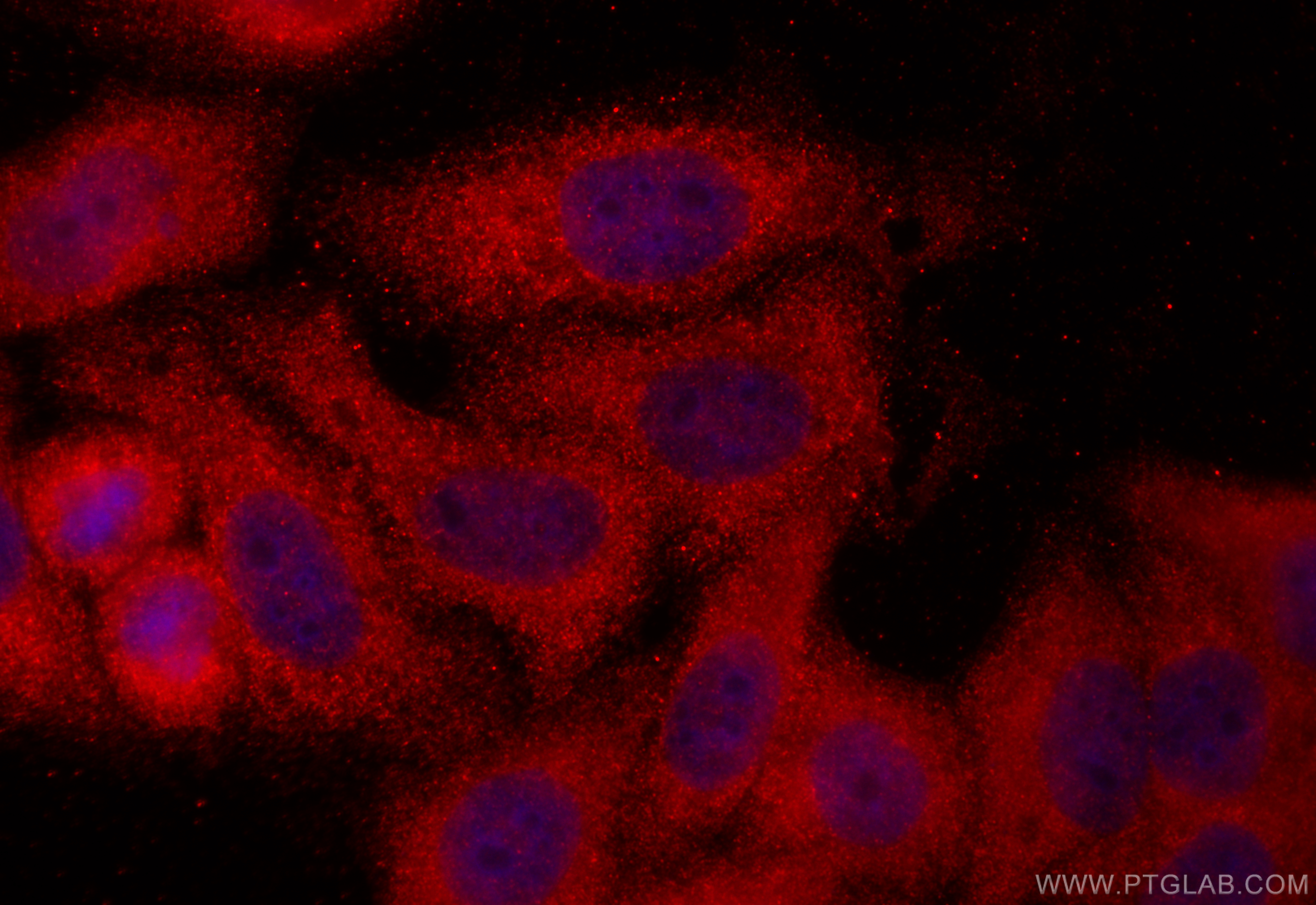
IF Staining of HepG2 using 18969-1-AP
Immunofluorescent analysis of (-20°C Ethanol) fixed HepG2 cells using CRMP4 antibody (18969-1-AP) at dilution of 1:200 and CoraLite®594-Conjugated Goat Anti-Rabbit IgG(H+L) (SA00013-4).
Immunofluorescent analysis of (-20°C Ethanol) fixed HepG2 cells using CRMP4 antibody (18969-1-AP) at dilution of 1:200 and CoraLite®594-Conjugated Goat Anti-Rabbit IgG(H+L) (SA00013-4).
The Proteintech guarantee covers Proteintech antibodies in any species and any application, including those not listed on the datasheet. If the antibody doesn’t perform, you can receive a hassle-free refund or credit note.
human liver cancer tissue, human kidney tissue Note: suggested antigen retrieval with TE buffer pH 9.0; (*) Alternatively, antigen retrieval may be performed with citrate buffer pH 6.0
Positive IF/ICC detected in
HepG2 cells
Recommended dilution
Application
Dilution
Western Blot (WB)
WB : 1:500-1:3000
Immunoprecipitation (IP)
IP : 0.5-4.0 ug for 1.0-3.0 mg of total protein lysate
Immunohistochemistry (IHC)
IHC : 1:100-1:400
Immunofluorescence (IF)/ICC
IF/ICC : 1:50-1:500
It is recommended that this reagent should be titrated in each testing system to obtain optimal results.
Sample-dependent, Check data in validation data gallery.
PBS with 0.02% sodium azide and 50% glycerol , pH 7.3
Storage Conditions
Store at -20°C. Stable for one year after shipment. Aliquoting is unnecessary for -20oC storage. 20ul sizes contain 0.1% BSA.
Background Information
DPYSL3(Dihydropyrimidinase-related protein 3) is also named as CRMP4, DRP3, ULIP, ULIP1 and belongs to the hydantoinase/dihydropyrimidinase subfamily.DPYSL3 is a developmentally regulated protein, strongly expressed in early embryonic post-mitotic neural cells, reaching a peak of expression around the first post-natal week. In the adult brain, DPYSL3 is present mainly in regions that retain neurogenesis.DPYSL3 protein is also found in synaptic sites at the adult neuromuscular junction where it may be involved in the maintenance of neuronal stability(PMID: 16135096).It has 2 isoforms produced by alternative splicing.
Proteomic analysis of the glutathione-deficient LEGSKO mouse lens reveals activation of EMT signaling, loss of lens specific markers, and changes in stress response proteins.
SH-SY5Y cells were subjected to SDS PAGE followed by western blot with 18969-1-AP (CRMP4 antibody) at dilution of 1:800 incubated at room temperature for 1.5 hours.
WB analysis of SH-SY5Y using 18969-1-AP
SH-SY5Y cells were subjected to SDS PAGE followed by western blot with 18969-1-AP (CRMP4 antibody) at dilution of 1:800 incubated at room temperature for 1.5 hours.
WB analysis of mouse thymus using 18969-1-AP
mouse thymus tissue were subjected to SDS PAGE followed by western blot with 18969-1-AP (CRMP4 antibody) at dilution of 1:800 incubated at room temperature for 1.5 hours.
WB analysis of Jurkat using 18969-1-AP
Jurkat cells were subjected to SDS PAGE followed by western blot with 18969-1-AP (CRMP4 antibody) at dilution of 1:800 incubated at room temperature for 1.5 hours.
WB analysis of mouse cerebellum using 18969-1-AP
mouse cerebellum tissue were subjected to SDS PAGE followed by western blot with 18969-1-AP (CRMP4 antibody) at dilution of 1:800 incubated at room temperature for 1.5 hours.
IHC Figures
IHC staining of human liver cancer using 18969-1-AP
Immunohistochemical analysis of paraffin-embedded human liver cancer tissue slide using 18969-1-AP (CRMP4 antibody at dilution of 1:200 (under 10x lens). Heat mediated antigen retrieval with Tris-EDTA buffer (pH 9.0).
IHC staining of human liver cancer using 18969-1-AP
Immunohistochemical analysis of paraffin-embedded human liver cancer tissue slide using 18969-1-AP (CRMP4 antibody at dilution of 1:200 (under 40x lens). Heat mediated antigen retrieval with Tris-EDTA buffer (pH 9.0).
IHC staining of human colon using 18969-1-AP
Immunohistochemical analysis of paraffin-embedded human normal colon slide using 18969-1-AP (CRMP4 antibody) at dilution of 1:300 (under 20x lens). Heat mediated antigen retrieval with Tris-EDTA buffer (pH 9.0).
IHC staining of human kidney using 18969-1-AP
Immunohistochemical analysis of paraffin-embedded human kidney using 18969-1-AP (CRMP4 antibody) at dilution of 1:100 (under 10x lens).
IHC staining of human kidney using 18969-1-AP
Immunohistochemical analysis of paraffin-embedded human kidney using 18969-1-AP (CRMP4 antibody) at dilution of 1:100 (under 40x lens).
IP Figures
IP experiment of SH-SY5Y using 18969-1-AP
IP result of anti-CRMP4 (IP:18969-1-AP, 3ug; Detection:18969-1-AP 1:700) with SH-SY5Y cells lysate 3000ug.
IF/ICC Figures
IF Staining of HepG2 using 18969-1-AP
Immunofluorescent analysis of (-20°C Ethanol) fixed HepG2 cells using CRMP4 antibody (18969-1-AP) at dilution of 1:200 and CoraLite®594-Conjugated Goat Anti-Rabbit IgG(H+L) (SA00013-4).
The species listed in Tested Reactivity are in-house verified and applicable species. For unlisted species, please refer to the homology analysis of the immunogen sequence and related species. For rabbit polyclonal antibodies, homology >70% is recommended. For mouse monoclonal antibodies and rabbit recombinant antibodies, homology >90% is recommended. Generally, the higher the homology, the greater the applicability. However, there will be certain differences in protein expression in different species, tissues or cells. Therefore, the homology analysis results are for reference only and do not serve as a guarantee.
At Proteintech, we pride ourselves on our antibody quality, customer service and transparency. As such, we are comparing our antibodies with other vendors, enabling easy identification and comparisons of key data to help you choose the suitable antibody for your needs.
We have selected the top cited antibodies from these vendors for you to compare.
Proteintech
CRMP4 Polyclonal antibody
Catalog Number
18969-1-AP
Citations
2
Dilutions
WB : 1:500-1:3000 IP : 0.5-4.0 ug for IP and 0.5-4.0 ug for 1.0-3.0 mg of total protein lysate for WB IHC : 1:100-1:400 IF/ICC : 1:50-1:500
Applications
WB, IHC, IF/ICC, IP, ELISA
Reactivity
human, mouse, rat
Product Guarantee
Covers any species including not listed on datasheet
Covers any applications including not listed on datasheet
 at dilution of 1:800 incubated at room temperature for 1.5 hours.")
 at dilution of 1:800 incubated at room temperature for 1.5 hours.")
 at dilution of 1:800 incubated at room temperature for 1.5 hours.")
 at dilution of 1:800 incubated at room temperature for 1.5 hours.")
 at dilution of 1:800 incubated at room temperature for 1.5 hours.")
 with SH-SY5Y cells lysate 3000ug.")
. Heat mediated antigen retrieval with Tris-EDTA buffer (pH 9.0).")
. Heat mediated antigen retrieval with Tris-EDTA buffer (pH 9.0).")
 at dilution of 1:300 (under 20x lens). Heat mediated antigen retrieval with Tris-EDTA buffer (pH 9.0).")
 at dilution of 1:100 (under 10x lens).")
 at dilution of 1:100 (under 40x lens).")
 fixed HepG2 cells using CRMP4 antibody (18969-1-AP) at dilution of 1:200 and CoraLite®594-Conjugated Goat Anti-Rabbit IgG(H+L) (SA00013-4).")