human brain tissue were subjected to SDS PAGE followed by western blot with 15557-1-AP (DIRAS2 antibody) at dilution of 1:500 incubated at room temperature for 1.5 hours.
human brain tissue were subjected to SDS PAGE followed by western blot with 15557-1-AP (DIRAS2 antibody) at dilution of 1:500 incubated at room temperature for 1.5 hours.
WB analysis using 15557-1-AP
Various lysates were subjected to SDS PAGE followed by western blot with 15557-1-AP (DIRAS2 antibody) at dilution of 1:1500 incubated at 4 degree celsius over night.
Various lysates were subjected to SDS PAGE followed by western blot with 15557-1-AP (DIRAS2 antibody) at dilution of 1:1500 incubated at 4 degree celsius over night.
WB analysis of mouse brain using 15557-1-AP
mouse brain tissue were subjected to SDS PAGE followed by western blot with 15557-1-AP (DIRAS2 Antibody) at dilution of 1:1000 incubated at room temperature for 1.5 hours.
mouse brain tissue were subjected to SDS PAGE followed by western blot with 15557-1-AP (DIRAS2 Antibody) at dilution of 1:1000 incubated at room temperature for 1.5 hours.
WB analysis of rat brain using 15557-1-AP
rat brain tissue were subjected to SDS PAGE followed by western blot with 15557-1-AP (DIRAS2 antibody) at dilution of 1:2000 incubated at room temperature for 1.5 hours.
rat brain tissue were subjected to SDS PAGE followed by western blot with 15557-1-AP (DIRAS2 antibody) at dilution of 1:2000 incubated at room temperature for 1.5 hours.
WB analysis of HeLa using 15557-1-AP
HeLa cells were subjected to SDS PAGE followed by western blot with 15557-1-AP (DIRAS2 antibody) at dilution of 1:500 incubated at room temperature for 1.5 hours.
HeLa cells were subjected to SDS PAGE followed by western blot with 15557-1-AP (DIRAS2 antibody) at dilution of 1:500 incubated at room temperature for 1.5 hours.
IP experiment of mouse brain using 15557-1-AP
IP result of anti-DIRAS2 (IP:15557-1-AP, 4ug; Detection:15557-1-AP 1:1000) with mouse brain tissue lysate 4400ug.
IP result of anti-DIRAS2 (IP:15557-1-AP, 4ug; Detection:15557-1-AP 1:1000) with mouse brain tissue lysate 4400ug.
IF Staining of HeLa using 15557-1-AP
Immunofluorescent analysis of (-20℃ Ethanol) fixed HeLa cells using 15557-1-AP (DIRAS2 antibody) at dilution of 1:50 and Alexa Fluor 488-conjugated AffiniPure Goat Anti-Rabbit IgG(H+L).
Immunofluorescent analysis of (-20℃ Ethanol) fixed HeLa cells using 15557-1-AP (DIRAS2 antibody) at dilution of 1:50 and Alexa Fluor 488-conjugated AffiniPure Goat Anti-Rabbit IgG(H+L).
IF Staining of HeLa using 15557-1-AP
Immunofluorescent analysis of Hela cells, using DIRAS2 antibody 15557-1-AP at 1:25 dilution and Rhodamine-labeled goat anti-rabbit IgG (red).
Immunofluorescent analysis of Hela cells, using DIRAS2 antibody 15557-1-AP at 1:25 dilution and Rhodamine-labeled goat anti-rabbit IgG (red).
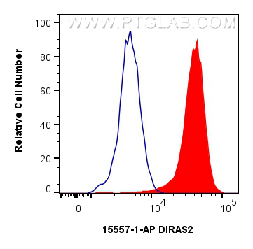
FC experiment of HeLa using 15557-1-AP
1x10^6 HeLa cells were intracellularly stained with 0.25 ug DIRAS2 Polyclonal antibody (15557-1-AP) and CoraLite®488-Conjugated Goat Anti-Rabbit IgG(H+L) (SA00013-2)(red), or 0.25 ug Rabbit IgG control Rabbit PolyAb (30000-0-AP) (blue). Cells were fixed with 4% PFA and permeabilized with Flow Cytometry Perm Buffer (PF00011-C).
1x10^6 HeLa cells were intracellularly stained with 0.25 ug DIRAS2 Polyclonal antibody (15557-1-AP) and CoraLite®488-Conjugated Goat Anti-Rabbit IgG(H+L) (SA00013-2)(red), or 0.25 ug Rabbit IgG control Rabbit PolyAb (30000-0-AP) (blue). Cells were fixed with 4% PFA and permeabilized with Flow Cytometry Perm Buffer (PF00011-C).
The Proteintech guarantee covers Proteintech antibodies in any species and any application, including those not listed on the datasheet. If the antibody doesn’t perform, you can receive a hassle-free refund or credit note.
Di-Ras2 promotes renal cell carcinoma formation by activating the mitogen-activated protein kinase pathway in the absence of von Hippel-Lindau protein.
eNAMPT/Ac-STAT3/DIRAS2 Axis Promotes Development and Cancer Stemness in Triple-Negative Breast Cancer by Enhancing Cytokine Crosstalk Between Tumor-Associated Macrophages and Cancer Cells
human brain tissue were subjected to SDS PAGE followed by western blot with 15557-1-AP (DIRAS2 antibody) at dilution of 1:500 incubated at room temperature for 1.5 hours.
WB analysis using 15557-1-AP
Various lysates were subjected to SDS PAGE followed by western blot with 15557-1-AP (DIRAS2 antibody) at dilution of 1:1500 incubated at 4 degree celsius over night.
WB analysis of mouse brain using 15557-1-AP
mouse brain tissue were subjected to SDS PAGE followed by western blot with 15557-1-AP (DIRAS2 Antibody) at dilution of 1:1000 incubated at room temperature for 1.5 hours.
WB analysis of rat brain using 15557-1-AP
rat brain tissue were subjected to SDS PAGE followed by western blot with 15557-1-AP (DIRAS2 antibody) at dilution of 1:2000 incubated at room temperature for 1.5 hours.
WB analysis of HeLa using 15557-1-AP
HeLa cells were subjected to SDS PAGE followed by western blot with 15557-1-AP (DIRAS2 antibody) at dilution of 1:500 incubated at room temperature for 1.5 hours.
IP Figures
IP experiment of mouse brain using 15557-1-AP
IP result of anti-DIRAS2 (IP:15557-1-AP, 4ug; Detection:15557-1-AP 1:1000) with mouse brain tissue lysate 4400ug.
IF/ICC Figures
IF Staining of HeLa using 15557-1-AP
Immunofluorescent analysis of (-20℃ Ethanol) fixed HeLa cells using 15557-1-AP (DIRAS2 antibody) at dilution of 1:50 and Alexa Fluor 488-conjugated AffiniPure Goat Anti-Rabbit IgG(H+L).
IF Staining of HeLa using 15557-1-AP
Immunofluorescent analysis of Hela cells, using DIRAS2 antibody 15557-1-AP at 1:25 dilution and Rhodamine-labeled goat anti-rabbit IgG (red).
FC (INTRA) Figures
FC experiment of HeLa using 15557-1-AP
1x10^6 HeLa cells were intracellularly stained with 0.25 ug DIRAS2 Polyclonal antibody (15557-1-AP) and CoraLite®488-Conjugated Goat Anti-Rabbit IgG(H+L) (SA00013-2)(red), or 0.25 ug Rabbit IgG control Rabbit PolyAb (30000-0-AP) (blue). Cells were fixed with 4% PFA and permeabilized with Flow Cytometry Perm Buffer (PF00011-C).
The species listed in Tested Reactivity are in-house verified and applicable species. For unlisted species, please refer to the homology analysis of the immunogen sequence and related species. For rabbit polyclonal antibodies, homology >70% is recommended. For mouse monoclonal antibodies and rabbit recombinant antibodies, homology >90% is recommended. Generally, the higher the homology, the greater the applicability. However, there will be certain differences in protein expression in different species, tissues or cells. Therefore, the homology analysis results are for reference only and do not serve as a guarantee.
At Proteintech, we pride ourselves on our antibody quality, customer service and transparency. As such, we are comparing our antibodies with other vendors, enabling easy identification and comparisons of key data to help you choose the suitable antibody for your needs.
We have selected the top cited antibodies from these vendors for you to compare.
Proteintech
KD/KO VALIDATED
DIRAS2 Polyclonal antibody
Catalog Number
15557-1-AP
Citations
5
Dilutions
WB : 1:500-1:3000 IP : 0.5-4.0 ug for IP and 0.5-4.0 ug for 1.0-3.0 mg of total protein lysate for WB IF/ICC : 1:50-1:500 FC (INTRA) : 0.25 ug per 10^6 cells in a 100 µl suspension
Applications
WB, IF/ICC, FC (Intra), IP, ELISA
Reactivity
human, mouse, rat
Product Guarantee
Covers any species including not listed on datasheet
Covers any applications including not listed on datasheet
 at dilution of 1:500 incubated at room temperature for 1.5 hours.")
 at dilution of 1:1500 incubated at 4 degree celsius over night.")
 at dilution of 1:1000 incubated at room temperature for 1.5 hours.")
 at dilution of 1:2000 incubated at room temperature for 1.5 hours.")
 at dilution of 1:500 incubated at room temperature for 1.5 hours.")
 with mouse brain tissue lysate 4400ug.")
 fixed HeLa cells using 15557-1-AP (DIRAS2 antibody) at dilution of 1:50 and Alexa Fluor 488-conjugated AffiniPure Goat Anti-Rabbit IgG(H+L).")
.")
 and CoraLite®488-Conjugated Goat Anti-Rabbit IgG(H+L) (SA00013-2)(red), or 0.25 ug Rabbit IgG control Rabbit PolyAb (30000-0-AP) (blue). Cells were fixed with 4% PFA and permeabilized with Flow Cytometry Perm Buffer (PF00011-C).")