Filter:
 at dilution of 1:600 incubated at room temperature for 1.5 hours.")
 at dilution of 1:600 incubated at room temperature for 1.5 hours.")
 at dilution of 1:600 incubated at room temperature for 1.5 hours.")
 at dilution of 1:200 (under 10x lens). Heat mediated antigen retrieval with Tris-EDTA buffer (pH 9.0).")
 at dilution of 1:200 (under 40x lens). Heat mediated antigen retrieval with Tris-EDTA buffer (pH 9.0).")
 at dilution of 1:4000 (under 10x lens). Heat mediated antigen retrieval with Tris-EDTA buffer (pH 9.0).")
 at dilution of 1:4000 (under 40x lens). Heat mediated antigen retrieval with Tris-EDTA buffer (pH 9.0).")
 at dilution of 1:4000 (under 10x lens). Heat mediated antigen retrieval with Tris-EDTA buffer (pH 9.0).")
 at dilution of 1:4000 (under 40x lens). Heat mediated antigen retrieval with Tris-EDTA buffer (pH 9.0).")
 fixed mouse skeletal muscle tissue using Caveolin-3 antibody (28358-1-AP) at dilution of 1:200 and CoraLite®488-Conjugated AffiniPure Goat Anti-Rabbit IgG(H+L).")
 fixed mouse skeletal muscle tissue using Caveolin-3 antibody (28358-1-AP) at dilution of 1:200 and CoraLite®488-Conjugated AffiniPure Goat Anti-Rabbit IgG(H+L).")
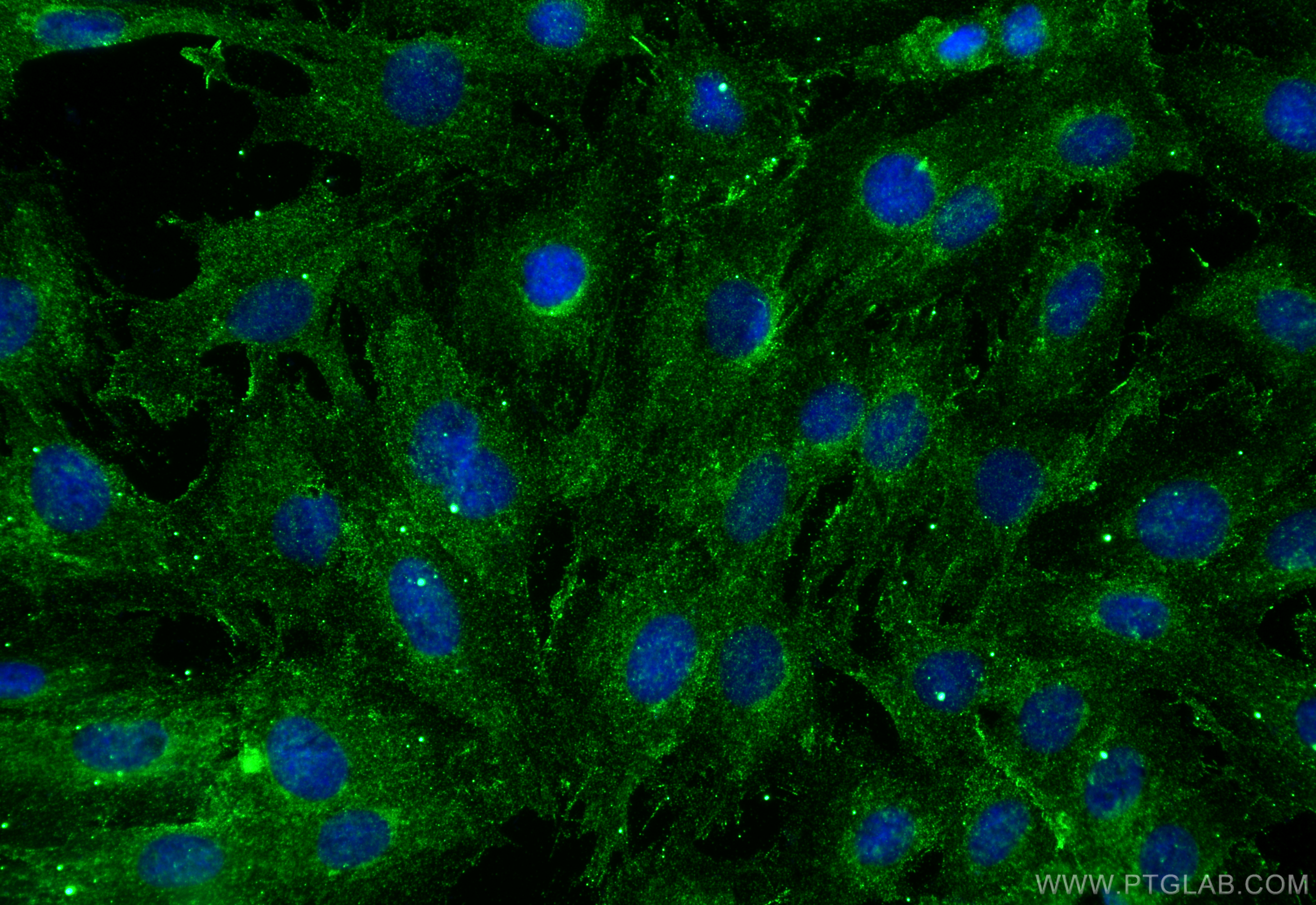
 fixed H9C2 cells using Caveolin-3 antibody (28358-1-AP) at dilution of 1:400 and Multi-rAb CoraLite ® Plus 488-Goat Anti-Rabbit Recombinant Secondary Antibody (H+L) (RGAR002).")
Tested Applications
| Positive WB detected in | mouse colon tissue, mouse heart tissue, mouse skeletal muscle tissue |
| Positive IHC detected in | mouse heart tissue, rat skeletal muscle tissue, mouse skeletal muscle tissue Note: suggested antigen retrieval with TE buffer pH 9.0; (*) Alternatively, antigen retrieval may be performed with citrate buffer pH 6.0 |
| Positive IF-P detected in | mouse skeletal muscle tissue |
| Positive IF/ICC detected in | H9C2 cells |
Recommended dilution
| Application | Dilution |
|---|---|
| Western Blot (WB) | WB : 1:500-1:1000 |
| Immunohistochemistry (IHC) | IHC : 1:50-1:500 |
| Immunofluorescence (IF)-P | IF-P : 1:50-1:500 |
| Immunofluorescence (IF)/ICC | IF/ICC : 1:200-1:800 |
| It is recommended that this reagent should be titrated in each testing system to obtain optimal results. | |
| Sample-dependent, Check data in validation data gallery. | |
Published Applications
| WB | See 3 publications below |
| IF | See 1 publications below |
Product Information
28358-1-AP targets Caveolin-3 in WB, IHC, IF/ICC, IF-P, ELISA applications and shows reactivity with human, mouse, rat samples.
| Tested Reactivity | human, mouse, rat |
| Cited Reactivity | human, mouse, rat |
| Host / Isotype | Rabbit / IgG |
| Class | Polyclonal |
| Type | Antibody |
| Immunogen |
CatNo: Ag28009 Product name: Recombinant human Caveolin-3 protein Source: e coli.-derived, PGEX-4T Tag: GST Domain: 1-40 aa of BC069368 Sequence: MMAEEHTDLEAQIVKDIHCKEIDLVNRDPKNINEDIVKVD Predict reactive species |
| Full Name | caveolin 3 |
| Calculated Molecular Weight | 151 aa, 17 kDa |
| Observed Molecular Weight | 20 kDa |
| GenBank Accession Number | BC069368 |
| Gene Symbol | Caveolin-3 |
| Gene ID (NCBI) | 859 |
| RRID | AB_2881120 |
| Conjugate | Unconjugated |
| Form | Liquid |
| Purification Method | Antigen affinity purification |
| UNIPROT ID | P56539 |
| Storage Buffer | PBS with 0.02% sodium azide and 50% glycerol, pH 7.3. |
| Storage Conditions | Store at -20°C. Stable for one year after shipment. Aliquoting is unnecessary for -20oC storage. 20ul sizes contain 0.1% BSA. |
Protocols
| Product Specific Protocols | |
|---|---|
| IF protocol for Caveolin-3 antibody 28358-1-AP | Download protocol |
| IHC protocol for Caveolin-3 antibody 28358-1-AP | Download protocol |
| WB protocol for Caveolin-3 antibody 28358-1-AP | Download protocol |
| Standard Protocols | |
|---|---|
| Click here to view our Standard Protocols |
Publications
| Species | Application | Title |
|---|---|---|
Oxid Med Cell Longev Trophoblasts Modulate the Ca2+ Oscillation and Contraction of Myometrial Smooth Muscle Cells by Small Extracellular Vesicle- (sEV-) Mediated Exporting of miR-25-3p during Premature Labor. | ||
Cell Metab Muscle-derived small extracellular vesicles induce liver fibrosis during overtraining | ||
Int Heart J Decrease in Caveolae-Gαq Interaction Mediates Pressure Overload-Induced Cardiac Remodeling in Rats |