MCF7 cells were subjected to SDS PAGE followed by western blot with 17532-1-AP (CTDSPL2 antibody) at dilution of 1:500 incubated at room temperature for 1.5 hours.
MCF7 cells were subjected to SDS PAGE followed by western blot with 17532-1-AP (CTDSPL2 antibody) at dilution of 1:500 incubated at room temperature for 1.5 hours.
WB analysis of mouse testis using 17532-1-AP
mouse testis tissue were subjected to SDS PAGE followed by western blot with 17532-1-AP (CTDSPL2 antibody) at dilution of 1:500 incubated at room temperature for 1.5 hours.
mouse testis tissue were subjected to SDS PAGE followed by western blot with 17532-1-AP (CTDSPL2 antibody) at dilution of 1:500 incubated at room temperature for 1.5 hours.
WB analysis of mouse eye using 17532-1-AP
mouse eye tissue were subjected to SDS PAGE followed by western blot with 17532-1-AP (CTDSPL2 antibody) at dilution of 1:600 incubated at room temperature for 1.5 hours.
mouse eye tissue were subjected to SDS PAGE followed by western blot with 17532-1-AP (CTDSPL2 antibody) at dilution of 1:600 incubated at room temperature for 1.5 hours.
WB analysis of HeLa using 17532-1-AP
HeLa cells were subjected to SDS PAGE followed by western blot with 17532-1-AP (CTDSPL2 antibody) at dilution of 1:500 incubated at room temperature for 1.5 hours.
HeLa cells were subjected to SDS PAGE followed by western blot with 17532-1-AP (CTDSPL2 antibody) at dilution of 1:500 incubated at room temperature for 1.5 hours.
WB analysis of human brain using 17532-1-AP
human brain tissue were subjected to SDS PAGE followed by western blot with 17532-1-AP (CTDSPL2 antibody) at dilution of 1:600 incubated at room temperature for 1.5 hours.
human brain tissue were subjected to SDS PAGE followed by western blot with 17532-1-AP (CTDSPL2 antibody) at dilution of 1:600 incubated at room temperature for 1.5 hours.
WB analysis of HeLa using 17532-1-AP
HeLa cells were subjected to SDS PAGE followed by western blot with 17532-1-AP (CTDSPL2 antibody) at dilution of 1:600 incubated at room temperature for 1.5 hours.
HeLa cells were subjected to SDS PAGE followed by western blot with 17532-1-AP (CTDSPL2 antibody) at dilution of 1:600 incubated at room temperature for 1.5 hours.
WB analysis of HepG2 using 17532-1-AP
HepG2 cells were subjected to SDS PAGE followed by western blot with 17532-1-AP (CTDSPL2 antibody) at dilution of 1:400 incubated at room temperature for 1.5 hours.
HepG2 cells were subjected to SDS PAGE followed by western blot with 17532-1-AP (CTDSPL2 antibody) at dilution of 1:400 incubated at room temperature for 1.5 hours.
WB analysis of Y79 using 17532-1-AP
Y79 cells were subjected to SDS PAGE followed by western blot with 17532-1-AP (CTDSPL2 antibody) at dilution of 1:400 incubated at room temperature for 1.5 hours.
Y79 cells were subjected to SDS PAGE followed by western blot with 17532-1-AP (CTDSPL2 antibody) at dilution of 1:400 incubated at room temperature for 1.5 hours.
IP experiment of mouse testis using 17532-1-AP
IP result of anti-CTDSPL2 (IP:17532-1-AP, 4ug; Detection:17532-1-AP 1:500) with mouse testis tissue lysate 4400ug.
Immunohistochemical analysis of paraffin-embedded human endometrial cancer using 17532-1-AP (CTDSPL2 antibody) at dilution of 1:100 (under 10x lens).
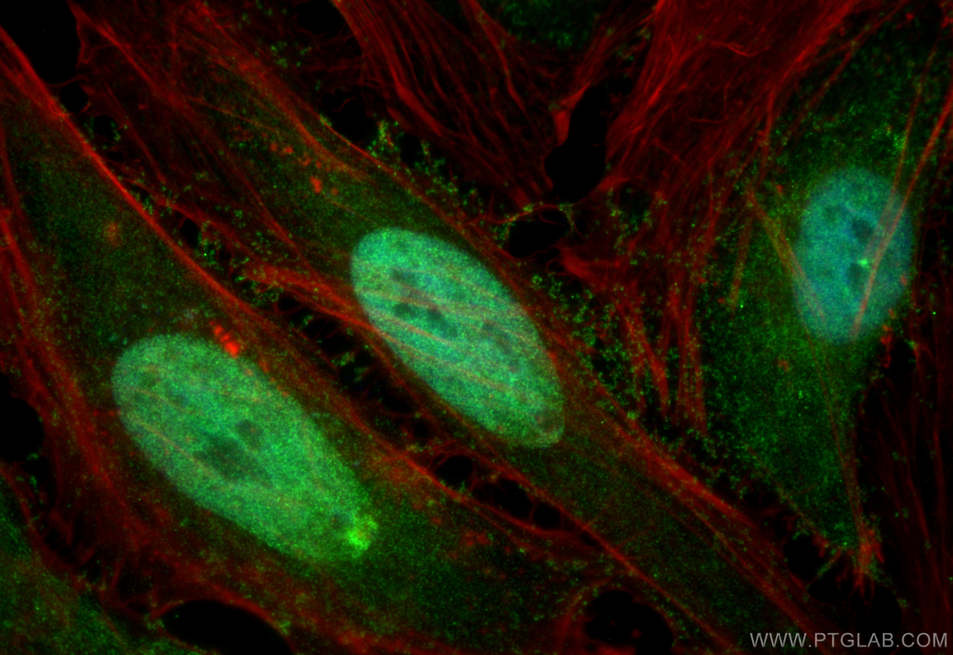
IF Staining of HeLa using 17532-1-AP
Immunofluorescent analysis of (4% PFA) fixed HeLa cells using CTDSPL2 antibody (17532-1-AP) at dilution of 1:200 and Multi-rAb CoraLite ® Plus 488-Goat Anti-Rabbit Recombinant Secondary Antibody (H+L) (RGAR002), CL594-phalloidin (red).
Immunofluorescent analysis of (4% PFA) fixed HeLa cells using CTDSPL2 antibody (17532-1-AP) at dilution of 1:200 and Multi-rAb CoraLite ® Plus 488-Goat Anti-Rabbit Recombinant Secondary Antibody (H+L) (RGAR002), CL594-phalloidin (red).
The Proteintech guarantee covers Proteintech antibodies in any species and any application, including those not listed on the datasheet. If the antibody doesn’t perform, you can receive a hassle-free refund or credit note.
MCF7 cells, HeLa cells, HepG2 cells, human brain tissue, mouse eye tissue, mouse testis tissue, Y79 cells
Positive IP detected in
mouse testis tissue
Positive IHC detected in
human endometrial cancer tissue, human testis tissue Note: suggested antigen retrieval with TE buffer pH 9.0; (*) Alternatively, antigen retrieval may be performed with citrate buffer pH 6.0
Positive IF/ICC detected in
HeLa cells
Recommended dilution
Application
Dilution
Western Blot (WB)
WB : 1:500-1:2400
Immunoprecipitation (IP)
IP : 0.5-4.0 ug for 1.0-3.0 mg of total protein lysate
Immunohistochemistry (IHC)
IHC : 1:20-1:200
Immunofluorescence (IF)/ICC
IF/ICC : 1:50-1:500
It is recommended that this reagent should be titrated in each testing system to obtain optimal results.
Sample-dependent, Check data in validation data gallery.
PBS with 0.02% sodium azide and 50% glycerol , pH 7.3
Storage Conditions
Store at -20°C. Stable for one year after shipment. Aliquoting is unnecessary for -20oC storage. 20ul sizes contain 0.1% BSA.
Background Information
CTD small phosphatase-like protein 2 (CTDSPL2) is also named as HSPC058 and HSPC129, belongs to the CTDSPL2 family. It is a putative RNA-polymerase II carboxy-terminal domain (CTD) phosphatase, whose specific CTD phosphatase activity was verified in vitro, and may regulate the dynamic phosphorylation of RNAP II CTD.( PMID: 17487459) It can obviously improve the expression of ε- and γ-globin genes in K562 cells and CD34+ cells derived from UCB.( PMID: 20932329) This antibody is a rabbit polyclonal antibody raised against a region of human CTDSPL2.
MCF7 cells were subjected to SDS PAGE followed by western blot with 17532-1-AP (CTDSPL2 antibody) at dilution of 1:500 incubated at room temperature for 1.5 hours.
WB analysis of mouse testis using 17532-1-AP
mouse testis tissue were subjected to SDS PAGE followed by western blot with 17532-1-AP (CTDSPL2 antibody) at dilution of 1:500 incubated at room temperature for 1.5 hours.
WB analysis of mouse eye using 17532-1-AP
mouse eye tissue were subjected to SDS PAGE followed by western blot with 17532-1-AP (CTDSPL2 antibody) at dilution of 1:600 incubated at room temperature for 1.5 hours.
WB analysis of HeLa using 17532-1-AP
HeLa cells were subjected to SDS PAGE followed by western blot with 17532-1-AP (CTDSPL2 antibody) at dilution of 1:500 incubated at room temperature for 1.5 hours.
WB analysis of human brain using 17532-1-AP
human brain tissue were subjected to SDS PAGE followed by western blot with 17532-1-AP (CTDSPL2 antibody) at dilution of 1:600 incubated at room temperature for 1.5 hours.
WB analysis of HeLa using 17532-1-AP
HeLa cells were subjected to SDS PAGE followed by western blot with 17532-1-AP (CTDSPL2 antibody) at dilution of 1:600 incubated at room temperature for 1.5 hours.
WB analysis of HepG2 using 17532-1-AP
HepG2 cells were subjected to SDS PAGE followed by western blot with 17532-1-AP (CTDSPL2 antibody) at dilution of 1:400 incubated at room temperature for 1.5 hours.
WB analysis of Y79 using 17532-1-AP
Y79 cells were subjected to SDS PAGE followed by western blot with 17532-1-AP (CTDSPL2 antibody) at dilution of 1:400 incubated at room temperature for 1.5 hours.
IHC Figures
IHC staining of human endometrial cancer using 17532-1-AP
Immunohistochemical analysis of paraffin-embedded human endometrial cancer using 17532-1-AP (CTDSPL2 antibody) at dilution of 1:100 (under 40x lens).
IHC staining of human testis using 17532-1-AP
Immunohistochemical analysis of paraffin-embedded human testis tissue slide using 17532-1-AP (CTDSPL2 Antibody) at dilution of 1:50 (under 40x lens).
IHC staining of human testis using 17532-1-AP
Immunohistochemical analysis of paraffin-embedded human testis tissue slide using 17532-1-AP (CTDSPL2 Antibody) at dilution of 1:50 (under 10x lens).
IHC staining of human endometrial cancer using 17532-1-AP
Immunohistochemical analysis of paraffin-embedded human endometrial cancer using 17532-1-AP (CTDSPL2 antibody) at dilution of 1:100 (under 10x lens).
IP Figures
IP experiment of mouse testis using 17532-1-AP
IP result of anti-CTDSPL2 (IP:17532-1-AP, 4ug; Detection:17532-1-AP 1:500) with mouse testis tissue lysate 4400ug.
IF/ICC Figures
IF Staining of HeLa using 17532-1-AP
Immunofluorescent analysis of (4% PFA) fixed HeLa cells using CTDSPL2 antibody (17532-1-AP) at dilution of 1:200 and Multi-rAb CoraLite ® Plus 488-Goat Anti-Rabbit Recombinant Secondary Antibody (H+L) (RGAR002), CL594-phalloidin (red).
The species listed in Tested Reactivity are in-house verified and applicable species. For unlisted species, please refer to the homology analysis of the immunogen sequence and related species. For rabbit polyclonal antibodies, homology >70% is recommended. For mouse monoclonal antibodies and rabbit recombinant antibodies, homology >90% is recommended. Generally, the higher the homology, the greater the applicability. However, there will be certain differences in protein expression in different species, tissues or cells. Therefore, the homology analysis results are for reference only and do not serve as a guarantee.
At Proteintech, we pride ourselves on our antibody quality, customer service and transparency. As such, we are comparing our antibodies with other vendors, enabling easy identification and comparisons of key data to help you choose the suitable antibody for your needs.
We have selected the top cited antibodies from these vendors for you to compare.
Proteintech
CTDSPL2 Polyclonal antibody
Catalog Number
17532-1-AP
Citations
3
Dilutions
WB : 1:500-1:2400 IP : 0.5-4.0 ug for IP and 0.5-4.0 ug for 1.0-3.0 mg of total protein lysate for WB IHC : 1:20-1:200 IF/ICC : 1:50-1:500
Applications
WB, IHC, IF/ICC, IP, ELISA
Reactivity
human, mouse, rat
Product Guarantee
Covers any species including not listed on datasheet
Covers any applications including not listed on datasheet
 at dilution of 1:500 incubated at room temperature for 1.5 hours.")
 at dilution of 1:500 incubated at room temperature for 1.5 hours.")
 at dilution of 1:600 incubated at room temperature for 1.5 hours.")
 at dilution of 1:500 incubated at room temperature for 1.5 hours.")
 at dilution of 1:600 incubated at room temperature for 1.5 hours.")
 at dilution of 1:600 incubated at room temperature for 1.5 hours.")
 at dilution of 1:400 incubated at room temperature for 1.5 hours.")
 at dilution of 1:400 incubated at room temperature for 1.5 hours.")
 with mouse testis tissue lysate 4400ug.")
 at dilution of 1:100 (under 40x lens).")
 at dilution of 1:50 (under 40x lens).")
 at dilution of 1:50 (under 10x lens).")
 at dilution of 1:100 (under 10x lens).")
 fixed HeLa cells using CTDSPL2 antibody (17532-1-AP) at dilution of 1:200 and Multi-rAb CoraLite ® Plus 488-Goat Anti-Rabbit Recombinant Secondary Antibody (H+L) (RGAR002), CL594-phalloidin (red).")