Tested Applications
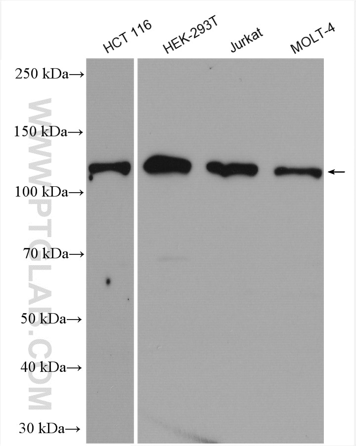
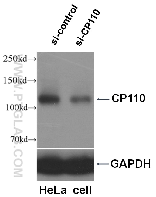
| Positive WB detected in | HCT 116 cells, HeLa cells, HEK-293T cells, Jurkat cells, MOLT-4 cells |
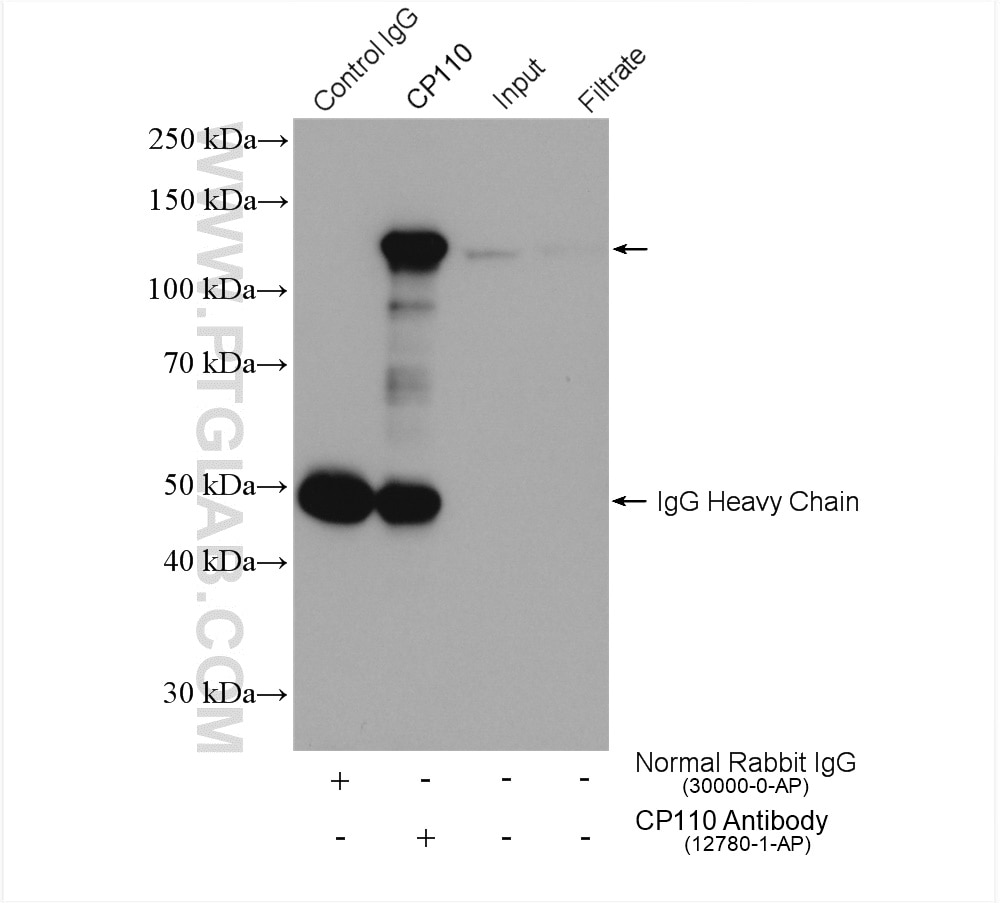
| Positive IP detected in | Jurkat cells |
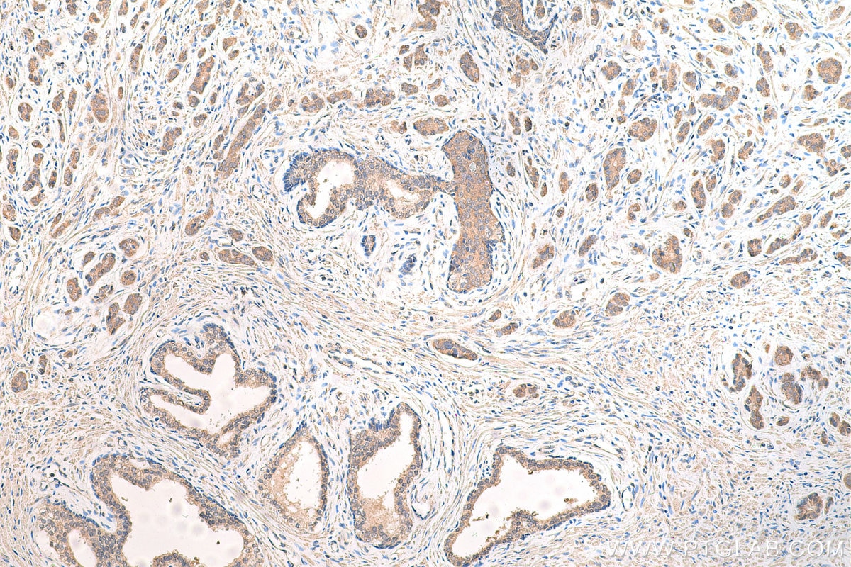
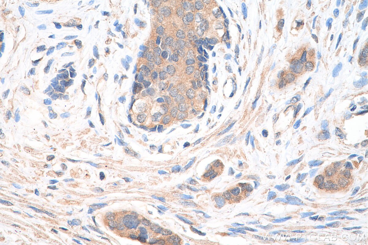
| Positive IHC detected in | human prostate cancer tissue Note: suggested antigen retrieval with TE buffer pH 9.0; (*) Alternatively, antigen retrieval may be performed with citrate buffer pH 6.0 |
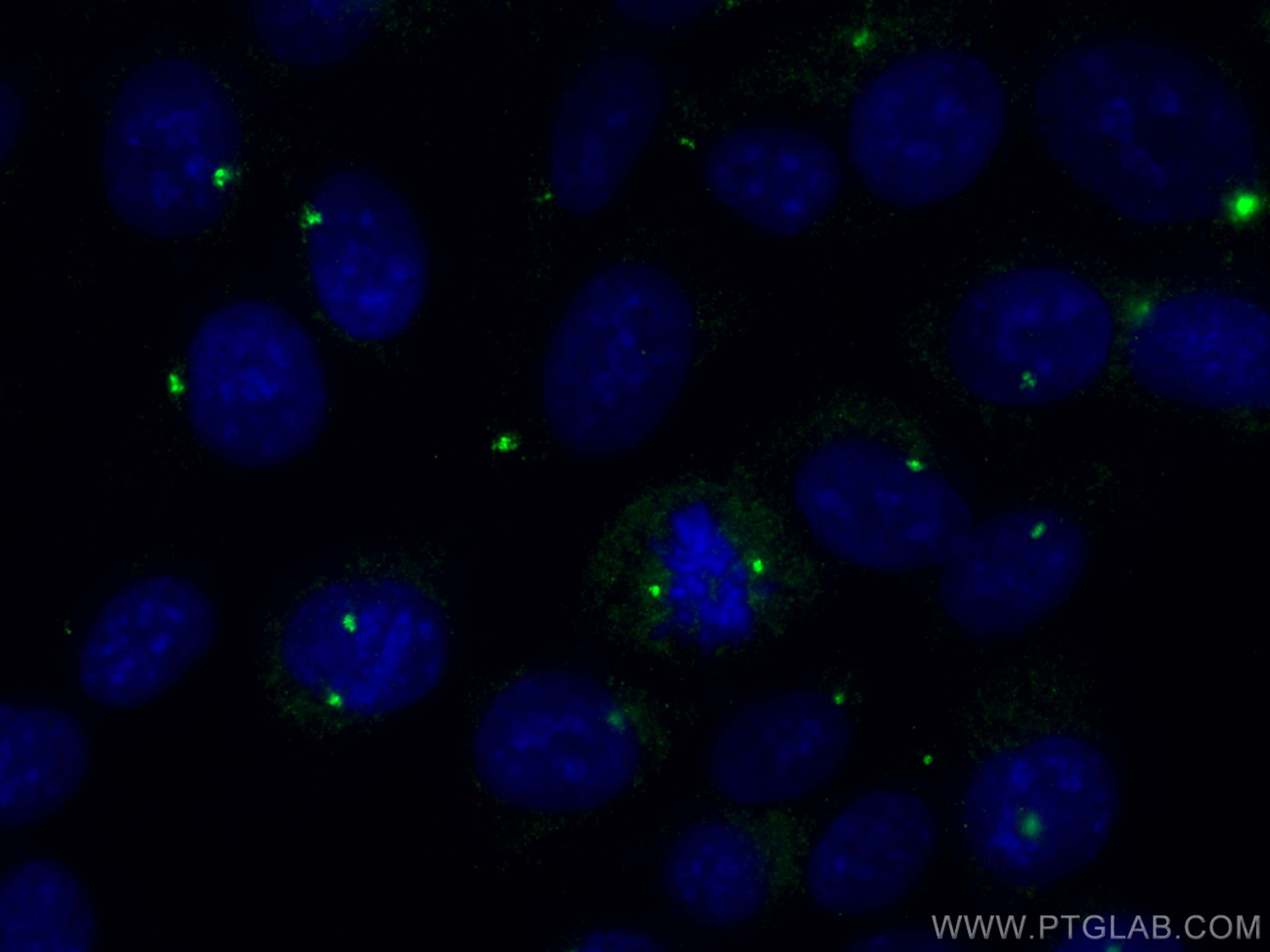
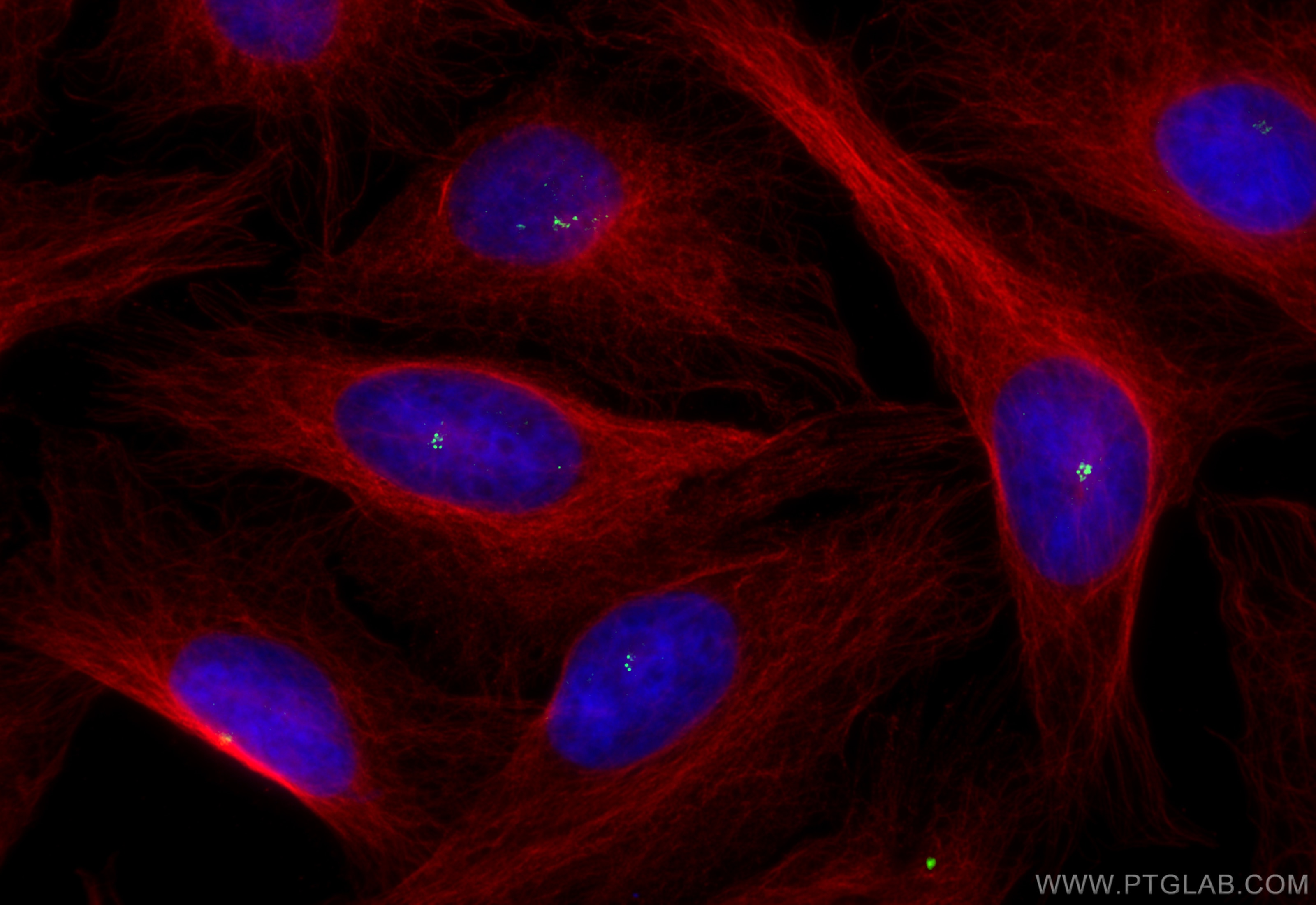
| Positive IF/ICC detected in | hTERT-RPE1 cells, U2OS cells |
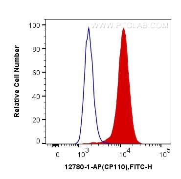
| Positive FC (Intra) detected in | HeLa cells |
Recommended dilution
| Application | Dilution |
|---|---|
| Western Blot (WB) | WB : 1:2000-1:12000 |
| Immunoprecipitation (IP) | IP : 0.5-4.0 ug for 1.0-3.0 mg of total protein lysate |
| Immunohistochemistry (IHC) | IHC : 1:250-1:1000 |
| Immunofluorescence (IF)/ICC | IF/ICC : 1:50-1:500 |
| Flow Cytometry (FC) (INTRA) | FC (INTRA) : 0.20 ug per 10^6 cells in a 100 µl suspension |
| It is recommended that this reagent should be titrated in each testing system to obtain optimal results. | |
| Sample-dependent, Check data in validation data gallery. | |
Published Applications
| KD/KO | See 7 publications below |
| WB | See 42 publications below |
| IHC | See 1 publications below |
| IF | See 134 publications below |
| IP | See 6 publications below |
Product Information
12780-1-AP targets CP110 in WB, IHC, IF/ICC, FC (Intra), IP, ELISA applications and shows reactivity with human samples.
| Tested Reactivity | human |
| Cited Reactivity | human, canine, zebrafish, drosophila, xenopus |
| Host / Isotype | Rabbit / IgG |
| Class | Polyclonal |
| Type | Antibody |
| Immunogen |
CatNo: Ag3489 Product name: Recombinant human CP110 protein Source: e coli.-derived, PGEX-4T Tag: GST Domain: 1-337 aa of BC036654 Sequence: MEEYEKFCEKSLARIQEASLSTESFLPAQSESISLIRFHGVAILSPLLNIEKRKEMQQEKQKALDVEARKQVNRKKALLTRVQEILDNVQVRKAPNASDFDQWEMETVYSNSEVRNLNVPATFPNSFPSHTEHSTAAKLDKIAGILPLDNEDQCKTDGIDLARDSEGFNSPKQCDSSNISHVENEAFPKTSSATPQETLISDGPFSVNEQQDLPLLAEVIPDPYVMSLQNLMKKSKEYIEREQSRRSLRGSMNRIVNESHLDKEHDAVEVADCVKEKGQLTGKHCVSVIPDKPSLNKSNVLLQGASTQASSMSMPVLASFSKVDIPIRTGHPTVLES Predict reactive species |
| Full Name | CP110 protein |
| Calculated Molecular Weight | 991 aa, 111 kDa |
| Observed Molecular Weight | 113 kDa |
| GenBank Accession Number | BC036654 |
| Gene Symbol | CP110 |
| Gene ID (NCBI) | 9738 |
| RRID | AB_10638480 |
| Conjugate | Unconjugated |
| Form | Liquid |
| Purification Method | Antigen affinity purification |
| UNIPROT ID | O43303 |
| Storage Buffer | PBS with 0.02% sodium azide and 50% glycerol, pH 7.3. |
| Storage Conditions | Store at -20°C. Stable for one year after shipment. Aliquoting is unnecessary for -20oC storage. 20ul sizes contain 0.1% BSA. |
Background Information
CP110, also named as CCP110 and KIAA0419, is an 110kDa protein. This gene is different to the gene CEP110 (geneID:11064; CNTRL). CP110 is a centriolar protein that positively regulates centrioleduplication while restricting centriole elongation and ciliogenesis. And it acts as a key negative regulator of ciliogenesis in collaboration with CEP97 by capping the mother centriole thereby preventing cilia formation.
Protocols
| Product Specific Protocols | |
|---|---|
| FC protocol for CP110 antibody 12780-1-AP | Download protocol |
| IF protocol for CP110 antibody 12780-1-AP | Download protocol |
| IHC protocol for CP110 antibody 12780-1-AP | Download protocol |
| IP protocol for CP110 antibody 12780-1-AP | Download protocol |
| WB protocol for CP110 antibody 12780-1-AP | Download protocol |
| Standard Protocols | |
|---|---|
| Click here to view our Standard Protocols |
Publications
| Species | Application | Title |
|---|---|---|
Science A liquid-like spindle domain promotes acentrosomal spindle assembly in mammalian oocytes. | ||
Nature USP33 regulates centrosome biogenesis via deubiquitination of the centriolar protein CP110. | ||
Nat Nanotechnol Intrinsic bioactivity of black phosphorus nanomaterials on mitotic centrosome destabilization through suppression of PLK1 kinase. | ||
Nat Cell Biol miR-129-3p controls cilia assembly by regulating CP110 and actin dynamics.
| ||
Nat Cell Biol The Cep63 paralogue Deup1 enables massive de novo centriole biogenesis for vertebrate multiciliogenesis. | ||
Nat Cell Biol CEP162 is an axoneme-recognition protein promoting ciliary transition zone assembly at the cilia base. |
Reviews
The reviews below have been submitted by verified Proteintech customers who received an incentive for providing their feedback.
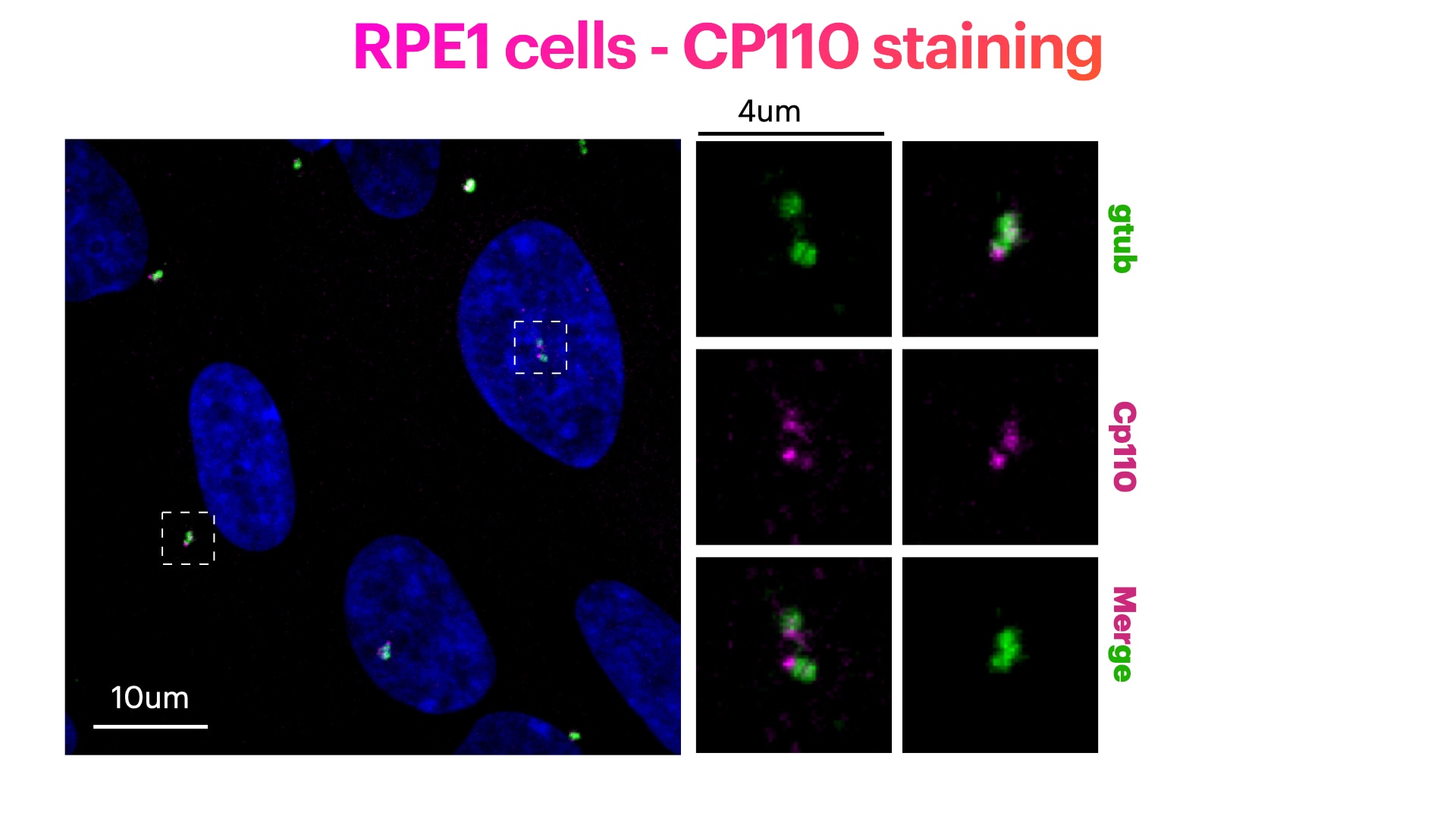
FH Elisa (Verified Customer) (01-05-2023) | RPE1 cells stained for Hoechst (DNA marker, in green), CP110 (in magenta) and gtub (pericentriolar matrix marker, in green). RPE1 cells were fixed in cold methanol for 10' at -20C. Cells were then rehydrated with PBS for 5'. Membrane permeabilization was then performed with 0.1% Triton + 0.1% Tween +0.01%SDS in PBS for 5'. Cells were finally incubated with blocking buffer (5% BSA+ 0.1% Tween in PBS) for 30' at RT. Primary antibody was diluted in blocking buffer 1:200 and incubated for 1h at room temperature. Alexa-488-Anti-rabbit was used as secondary antibody (1:600 dilution) (1h at room temperature).
 |
FH Sarah (Verified Customer) (02-16-2021) | Works very well for detection of CP110 protein using western blotting and immunfluorescence
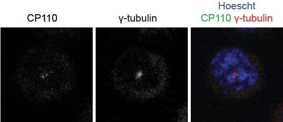 |
FH EK (Verified Customer) (05-04-2020) | works well for immunofluorescence detection of basal body localization
|

