Various lysates were subjected to SDS PAGE followed by western blot with 10256-1-AP (COPS6 antibody) at dilution of 1:1500 incubated at room temperature for 1.5 hours.
Various lysates were subjected to SDS PAGE followed by western blot with 10256-1-AP (COPS6 antibody) at dilution of 1:1500 incubated at room temperature for 1.5 hours.
WB analysis of 3T3-L1 using 10256-1-AP
3T3-L1 cells were subjected to SDS PAGE followed by western blot with 10256-1-AP (COPS6 antibody) at dilution of 1:600 incubated at room temperature for 1.5 hours.
3T3-L1 cells were subjected to SDS PAGE followed by western blot with 10256-1-AP (COPS6 antibody) at dilution of 1:600 incubated at room temperature for 1.5 hours.
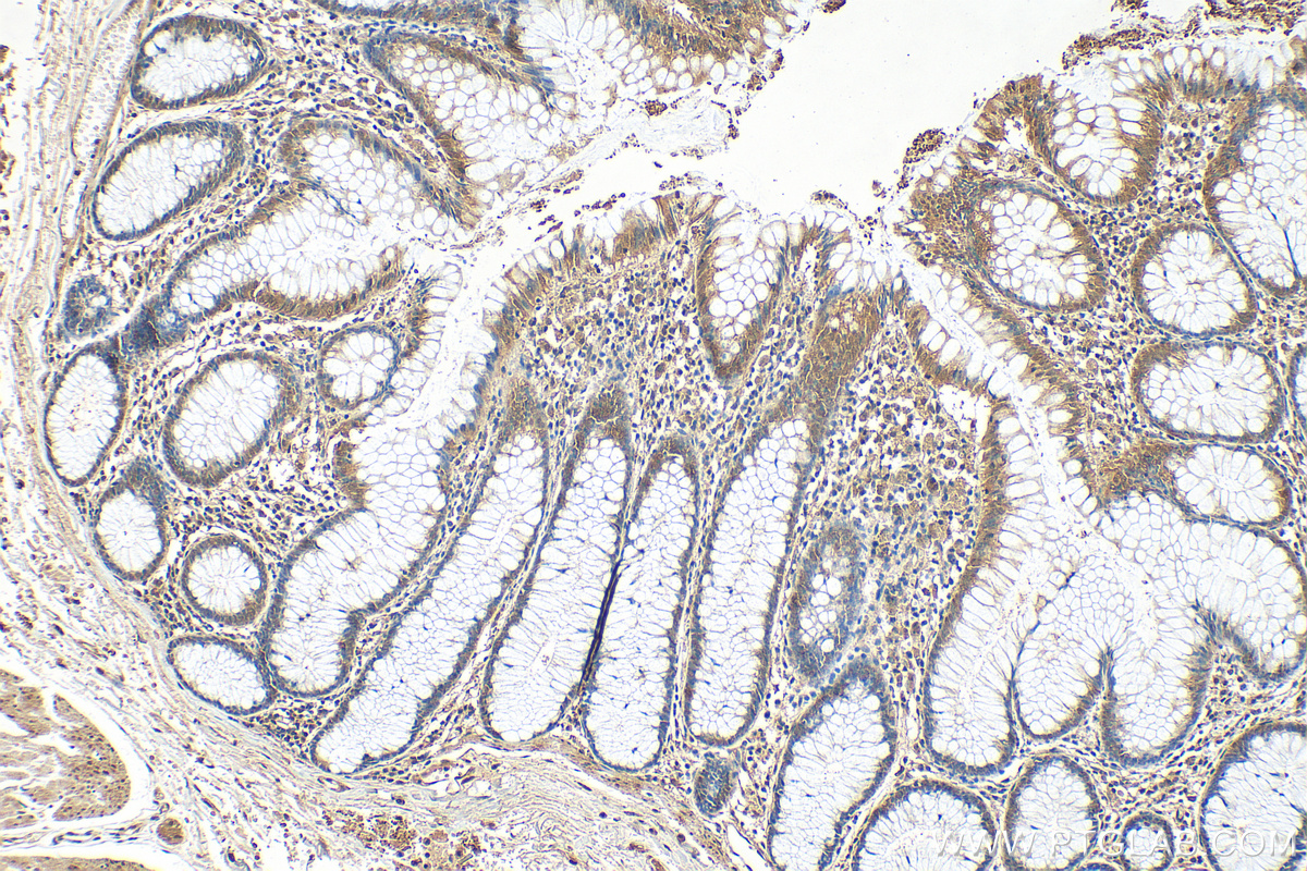
IHC staining of human colon using 10256-1-AP
Immunohistochemical analysis of paraffin-embedded human colon tissue slide using 10256-1-AP (COPS6 antibody) at dilution of 1:200 (under 10x lens). Heat mediated antigen retrieval with Tris-EDTA buffer (pH 9.0).
Immunohistochemical analysis of paraffin-embedded human colon tissue slide using 10256-1-AP (COPS6 antibody) at dilution of 1:200 (under 10x lens). Heat mediated antigen retrieval with Tris-EDTA buffer (pH 9.0).
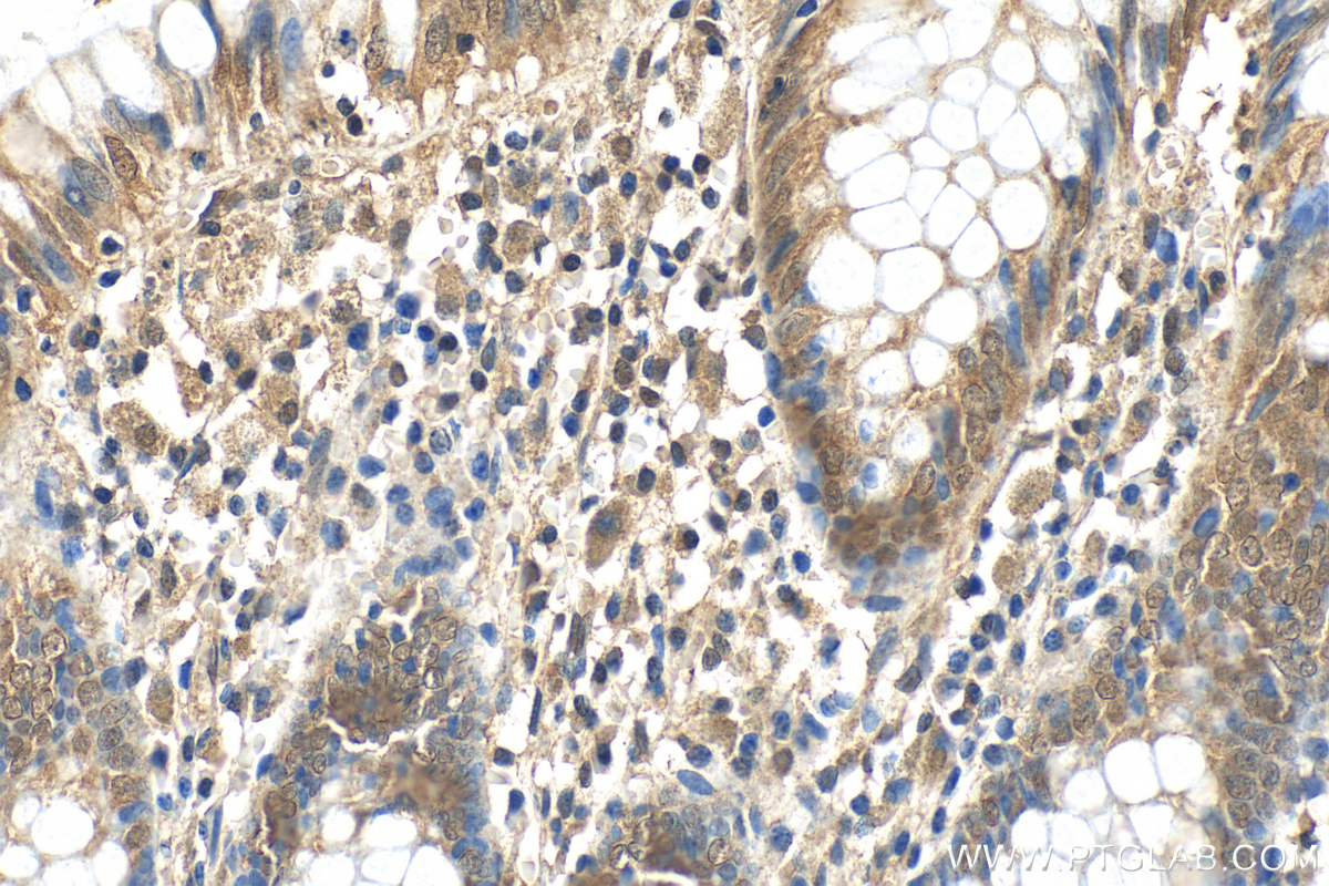
IHC staining of human colon using 10256-1-AP
Immunohistochemical analysis of paraffin-embedded human colon tissue slide using 10256-1-AP (COPS6 antibody) at dilution of 1:200 (under 40x lens). Heat mediated antigen retrieval with Tris-EDTA buffer (pH 9.0).
Immunohistochemical analysis of paraffin-embedded human colon tissue slide using 10256-1-AP (COPS6 antibody) at dilution of 1:200 (under 40x lens). Heat mediated antigen retrieval with Tris-EDTA buffer (pH 9.0).
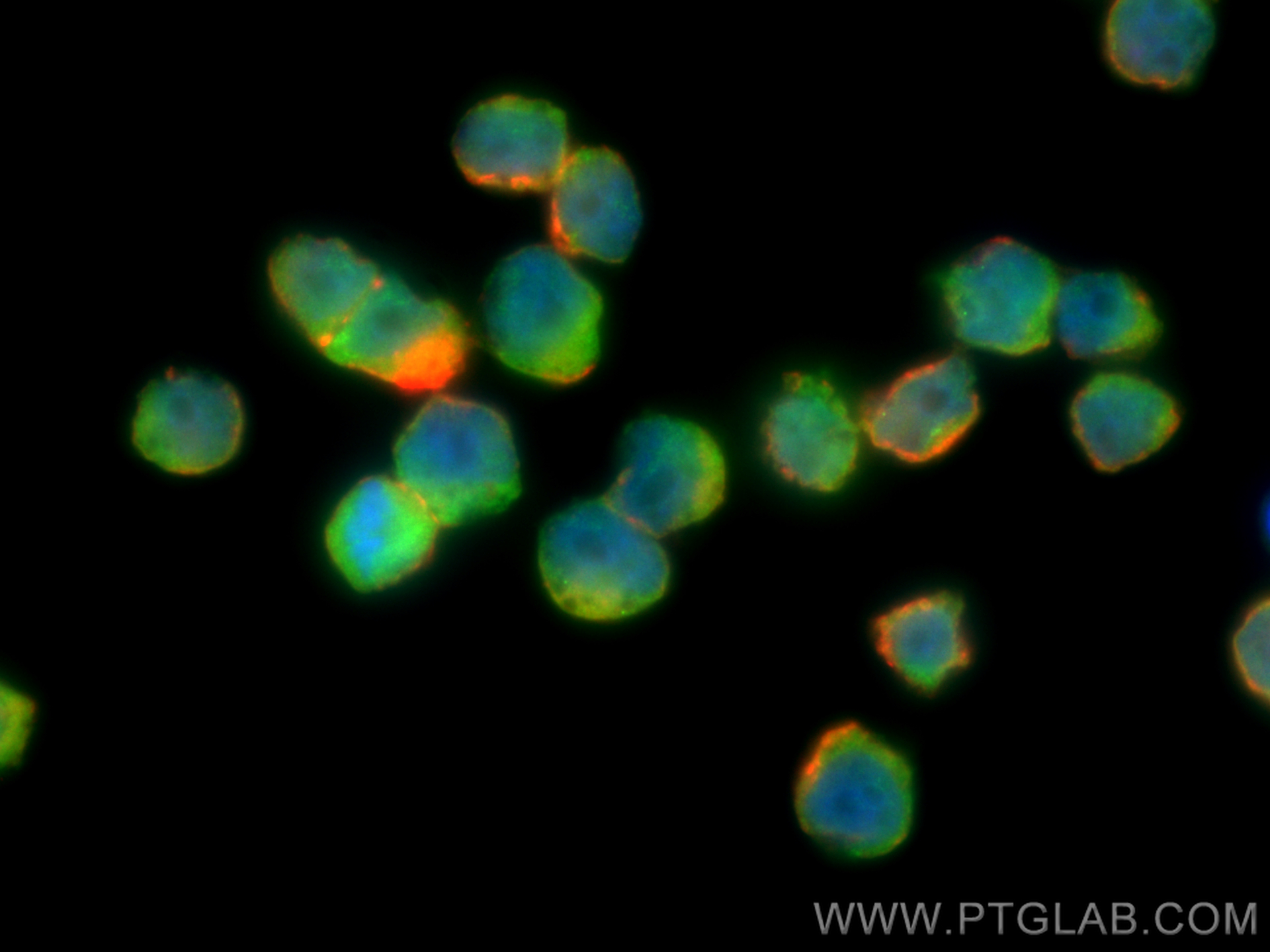
IF Staining of COLO 320 using 10256-1-AP
Immunofluorescent analysis of (4% PFA) fixed COLO 320 cells using COPS6 antibody (10256-1-AP) at dilution of 1:400 and Multi-rAb CoraLite ® Plus 488-Goat Anti-Rabbit Recombinant Secondary Antibody (H+L) (RGAR002), CL594-Phalloidin (red).
Immunofluorescent analysis of (4% PFA) fixed COLO 320 cells using COPS6 antibody (10256-1-AP) at dilution of 1:400 and Multi-rAb CoraLite ® Plus 488-Goat Anti-Rabbit Recombinant Secondary Antibody (H+L) (RGAR002), CL594-Phalloidin (red).
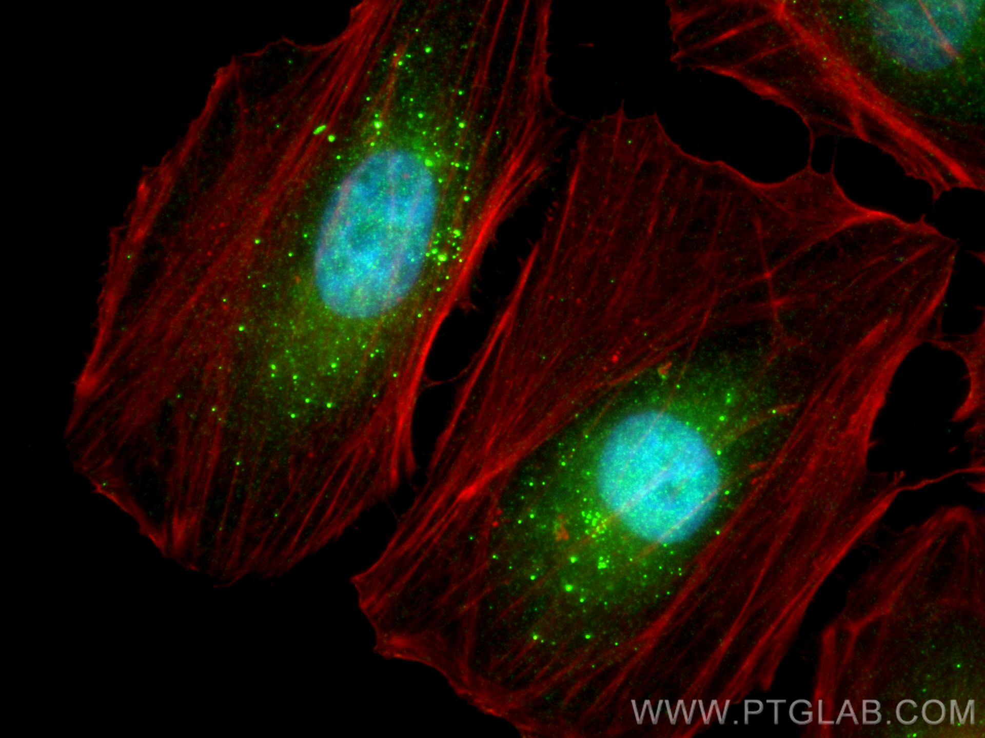
IF Staining of HeLa using 10256-1-AP
Immunofluorescent analysis of (4% PFA) fixed HeLa cells using COPS6 antibody (10256-1-AP) at dilution of 1:400 and Multi-rAb CoraLite ® Plus 488-Goat Anti-Rabbit Recombinant Secondary Antibody (H+L) (RGAR002), CL594-Phalloidin (red).
Immunofluorescent analysis of (4% PFA) fixed HeLa cells using COPS6 antibody (10256-1-AP) at dilution of 1:400 and Multi-rAb CoraLite ® Plus 488-Goat Anti-Rabbit Recombinant Secondary Antibody (H+L) (RGAR002), CL594-Phalloidin (red).
The Proteintech guarantee covers Proteintech antibodies in any species and any application, including those not listed on the datasheet. If the antibody doesn’t perform, you can receive a hassle-free refund or credit note.
human colon tissue Note: suggested antigen retrieval with TE buffer pH 9.0; (*) Alternatively, antigen retrieval may be performed with citrate buffer pH 6.0
Positive IF/ICC detected in
HeLa cells, COLO 320 cells
Recommended dilution
Application
Dilution
Western Blot (WB)
WB : 1:500-1:3000
Immunohistochemistry (IHC)
IHC : 1:50-1:500
Immunofluorescence (IF)/ICC
IF/ICC : 1:200-1:800
It is recommended that this reagent should be titrated in each testing system to obtain optimal results.
Sample-dependent, Check data in validation data gallery.
PBS with 0.02% sodium azide and 50% glycerol , pH 7.3
Storage Conditions
Store at -20°C. Stable for one year after shipment. Aliquoting is unnecessary for -20oC storage. 20ul sizes contain 0.1% BSA.
Background Information
The COPS6 protein, also named mov34, is a 34 kDa subunit of COP9 signalosome, a highly conserved eight-subunit protein complex that functions as an important regulator in multiple signaling pathways (PMID: 38308184). This protein belongs to translation initiation factor 3 (eIF3) superfamily. It is involved in the regulation of cell cycle and acts as the regulatory subunit of the 26S proteasome.
Various lysates were subjected to SDS PAGE followed by western blot with 10256-1-AP (COPS6 antibody) at dilution of 1:1500 incubated at room temperature for 1.5 hours.
WB analysis of 3T3-L1 using 10256-1-AP
3T3-L1 cells were subjected to SDS PAGE followed by western blot with 10256-1-AP (COPS6 antibody) at dilution of 1:600 incubated at room temperature for 1.5 hours.
IHC Figures
IHC staining of human colon using 10256-1-AP
Immunohistochemical analysis of paraffin-embedded human colon tissue slide using 10256-1-AP (COPS6 antibody) at dilution of 1:200 (under 10x lens). Heat mediated antigen retrieval with Tris-EDTA buffer (pH 9.0).
IHC staining of human colon using 10256-1-AP
Immunohistochemical analysis of paraffin-embedded human colon tissue slide using 10256-1-AP (COPS6 antibody) at dilution of 1:200 (under 40x lens). Heat mediated antigen retrieval with Tris-EDTA buffer (pH 9.0).
IF/ICC Figures
IF Staining of COLO 320 using 10256-1-AP
Immunofluorescent analysis of (4% PFA) fixed COLO 320 cells using COPS6 antibody (10256-1-AP) at dilution of 1:400 and Multi-rAb CoraLite ® Plus 488-Goat Anti-Rabbit Recombinant Secondary Antibody (H+L) (RGAR002), CL594-Phalloidin (red).
IF Staining of HeLa using 10256-1-AP
Immunofluorescent analysis of (4% PFA) fixed HeLa cells using COPS6 antibody (10256-1-AP) at dilution of 1:400 and Multi-rAb CoraLite ® Plus 488-Goat Anti-Rabbit Recombinant Secondary Antibody (H+L) (RGAR002), CL594-Phalloidin (red).
The species listed in Tested Reactivity are in-house verified and applicable species. For unlisted species, please refer to the homology analysis of the immunogen sequence and related species. For rabbit polyclonal antibodies, homology >70% is recommended. For mouse monoclonal antibodies and rabbit recombinant antibodies, homology >90% is recommended. Generally, the higher the homology, the greater the applicability. However, there will be certain differences in protein expression in different species, tissues or cells. Therefore, the homology analysis results are for reference only and do not serve as a guarantee.
At Proteintech, we pride ourselves on our antibody quality, customer service and transparency. As such, we are comparing our antibodies with other vendors, enabling easy identification and comparisons of key data to help you choose the suitable antibody for your needs.
We have selected the top cited antibodies from these vendors for you to compare.
 at dilution of 1:1500 incubated at room temperature for 1.5 hours.")
 at dilution of 1:600 incubated at room temperature for 1.5 hours.")
 at dilution of 1:200 (under 10x lens). Heat mediated antigen retrieval with Tris-EDTA buffer (pH 9.0).")
 at dilution of 1:200 (under 40x lens). Heat mediated antigen retrieval with Tris-EDTA buffer (pH 9.0).")
 fixed COLO 320 cells using COPS6 antibody (10256-1-AP) at dilution of 1:400 and Multi-rAb CoraLite ® Plus 488-Goat Anti-Rabbit Recombinant Secondary Antibody (H+L) (RGAR002), CL594-Phalloidin (red).")
 fixed HeLa cells using COPS6 antibody (10256-1-AP) at dilution of 1:400 and Multi-rAb CoraLite ® Plus 488-Goat Anti-Rabbit Recombinant Secondary Antibody (H+L) (RGAR002), CL594-Phalloidin (red).")