Tested Applications
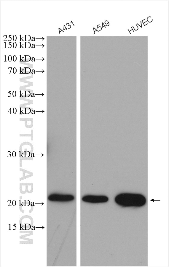
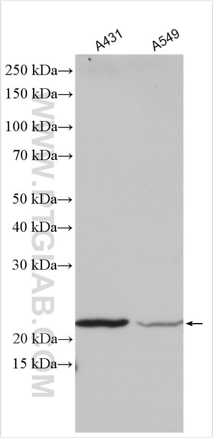
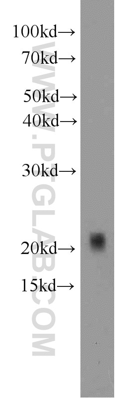
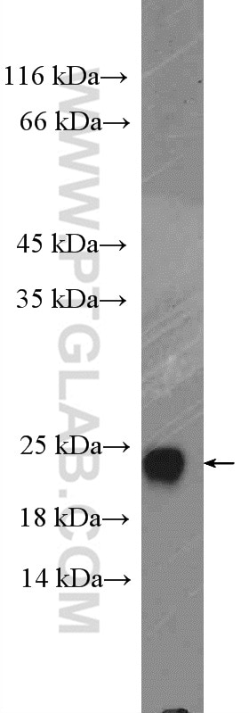
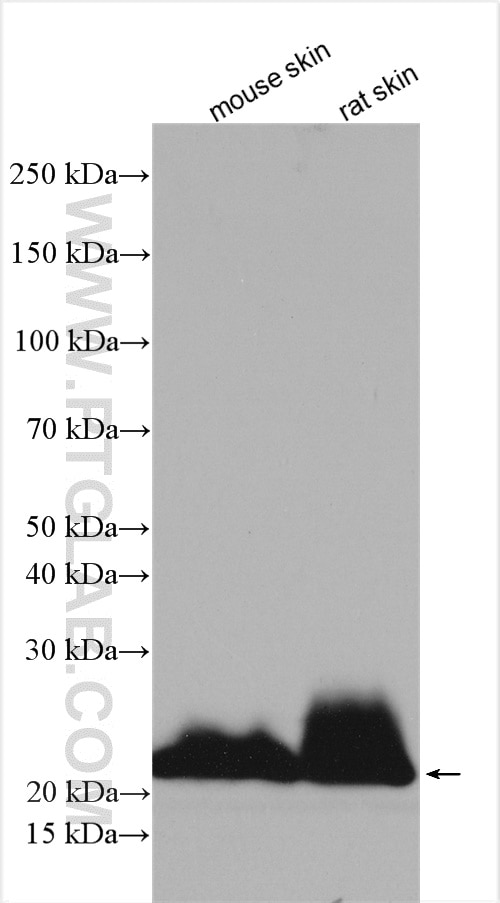
| Positive WB detected in | A431 cells, mouse liver tissue, mouse skin tissue, mouse thymus tissue, A549 cells, HUVEC cells, rat skin tissue |
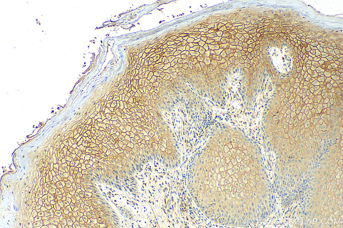
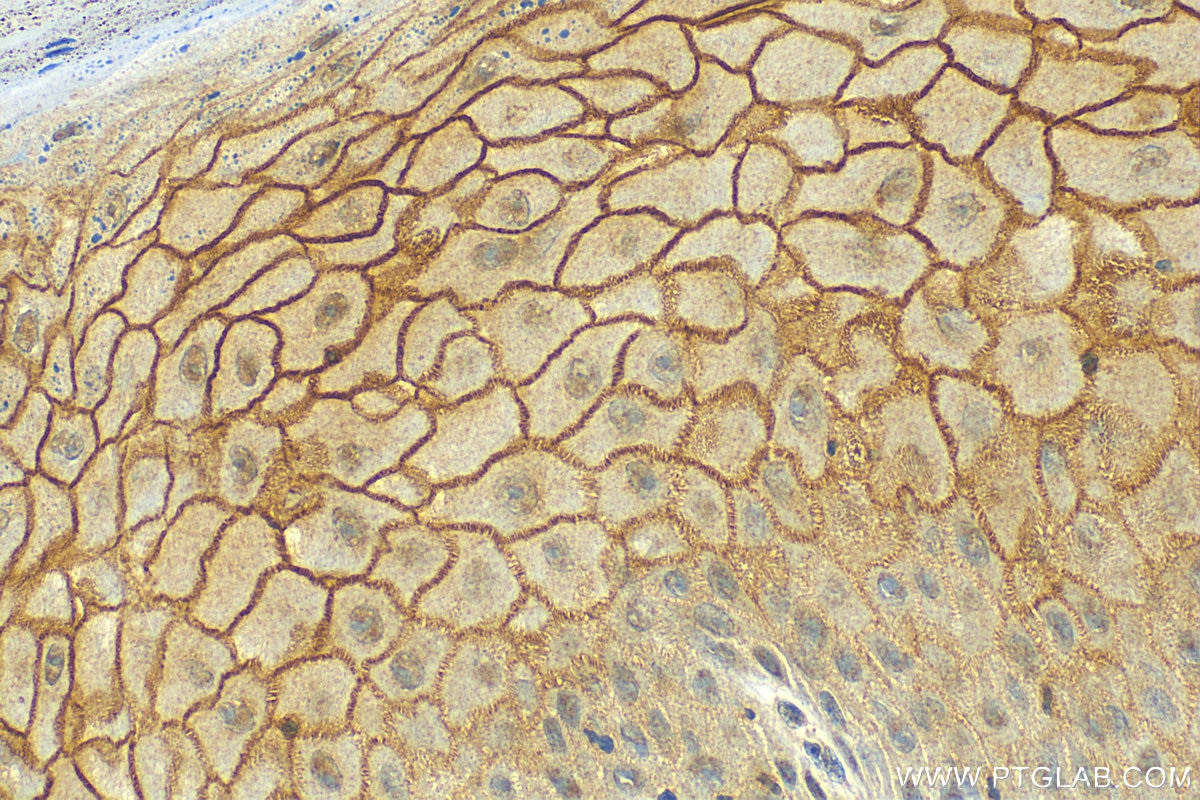
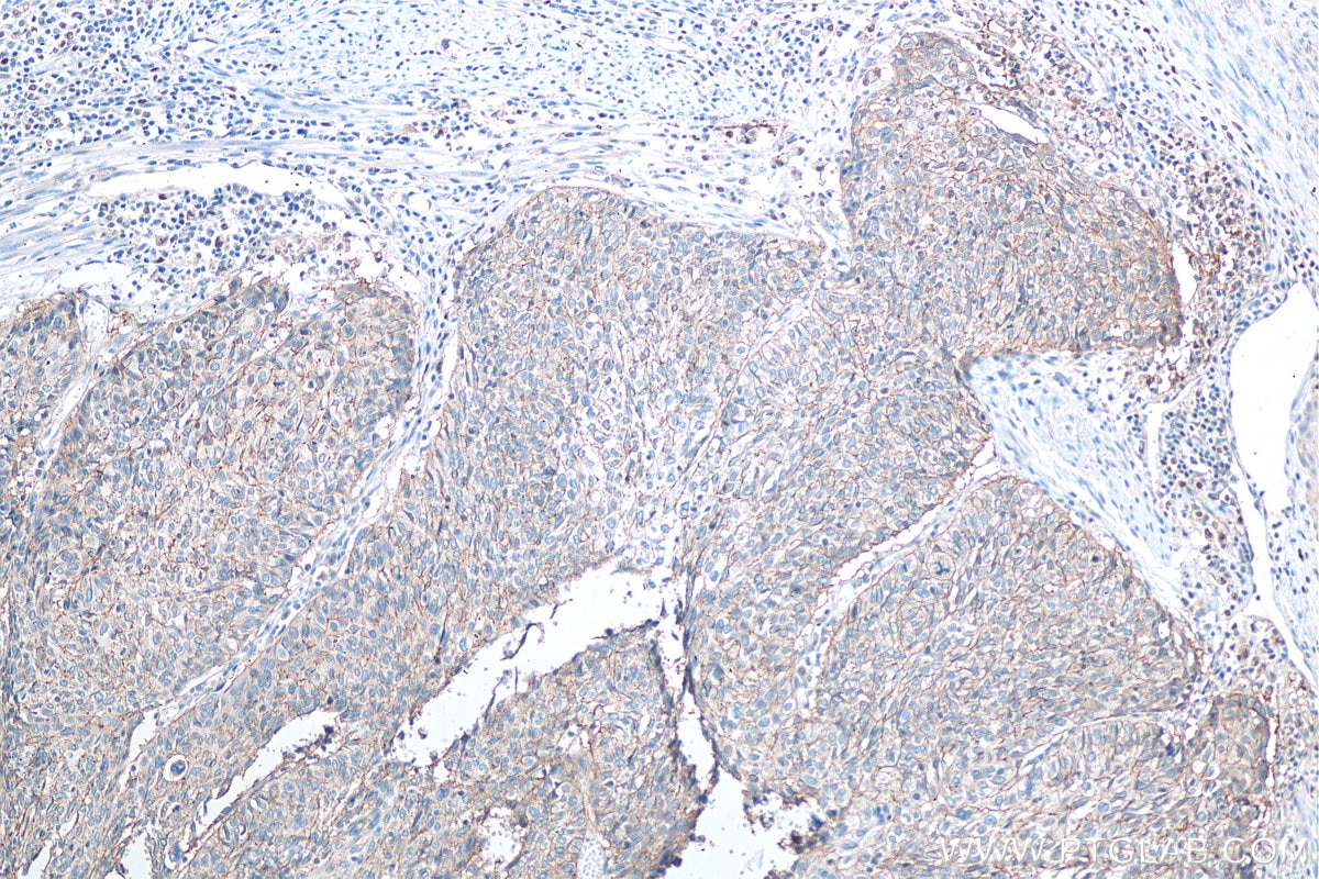
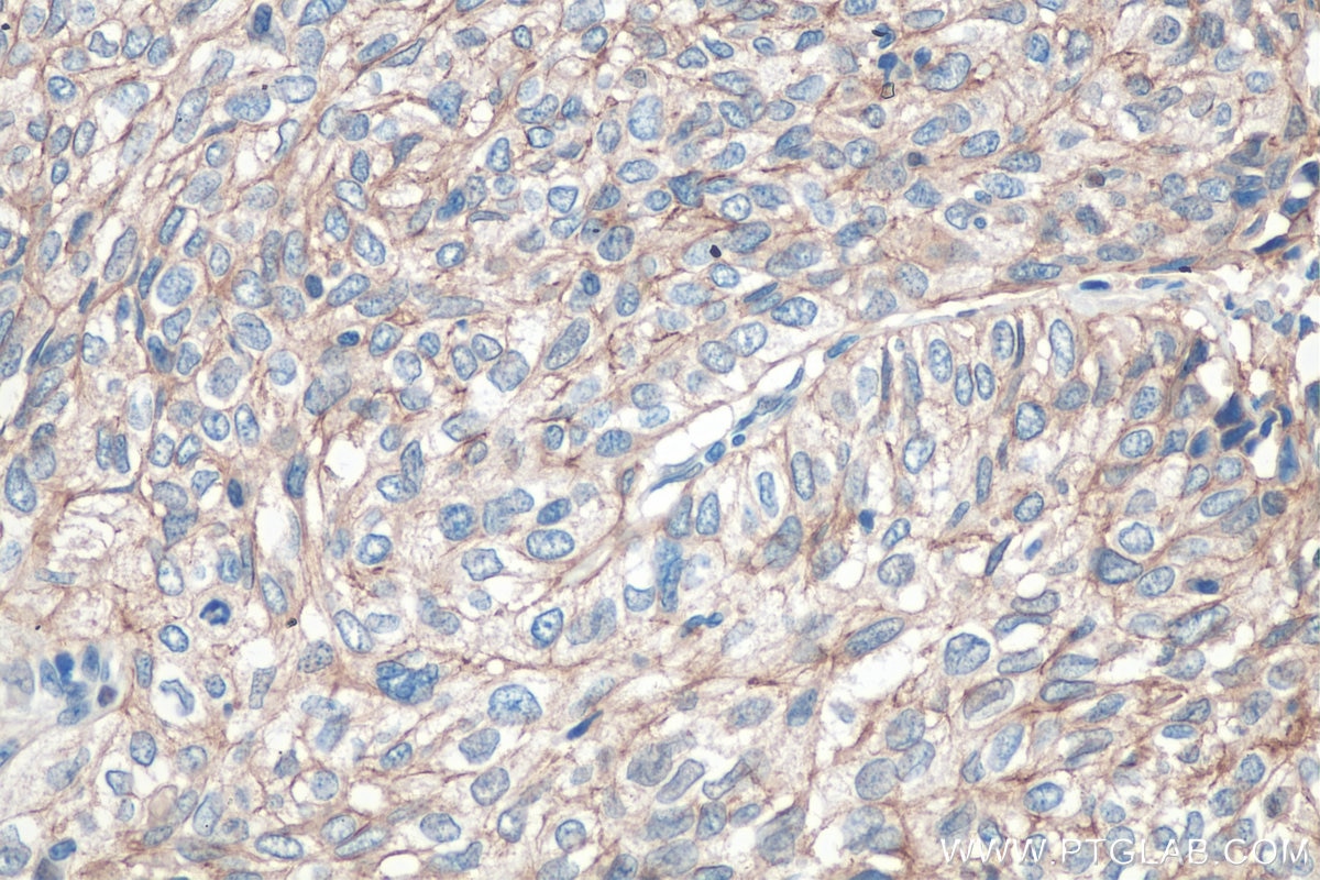
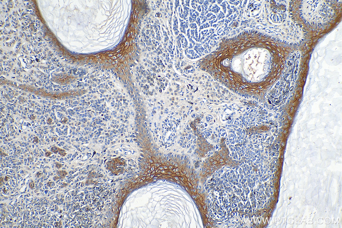
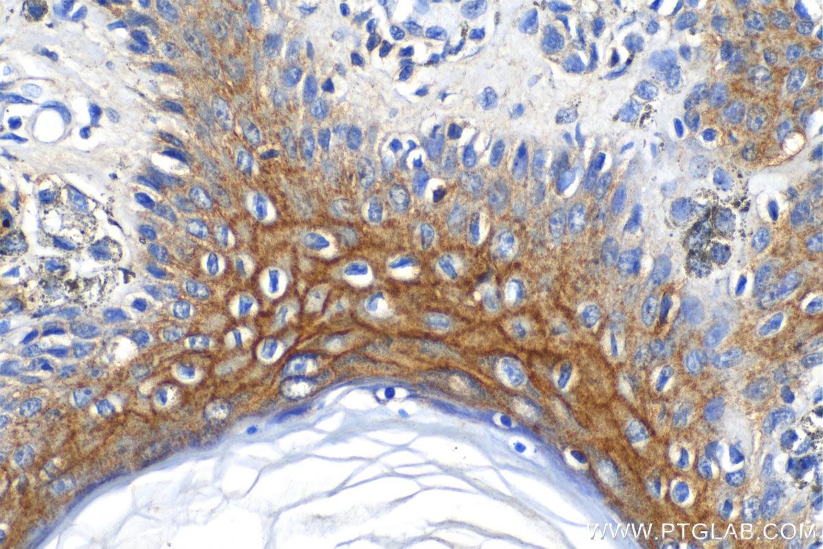
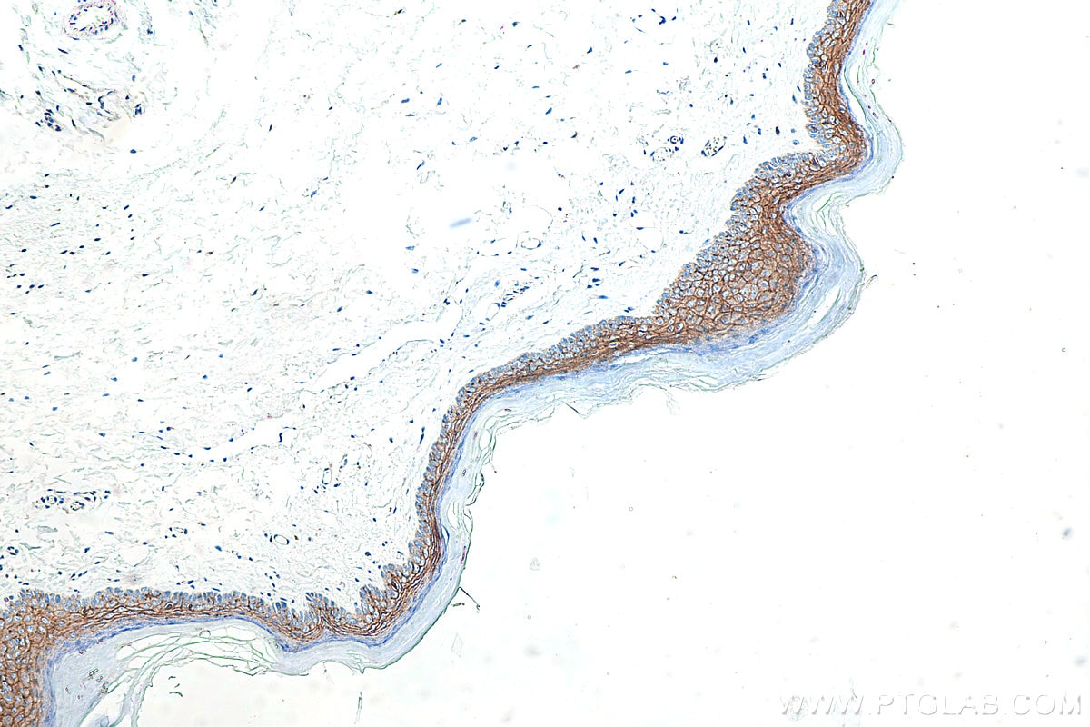
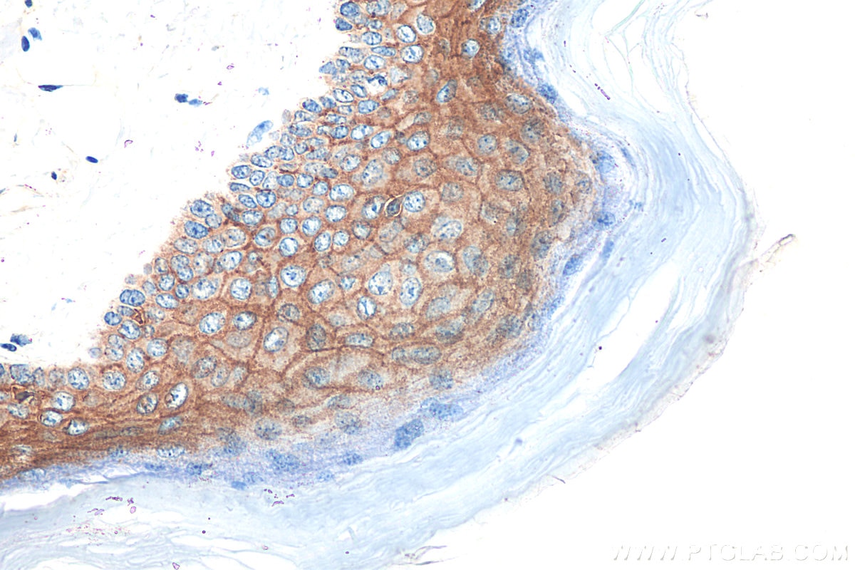
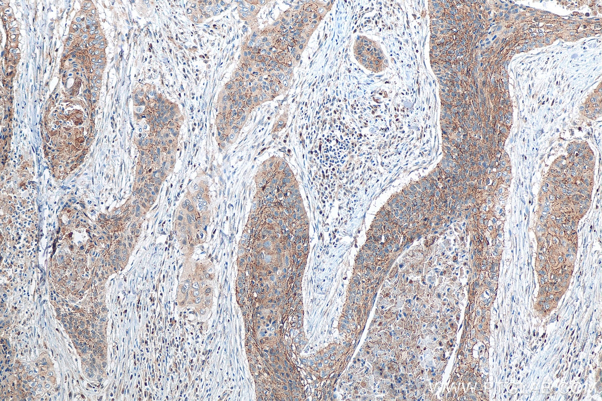
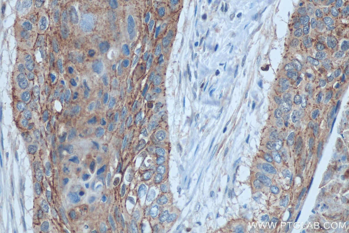
| Positive IHC detected in | human cervical cancer tissue, human skin cancer tissue, mouse skin tissue, human oesophagus cancer tissue, human malignant melanoma tissue Note: suggested antigen retrieval with TE buffer pH 9.0; (*) Alternatively, antigen retrieval may be performed with citrate buffer pH 6.0 |
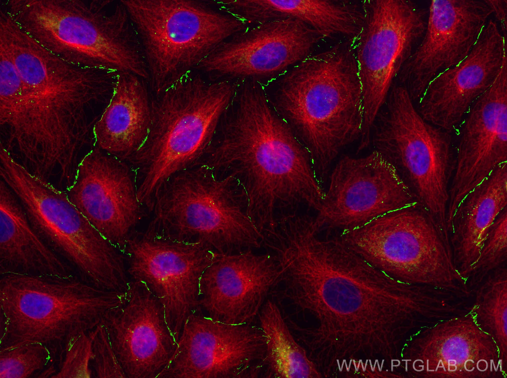
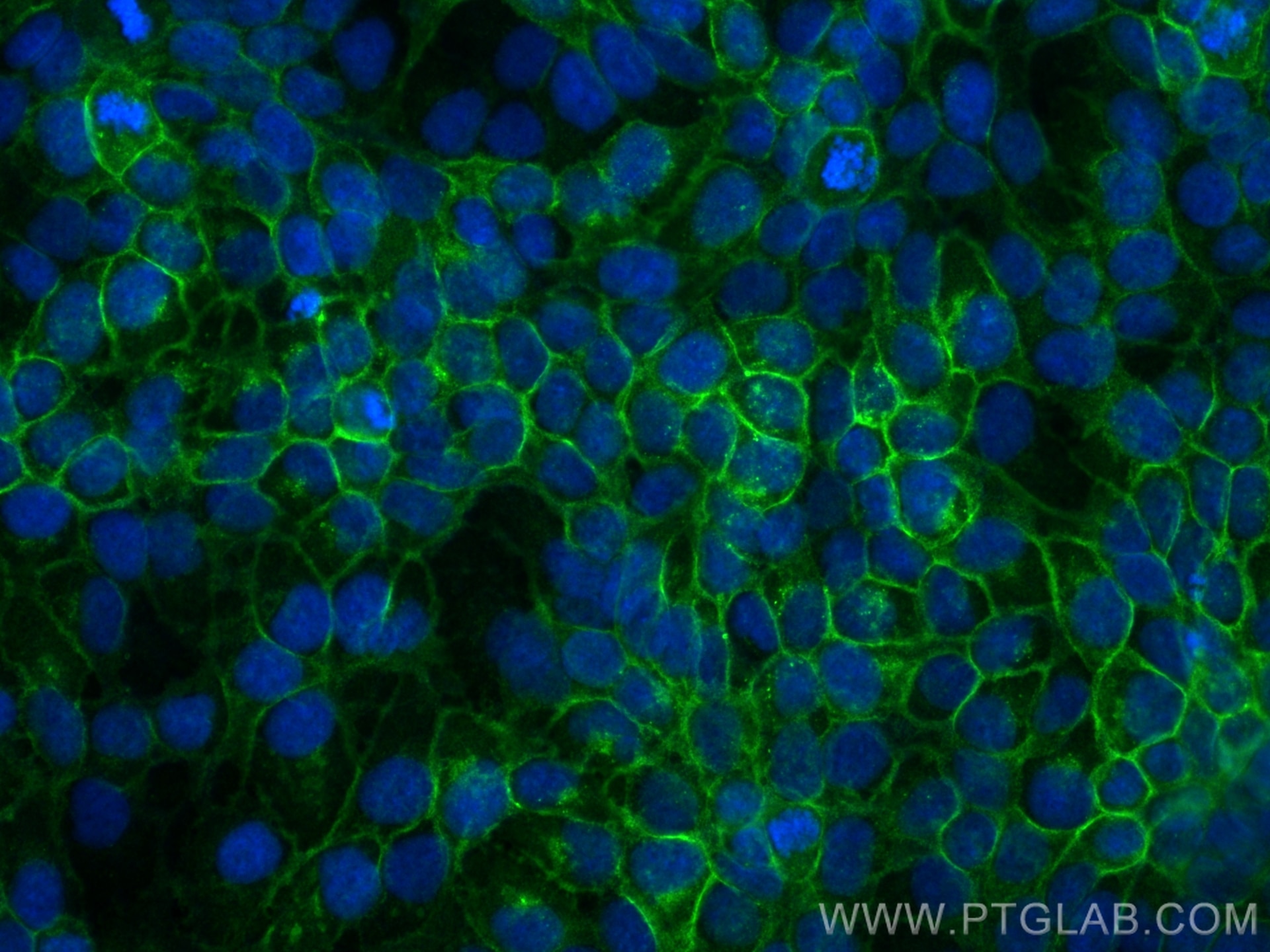
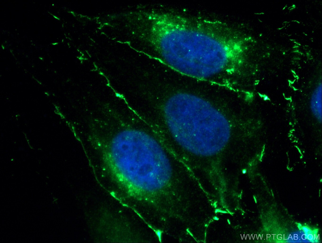
| Positive IF/ICC detected in | HUVEC cells, MDCK cells, HaCaT cells |
Freshly prepared samples are recommended.
Recommended dilution
| Application | Dilution |
|---|---|
| Western Blot (WB) | WB : 1:1000-1:8000 |
| Immunohistochemistry (IHC) | IHC : 1:50-1:500 |
| Immunofluorescence (IF)/ICC | IF/ICC : 1:1000-1:4000 |
| It is recommended that this reagent should be titrated in each testing system to obtain optimal results. | |
| Sample-dependent, Check data in validation data gallery. | |
Published Applications
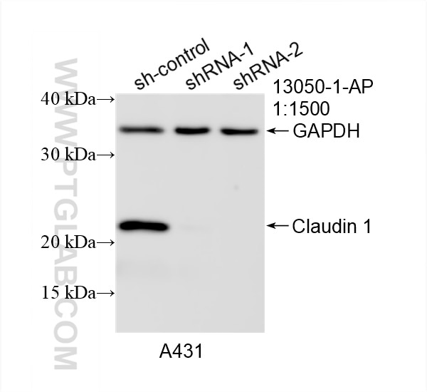
| KD/KO | See 2 publications below |
| WB | See 266 publications below |
| IHC | See 58 publications below |
| IF | See 75 publications below |
Product Information
13050-1-AP targets Claudin 1 in WB, IHC, IF/ICC, ELISA applications and shows reactivity with human, mouse, rat, canine samples.
| Tested Reactivity | human, mouse, rat, canine |
| Cited Reactivity | human, mouse, rat, pig, canine, chicken, bovine, sheep, goat |
| Host / Isotype | Rabbit / IgG |
| Class | Polyclonal |
| Type | Antibody |
| Immunogen |
CatNo: Ag3713 Product name: Recombinant human Claudin 1 protein Source: e coli.-derived, PGEX-4T Tag: GST Domain: 1-211 aa of BC012471 Sequence: MANAGLQLLGFILAFLGWIGAIVSTALPQWRIYSYAGDNIVTAQAMYEGLWMSCVSQSTGQIQCKVFDSLLNLSSTLQATRALMVVGILLGVIAIFVATVGMKCMKCLEDDEVQKMRMAVIGGAIFLLAGLAILVATAWYGNRIVQEFYDPMTPVNARYEFGQALFTGWAAASLCLLGGALLCCSCPRKTTSYPTPRPYPKPAPSSGKDYV Predict reactive species |
| Full Name | claudin 1 |
| Calculated Molecular Weight | 211 aa, 23 kDa |
| Observed Molecular Weight | 20-23 kDa |
| GenBank Accession Number | BC012471 |
| Gene Symbol | Claudin 1 |
| Gene ID (NCBI) | 9076 |
| RRID | AB_2079881 |
| Conjugate | Unconjugated |
| Form | Liquid |
| Purification Method | Antigen affinity purification |
| UNIPROT ID | O95832 |
| Storage Buffer | PBS with 0.02% sodium azide and 50% glycerol, pH 7.3. |
| Storage Conditions | Store at -20°C. Stable for one year after shipment. Aliquoting is unnecessary for -20oC storage. 20ul sizes contain 0.1% BSA. |
Background Information
Claudins are a family of proteins that are the most important components of the tight junctions, where they establish the paracellular barrier that controls the flow of molecules in the intercellular space between the cells of an epithelium. 23 claudins have been identified. They are small (20-27 kilodalton (kDa)) proteins with similar structures. They have four transmembrane domains, with the N-terminus and the C-terminus in the cytoplasm. Claudin-1 is an integral membrane protein expressed primarily in keratinocytes and normal mammary epithelial cells. Claudin 1 forms tight junctions with other claudin proteins and plays an important role in the intestinal epithelial barrier.
Protocols
| Product Specific Protocols | |
|---|---|
| IF protocol for Claudin 1 antibody 13050-1-AP | Download protocol |
| IHC protocol for Claudin 1 antibody 13050-1-AP | Download protocol |
| WB protocol for Claudin 1 antibody 13050-1-AP | Download protocol |
| Standard Protocols | |
|---|---|
| Click here to view our Standard Protocols |
Publications
| Species | Application | Title |
|---|---|---|
Cell Host Microbe Gut microbiome dysbiosis contributes to abdominal aortic aneurysm by promoting neutrophil extracellular trap formation | ||
Microbiome Dietary emulsifier carboxymethylcellulose-induced gut dysbiosis and SCFA reduction aggravate acute pancreatitis through classical monocyte activation | ||
J Nanobiotechnology Orally biomimetic metal-phenolic nanozyme with quadruple safeguards for intestinal homeostasis to ameliorate ulcerative colitis | ||
Clin Cancer Res ZIP4 Promotes Pancreatic Cancer Progression by Repressing ZO-1 and claudin-1 through a ZEB1-Dependent Transcriptional Mechanism. | ||
EMBO Rep O-glycan initiation directs distinct biological pathways and controls epithelial differentiation. | ||
Int J Biol Macromol Glucose-modified BSA/procyanidin C1 NPs penetrate the blood-brain barrier and alleviate neuroinflammation in Alzheimer's disease models |
Reviews
The reviews below have been submitted by verified Proteintech customers who received an incentive for providing their feedback.
FH Iram (Verified Customer) (12-19-2025) | Antibody works well for western and IHC
|
FH Yogain (Verified Customer) (12-19-2025) | Antibody works really well for western blot.
|
FH Iram (Verified Customer) (12-19-2025) | Works well for IHC and IB
|
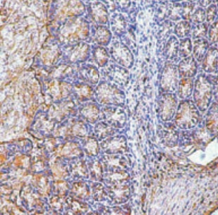
FH James (Verified Customer) (01-06-2022) | Works good. Nice image
 |
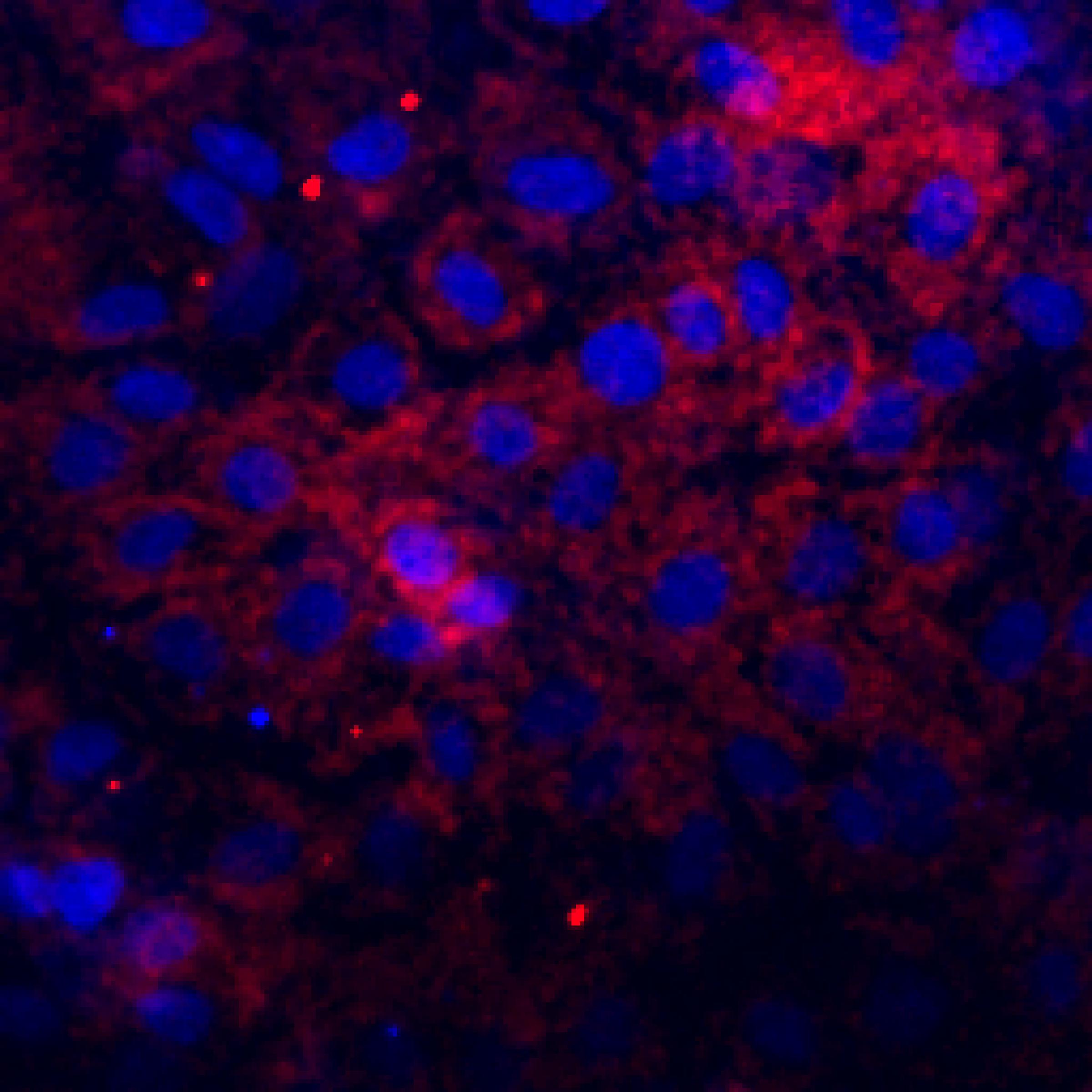
FH Joshua (Verified Customer) (07-27-2019) | Cells differentiated at air-liquid interface. Cells fixed in 4% paraformaldehyde and stained at 4C overnight. Moderate staining with background. Staining is a mix of junctional (bright) and cytosolic (dim).
 |

