Various lysates were subjected to SDS PAGE followed by western blot with 68030-1-Ig (CISD1 antibody) at dilution of 1:20000 incubated at room temperature for 1.5 hours.
Various lysates were subjected to SDS PAGE followed by western blot with 68030-1-Ig (CISD1 antibody) at dilution of 1:20000 incubated at room temperature for 1.5 hours.
IHC staining of human pancreas cancer using 68030-1-Ig
Immunohistochemical analysis of paraffin-embedded human pancreas cancer tissue slide using 68030-1-Ig (CISD1 antibody) at dilution of 1:2000 (under 10x lens). Heat mediated antigen retrieval with Tris-EDTA buffer (pH 9.0).
Immunohistochemical analysis of paraffin-embedded human pancreas cancer tissue slide using 68030-1-Ig (CISD1 antibody) at dilution of 1:2000 (under 10x lens). Heat mediated antigen retrieval with Tris-EDTA buffer (pH 9.0).
IHC staining of human pancreas cancer using 68030-1-Ig
Immunohistochemical analysis of paraffin-embedded human pancreas cancer tissue slide using 68030-1-Ig (CISD1 antibody) at dilution of 1:2000 (under 40x lens). Heat mediated antigen retrieval with Tris-EDTA buffer (pH 9.0).
Immunohistochemical analysis of paraffin-embedded human pancreas cancer tissue slide using 68030-1-Ig (CISD1 antibody) at dilution of 1:2000 (under 40x lens). Heat mediated antigen retrieval with Tris-EDTA buffer (pH 9.0).
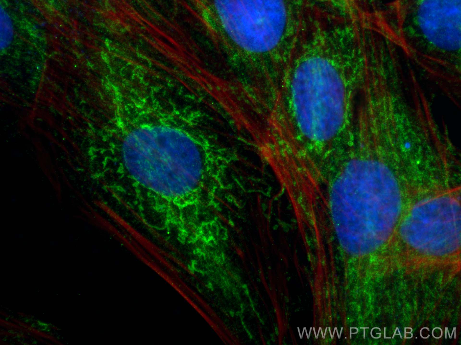
IF Staining of H9C2 using 68030-1-Ig
Immunofluorescent analysis of (4% PFA) fixed H9C2 cells using CISD1 antibody (68030-1-Ig, Clone: 1D7E3 ) at dilution of 1:400 and CoraLite®488-Conjugated AffiniPure Goat Anti-Mouse IgG(H+L) (SA00013-1), CL594-phalloidin (red).
The Proteintech guarantee covers Proteintech antibodies in any species and any application, including those not listed on the datasheet. If the antibody doesn’t perform, you can receive a hassle-free refund or credit note.
LNCaP cells, HeLa cells, HEK-293 cells, pig brain tissue, rabbit brain tissue, rat brian tissue, mouse brain tissue, chicken brain tissue, mouse cerebellum tissue
Positive IHC detected in
human pancreas cancer tissue Note: suggested antigen retrieval with TE buffer pH 9.0; (*) Alternatively, antigen retrieval may be performed with citrate buffer pH 6.0
Positive IF/ICC detected in
H9C2 cells
Recommended dilution
Application
Dilution
Western Blot (WB)
WB : 1:5000-1:50000
Immunohistochemistry (IHC)
IHC : 1:1000-1:4000
Immunofluorescence (IF)/ICC
IF/ICC : 1:200-1:800
It is recommended that this reagent should be titrated in each testing system to obtain optimal results.
Sample-dependent, Check data in validation data gallery.
Product Information
68030-1-Ig targets CISD1 in WB, IHC, IF/ICC, ELISA applications and shows reactivity with human, mouse, rat, pig, rabbit, chicken samples.
PBS with 0.02% sodium azide and 50% glycerol, pH 7.3.
Storage Conditions
Store at -20°C. Stable for one year after shipment. Aliquoting is unnecessary for -20oC storage. 20ul sizes contain 0.1% BSA.
Background Information
MitoNEET, also named CISD1, belongs to a previously uncharacterized ancient family of proteins for which the hallmark is the presence of a unique 39 amino acid CDGSH domain. It is a single-pass type III membrane protein, located in mitochondrion outer membrane and may play a role in regulating maximal capacity for electron transport and oxidative phosphorylation. MitoNEET is a recently identified drug target for a commonly prescribed diabetes drug, Pioglitazone. This antibody recognizing MitoNEET (calculated 12 kDa) as a 17 kDa protein may be due to its posttranslational modification or metal binding activity.
Various lysates were subjected to SDS PAGE followed by western blot with 68030-1-Ig (CISD1 antibody) at dilution of 1:20000 incubated at room temperature for 1.5 hours.
IHC Figures
IHC staining of human pancreas cancer using 68030-1-Ig
Immunohistochemical analysis of paraffin-embedded human pancreas cancer tissue slide using 68030-1-Ig (CISD1 antibody) at dilution of 1:2000 (under 10x lens). Heat mediated antigen retrieval with Tris-EDTA buffer (pH 9.0).
IHC staining of human pancreas cancer using 68030-1-Ig
Immunohistochemical analysis of paraffin-embedded human pancreas cancer tissue slide using 68030-1-Ig (CISD1 antibody) at dilution of 1:2000 (under 40x lens). Heat mediated antigen retrieval with Tris-EDTA buffer (pH 9.0).
IF/ICC Figures
IF Staining of H9C2 using 68030-1-Ig
Immunofluorescent analysis of (4% PFA) fixed H9C2 cells using CISD1 antibody (68030-1-Ig, Clone: 1D7E3 ) at dilution of 1:400 and CoraLite®488-Conjugated AffiniPure Goat Anti-Mouse IgG(H+L) (SA00013-1), CL594-phalloidin (red).
The species listed in Tested Reactivity are in-house verified and applicable species. For unlisted species, please refer to the homology analysis of the immunogen sequence and related species. For rabbit polyclonal antibodies, homology >70% is recommended. For mouse monoclonal antibodies and rabbit recombinant antibodies, homology >90% is recommended. Generally, the higher the homology, the greater the applicability. However, there will be certain differences in protein expression in different species, tissues or cells. Therefore, the homology analysis results are for reference only and do not serve as a guarantee.
At Proteintech, we pride ourselves on our antibody quality, customer service and transparency. As such, we are comparing our antibodies with other vendors, enabling easy identification and comparisons of key data to help you choose the suitable antibody for your needs.
We have selected the top cited antibodies from these vendors for you to compare.
 at dilution of 1:20000 incubated at room temperature for 1.5 hours.")
 at dilution of 1:2000 (under 10x lens). Heat mediated antigen retrieval with Tris-EDTA buffer (pH 9.0).")
 at dilution of 1:2000 (under 40x lens). Heat mediated antigen retrieval with Tris-EDTA buffer (pH 9.0).")
 fixed H9C2 cells using CISD1 antibody (68030-1-Ig, Clone: 1D7E3 ) at dilution of 1:400 and CoraLite®488-Conjugated AffiniPure Goat Anti-Mouse IgG(H+L) (SA00013-1), CL594-phalloidin (red).")