Various lysates were subjected to SDS PAGE followed by western blot with 30380-1-AP (CDX2 antibody) at dilution of 1:5000 incubated at room temperature for 1.5 hours.
Various lysates were subjected to SDS PAGE followed by western blot with 30380-1-AP (CDX2 antibody) at dilution of 1:5000 incubated at room temperature for 1.5 hours.
IHC staining of human colon cancer using 30380-1-AP
Immunohistochemical analysis of paraffin-embedded human colon cancer tissue slide using 30380-1-AP (CDX2 antibody) at dilution of 1:200 (under 10x lens). Heat mediated antigen retrieval with Tris-EDTA buffer (pH 9.0).
Immunohistochemical analysis of paraffin-embedded human colon cancer tissue slide using 30380-1-AP (CDX2 antibody) at dilution of 1:200 (under 10x lens). Heat mediated antigen retrieval with Tris-EDTA buffer (pH 9.0).
IHC staining of human colon cancer using 30380-1-AP
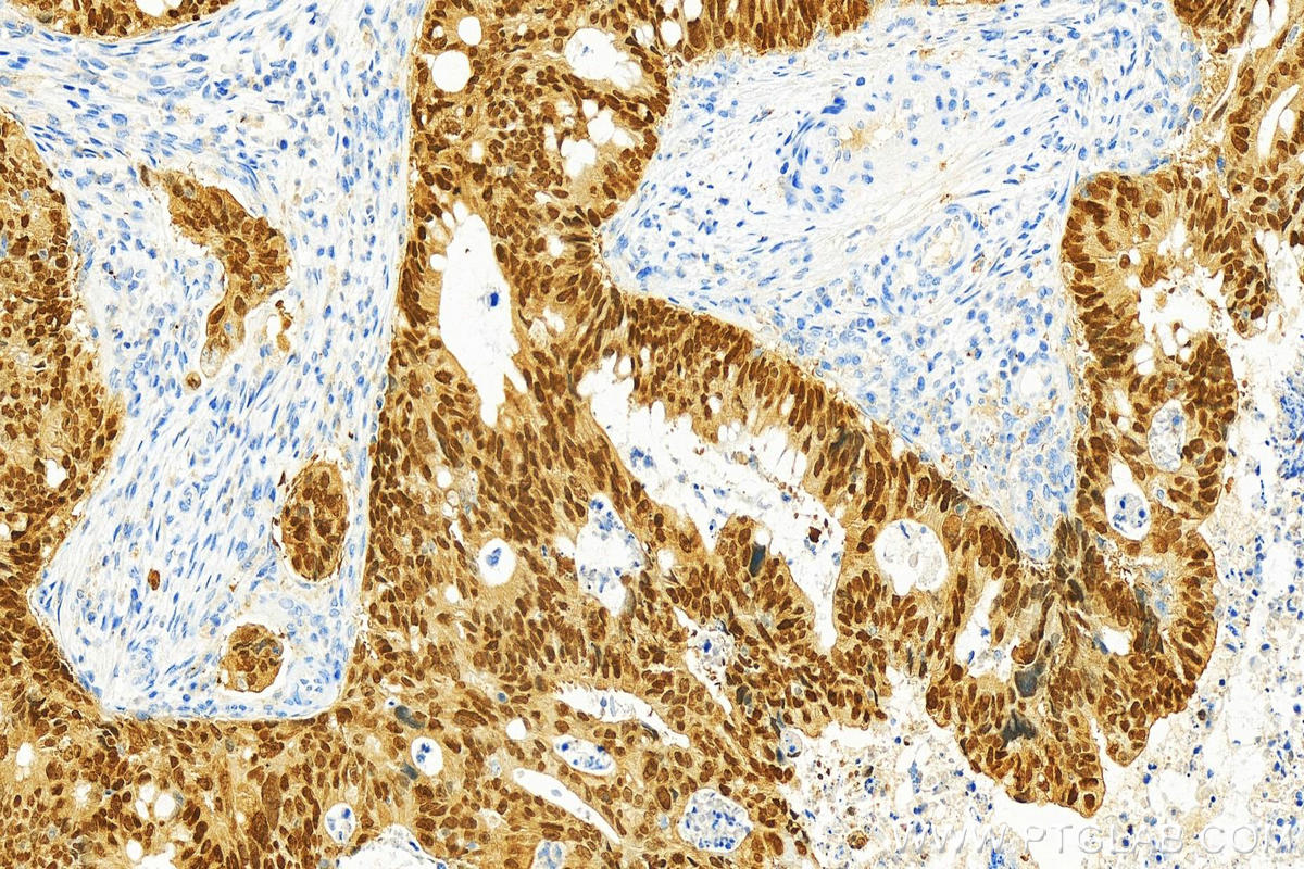
Immunohistochemical analysis of paraffin-embedded human colon cancer tissue slide using 30380-1-AP (CDX2 antibody) at dilution of 1:200 (under 40x lens). Heat mediated antigen retrieval with Tris-EDTA buffer (pH 9.0).
Immunohistochemical analysis of paraffin-embedded human colon cancer tissue slide using 30380-1-AP (CDX2 antibody) at dilution of 1:200 (under 40x lens). Heat mediated antigen retrieval with Tris-EDTA buffer (pH 9.0).
IHC staining of mouse colon using 30380-1-AP
Immunohistochemical analysis of paraffin-embedded mouse colon tissue slide using 30380-1-AP (CDX2 antibody) at dilution of 1:200 (under 10x lens). Heat mediated antigen retrieval with Tris-EDTA buffer (pH 9.0).
Immunohistochemical analysis of paraffin-embedded mouse colon tissue slide using 30380-1-AP (CDX2 antibody) at dilution of 1:200 (under 10x lens). Heat mediated antigen retrieval with Tris-EDTA buffer (pH 9.0).
IHC staining of rat colon using 30380-1-AP
Immunohistochemical analysis of paraffin-embedded rat colon tissue slide using 30380-1-AP (CDX2 antibody) at dilution of 1:200 (under 40x lens). Heat mediated antigen retrieval with Tris-EDTA buffer (pH 9.0).
Immunohistochemical analysis of paraffin-embedded rat colon tissue slide using 30380-1-AP (CDX2 antibody) at dilution of 1:200 (under 40x lens). Heat mediated antigen retrieval with Tris-EDTA buffer (pH 9.0).
IHC staining of human colon cancer using 30380-1-AP
Immunohistochemical analysis of paraffin-embedded colon cancer slide using 30380-1-AP (CDX2 antibody) at dilution of 1:1000 (under 20x lens). Heat mediated antigen retrieval with Tris-EDTA buffer (pH 9.0).
Immunohistochemical analysis of paraffin-embedded colon cancer slide using 30380-1-AP (CDX2 antibody) at dilution of 1:1000 (under 20x lens). Heat mediated antigen retrieval with Tris-EDTA buffer (pH 9.0).
IHC staining of human colon cancer using 30380-1-AP
Immunohistochemical analysis of paraffin-embedded human colon cancer tissue slide using 30380-1-AP (CDX2 antibody) at dilution of 1:200 (under 40x lens). Heat mediated antigen retrieval with Tris-EDTA buffer (pH 9.0).
Immunohistochemical analysis of paraffin-embedded human colon cancer tissue slide using 30380-1-AP (CDX2 antibody) at dilution of 1:200 (under 40x lens). Heat mediated antigen retrieval with Tris-EDTA buffer (pH 9.0).
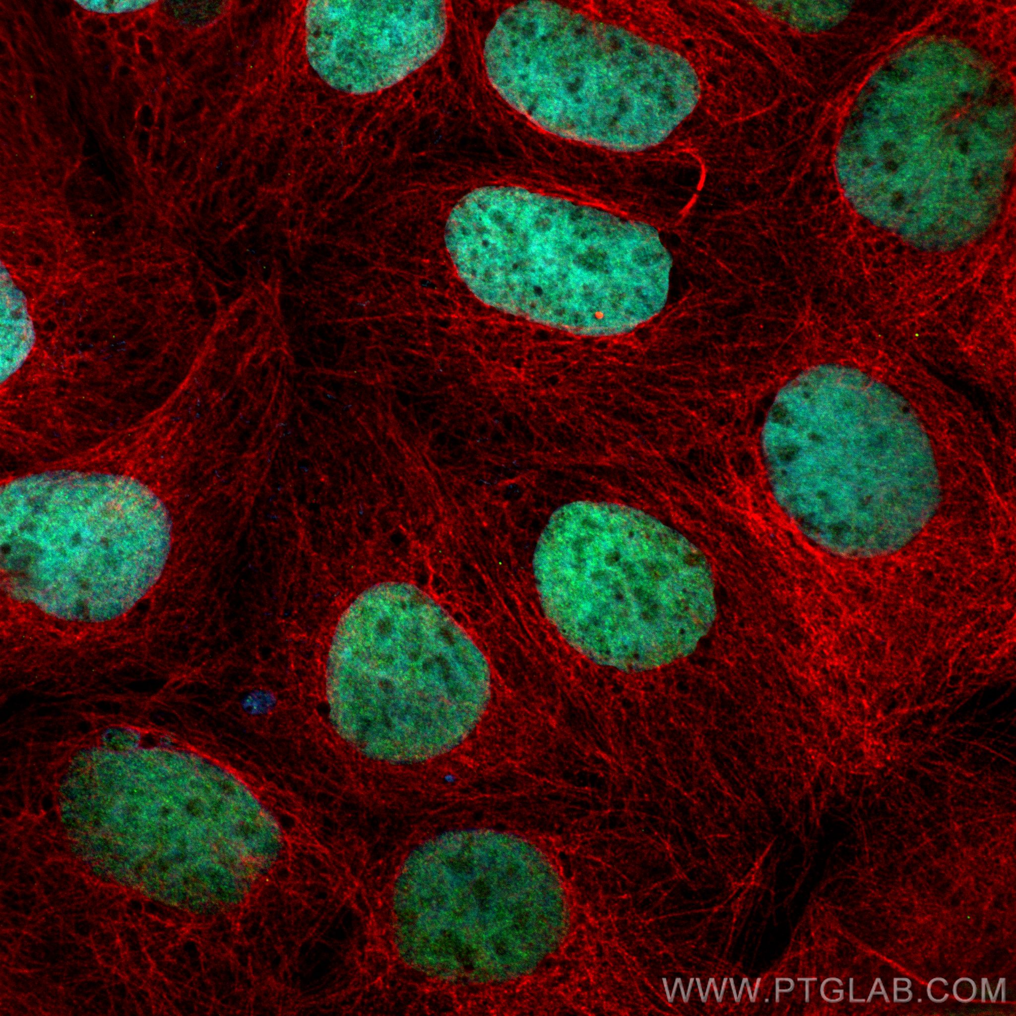
IF Staining of Caco-2 using 30380-1-AP
Immunofluorescent analysis of (4% PFA) fixed Caco-2 cells using CDX2 antibody (30380-1-AP) at dilution of 1:400 and CoraLite®488-Conjugated AffiniPure Goat Anti-Rabbit IgG(H+L) (SA00013-2), Alpha Tubulin antibody (66031-1-Ig, Clone: 1E4C11, red).
The Proteintech guarantee covers Proteintech antibodies in any species and any application, including those not listed on the datasheet. If the antibody doesn’t perform, you can receive a hassle-free refund or credit note.
human colon cancer tissue, mouse colon tissue, rat colon tissue Note: suggested antigen retrieval with TE buffer pH 9.0; (*) Alternatively, antigen retrieval may be performed with citrate buffer pH 6.0
Positive IF/ICC detected in
Caco-2 cells
Recommended dilution
Application
Dilution
Western Blot (WB)
WB : 1:2000-1:10000
Immunohistochemistry (IHC)
IHC : 1:50-1:500
Immunofluorescence (IF)/ICC
IF/ICC : 1:200-1:800
It is recommended that this reagent should be titrated in each testing system to obtain optimal results.
Sample-dependent, Check data in validation data gallery.
Product Information
30380-1-AP targets CDX2 in WB, IHC, IF/ICC, ELISA applications and shows reactivity with human, mouse, rat, pig samples.
PBS with 0.02% sodium azide and 50% glycerol, pH 7.3.
Storage Conditions
Store at -20°C. Stable for one year after shipment. Aliquoting is unnecessary for -20oC storage. 20ul sizes contain 0.1% BSA.
Background Information
CDX2, also named as Homeobox protein CDX-2, is a 313 amino acid protein, which contains one homeobox DNA-binding domain and belongs to the Caudal homeobox family. CDX2 localizes in the nucleus and is involved in the transcriptional regulation of multiple genes expression in the intestinal epithelium. The relative expression of CDX1 to CDX2 protein may be important in the anterior to posterior patterning of the intestinal epithelium and in defining patterns of proliferation and differentiation along the crypt-villus axis. Both Cdx1 and Cdx2 genes must be expressed to reduce tumorigenic potential, to increase sensitivity to apoptosis, and to reduce cell migration, suggesting that the two genes control the normal phenotype by independent pathways.
Various lysates were subjected to SDS PAGE followed by western blot with 30380-1-AP (CDX2 antibody) at dilution of 1:5000 incubated at room temperature for 1.5 hours.
IHC Figures
IHC staining of human colon cancer using 30380-1-AP
Immunohistochemical analysis of paraffin-embedded human colon cancer tissue slide using 30380-1-AP (CDX2 antibody) at dilution of 1:200 (under 10x lens). Heat mediated antigen retrieval with Tris-EDTA buffer (pH 9.0).
IHC staining of human colon cancer using 30380-1-AP
Immunohistochemical analysis of paraffin-embedded human colon cancer tissue slide using 30380-1-AP (CDX2 antibody) at dilution of 1:200 (under 40x lens). Heat mediated antigen retrieval with Tris-EDTA buffer (pH 9.0).
IHC staining of mouse colon using 30380-1-AP
Immunohistochemical analysis of paraffin-embedded mouse colon tissue slide using 30380-1-AP (CDX2 antibody) at dilution of 1:200 (under 10x lens). Heat mediated antigen retrieval with Tris-EDTA buffer (pH 9.0).
IHC staining of rat colon using 30380-1-AP
Immunohistochemical analysis of paraffin-embedded rat colon tissue slide using 30380-1-AP (CDX2 antibody) at dilution of 1:200 (under 40x lens). Heat mediated antigen retrieval with Tris-EDTA buffer (pH 9.0).
IHC staining of human colon cancer using 30380-1-AP
Immunohistochemical analysis of paraffin-embedded colon cancer slide using 30380-1-AP (CDX2 antibody) at dilution of 1:1000 (under 20x lens). Heat mediated antigen retrieval with Tris-EDTA buffer (pH 9.0).
IHC staining of human colon cancer using 30380-1-AP
Immunohistochemical analysis of paraffin-embedded human colon cancer tissue slide using 30380-1-AP (CDX2 antibody) at dilution of 1:200 (under 40x lens). Heat mediated antigen retrieval with Tris-EDTA buffer (pH 9.0).
IF/ICC Figures
IF Staining of Caco-2 using 30380-1-AP
Immunofluorescent analysis of (4% PFA) fixed Caco-2 cells using CDX2 antibody (30380-1-AP) at dilution of 1:400 and CoraLite®488-Conjugated AffiniPure Goat Anti-Rabbit IgG(H+L) (SA00013-2), Alpha Tubulin antibody (66031-1-Ig, Clone: 1E4C11, red).
The species listed in Tested Reactivity are in-house verified and applicable species. For unlisted species, please refer to the homology analysis of the immunogen sequence and related species. For rabbit polyclonal antibodies, homology >70% is recommended. For mouse monoclonal antibodies and rabbit recombinant antibodies, homology >90% is recommended. Generally, the higher the homology, the greater the applicability. However, there will be certain differences in protein expression in different species, tissues or cells. Therefore, the homology analysis results are for reference only and do not serve as a guarantee.
At Proteintech, we pride ourselves on our antibody quality, customer service and transparency. As such, we are comparing our antibodies with other vendors, enabling easy identification and comparisons of key data to help you choose the suitable antibody for your needs.
We have selected the top cited antibodies from these vendors for you to compare.
 at dilution of 1:5000 incubated at room temperature for 1.5 hours.")
 at dilution of 1:200 (under 10x lens). Heat mediated antigen retrieval with Tris-EDTA buffer (pH 9.0).")
 at dilution of 1:200 (under 40x lens). Heat mediated antigen retrieval with Tris-EDTA buffer (pH 9.0).")
 at dilution of 1:200 (under 10x lens). Heat mediated antigen retrieval with Tris-EDTA buffer (pH 9.0).")
 at dilution of 1:200 (under 40x lens). Heat mediated antigen retrieval with Tris-EDTA buffer (pH 9.0).")
 at dilution of 1:1000 (under 20x lens). Heat mediated antigen retrieval with Tris-EDTA buffer (pH 9.0).")
 at dilution of 1:200 (under 40x lens). Heat mediated antigen retrieval with Tris-EDTA buffer (pH 9.0).")
 fixed Caco-2 cells using CDX2 antibody (30380-1-AP) at dilution of 1:400 and CoraLite®488-Conjugated AffiniPure Goat Anti-Rabbit IgG(H+L) (SA00013-2), Alpha Tubulin antibody (66031-1-Ig, Clone: 1E4C11, red).")