Tested Applications
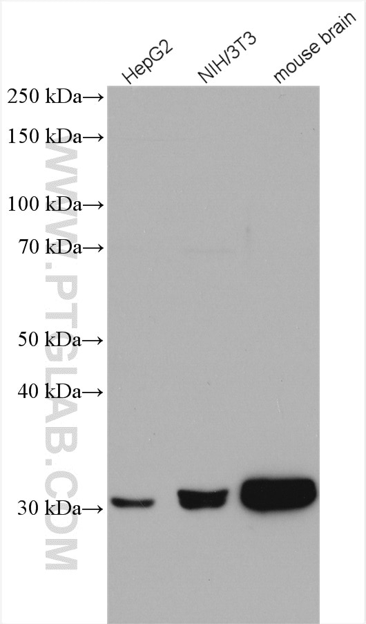
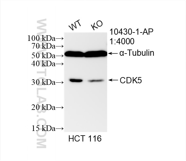
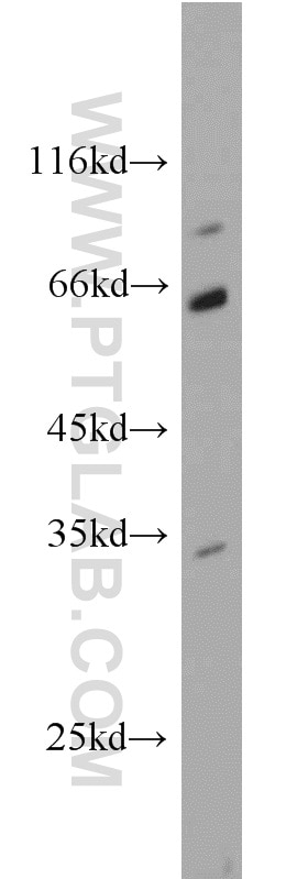
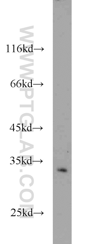
| Positive WB detected in | HepG2 cells, HCT 116 cells, mouse pancreas tissue, NIH/3T3 cells, WB result of CDK5 antibody (10430-1-AP; 1:1000; room temperature for 1.5 hours) with wild-type and CDK5 knockout HCT 116 cells., mouse brain tissue |
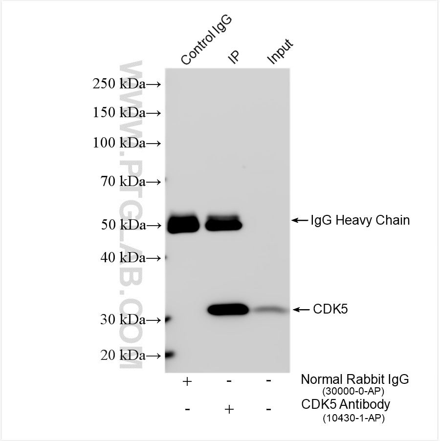
| Positive IP detected in | mouse brain tissue |
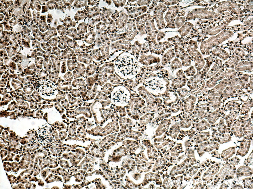
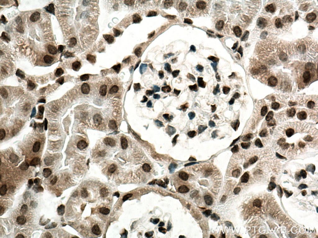
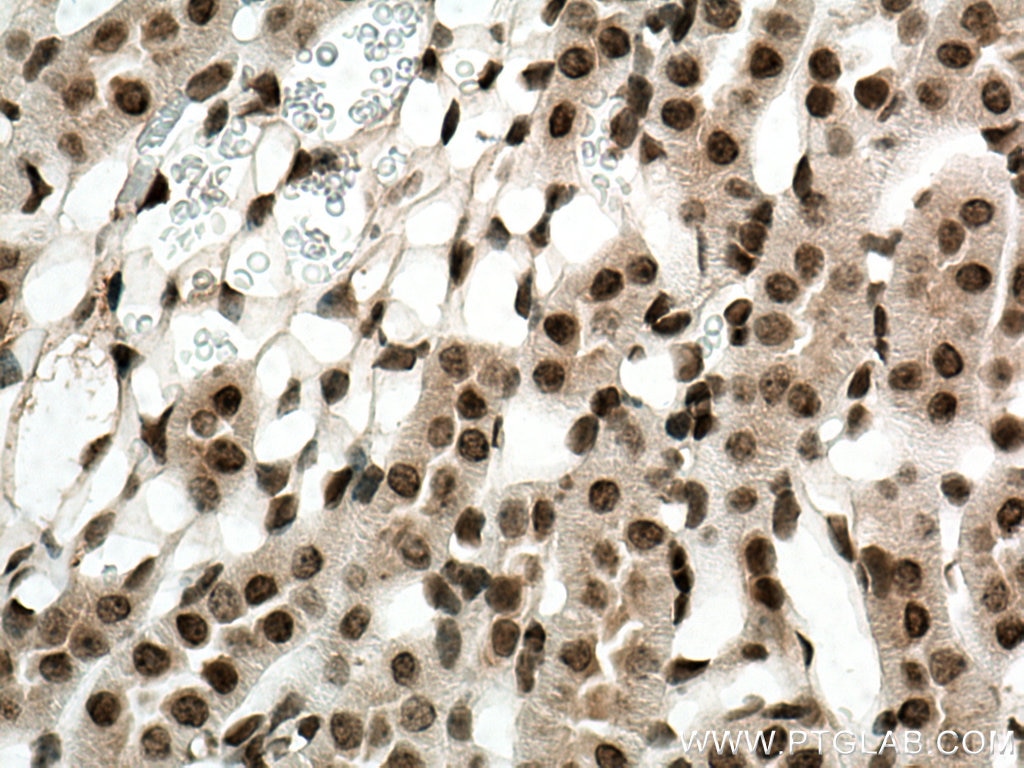
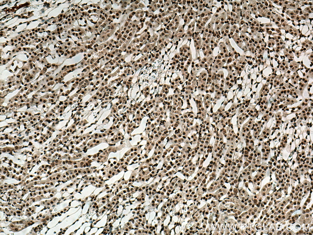
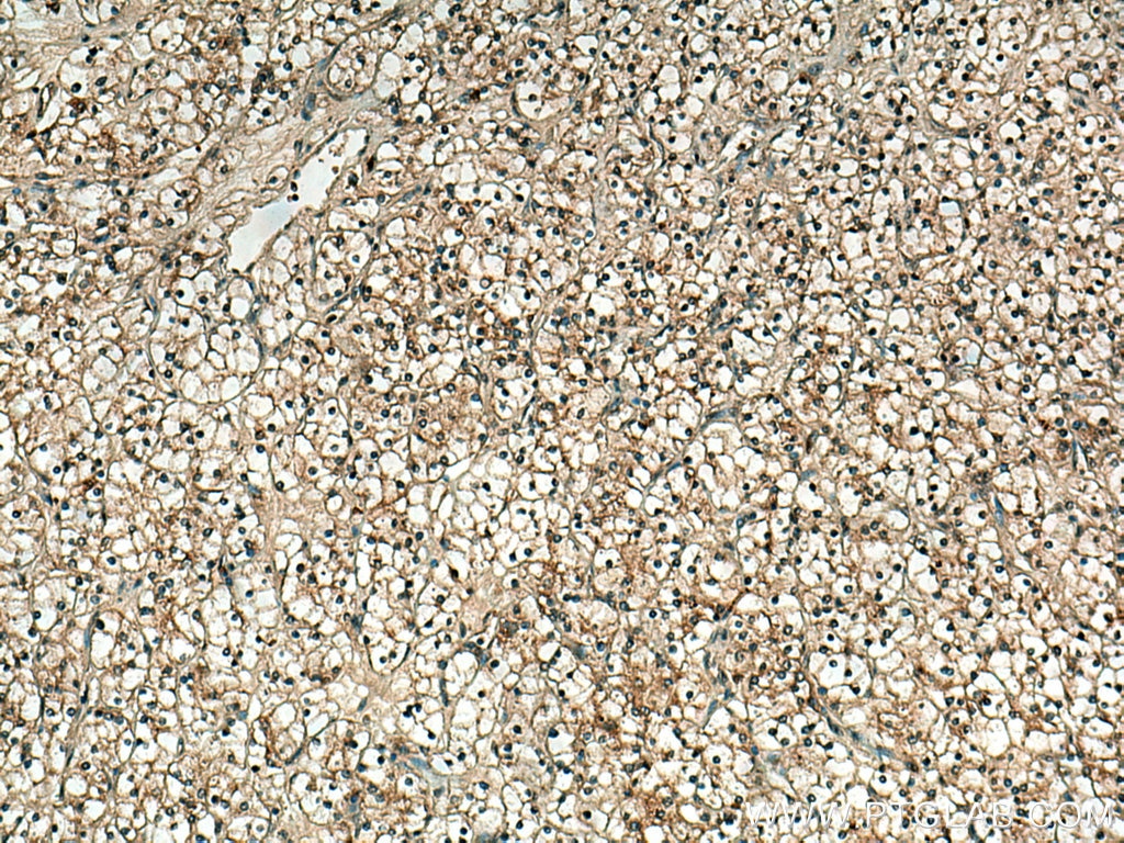
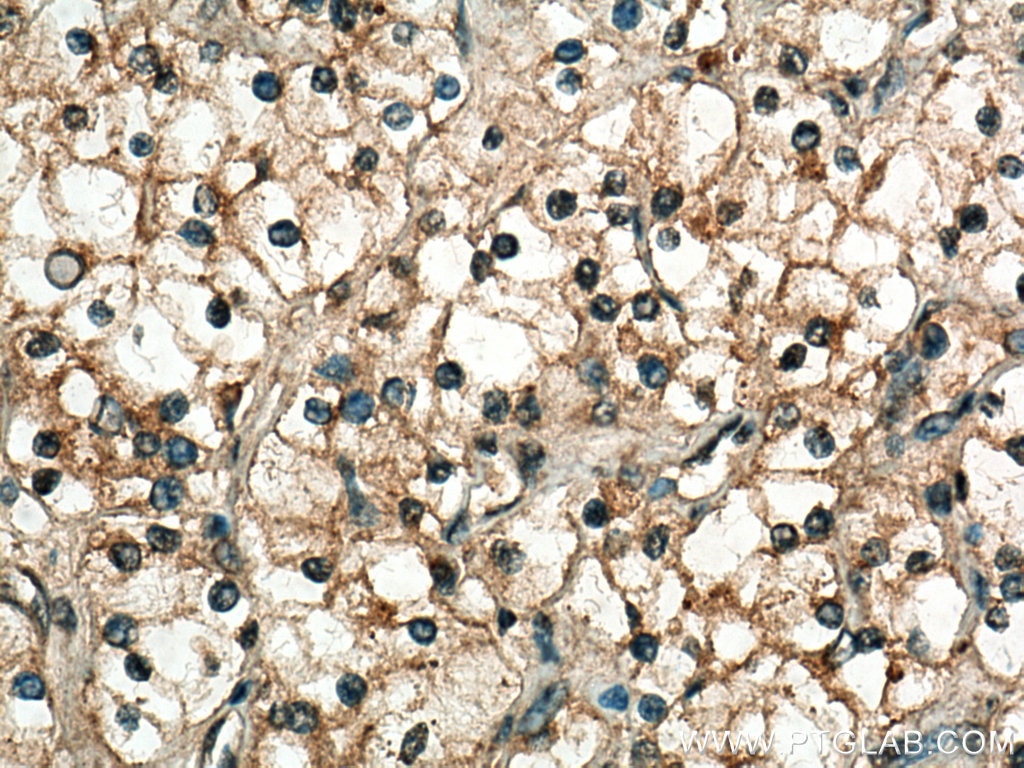
| Positive IHC detected in | mouse kidney tissue, human renal cell carcinoma tissue Note: suggested antigen retrieval with TE buffer pH 9.0; (*) Alternatively, antigen retrieval may be performed with citrate buffer pH 6.0 |
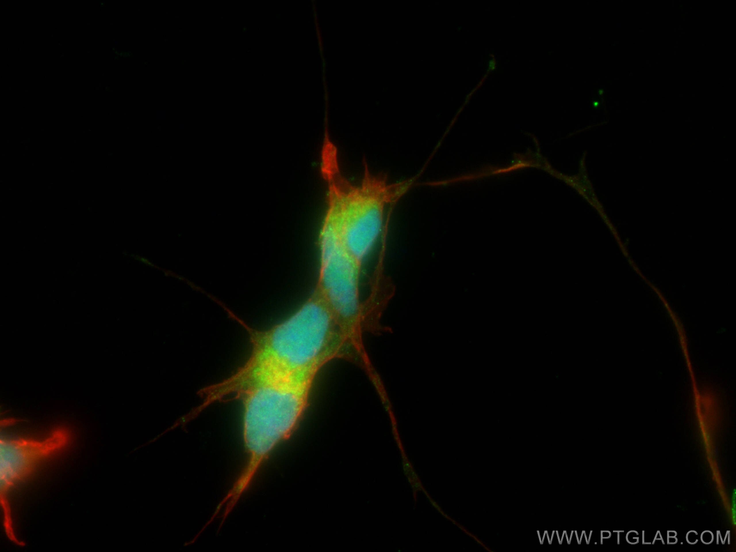
| Positive IF/ICC detected in | SH-SY5Y cells |
Recommended dilution
| Application | Dilution |
|---|---|
| Western Blot (WB) | WB : 1:500-1:2000 |
| Immunoprecipitation (IP) | IP : 0.5-4.0 ug for 1.0-3.0 mg of total protein lysate |
| Immunohistochemistry (IHC) | IHC : 1:50-1:500 |
| Immunofluorescence (IF)/ICC | IF/ICC : 1:200-1:800 |
| It is recommended that this reagent should be titrated in each testing system to obtain optimal results. | |
| Sample-dependent, Check data in validation data gallery. | |
Published Applications
| KD/KO | See 2 publications below |
| WB | See 19 publications below |
| IHC | See 1 publications below |
| IF | See 1 publications below |
| IP | See 1 publications below |
Product Information
10430-1-AP targets CDK5 in WB, IHC, IF/ICC, IP, ELISA applications and shows reactivity with human, mouse, rat samples.
| Tested Reactivity | human, mouse, rat |
| Cited Reactivity | human, mouse, rat |
| Host / Isotype | Rabbit / IgG |
| Class | Polyclonal |
| Type | Antibody |
| Immunogen |
CatNo: Ag0686 Product name: Recombinant human CDK5 protein Source: e coli.-derived, PGEX-4T Tag: GST Domain: 92-273 aa of BC005115 Sequence: DSCNGDLDPEIVKSFLFQLLKGLGFCHSRNVLHRDLKPQNLLINRNGELKLADFGLARAFGIPVRCYSAEVVTLWYRPPDVLFGAKLYSTSIDMWSAGCIFAELANAGRPLFPGNDVDDQLKRIFRLLGTPTEEQWPSMTKLPDYKPYPMYPATTSLVNVVPKLNATGRDLLQNLLKCNPVQ Predict reactive species |
| Full Name | cyclin-dependent kinase 5 |
| Calculated Molecular Weight | 31 kDa, 33 kDa |
| Observed Molecular Weight | 33 kDa |
| GenBank Accession Number | BC005115 |
| Gene Symbol | CDK5 |
| Gene ID (NCBI) | 1020 |
| RRID | AB_2078859 |
| Conjugate | Unconjugated |
| Form | Liquid |
| Purification Method | Antigen affinity purification |
| UNIPROT ID | Q00535 |
| Storage Buffer | PBS with 0.02% sodium azide and 50% glycerol, pH 7.3. |
| Storage Conditions | Store at -20°C. Stable for one year after shipment. Aliquoting is unnecessary for -20oC storage. 20ul sizes contain 0.1% BSA. |
Background Information
Cyclin-dependent kinase 5 (CDK5), belongs to the cyclin-dependent kinase family, is a proline-directed serine/threonine-protein kinase that essential for neuronal cell cycle arrest and differentiation and may be involved in apoptotic cell death in neuronal diseases by triggering abortive cell cycle re-entry. CDK5 predominantly expressed in neurons where it phosphorylates both high molecular weight neurofilaments and microtubule-associated protein tau.
Protocols
| Product Specific Protocols | |
|---|---|
| IF protocol for CDK5 antibody 10430-1-AP | Download protocol |
| IHC protocol for CDK5 antibody 10430-1-AP | Download protocol |
| IP protocol for CDK5 antibody 10430-1-AP | Download protocol |
| WB protocol for CDK5 antibody 10430-1-AP | Download protocol |
| Standard Protocols | |
|---|---|
| Click here to view our Standard Protocols |
Publications
| Species | Application | Title |
|---|---|---|
Signal Transduct Target Ther SARS-CoV-2 hijacks cellular kinase CDK2 to promote viral RNA synthesis | ||
Neuropharmacology Inhibition of Cdk5 rejuvenates inhibitory circuits and restores experience-dependent plasticity in adult visual cortex.
| ||
Cells Phosphorylation-Induced Ubiquitination and Degradation of PXR through CDK2-TRIM21 Axis.
| ||
Am J Transl Res Hypoxia induces HT-22 neuronal cell death via Orai1/CDK5 pathway-mediated Tau hyperphosphorylation. | ||
J Cell Biochem Nestin protects mouse podocytes against high glucose-induced apoptosis by a Cdk5-dependent mechanism. | ||
BMC Neurol The LPA-CDK5-tau pathway mediates neuronal injury in an in vitro model of ischemia-reperfusion insult. |
Reviews
The reviews below have been submitted by verified Proteintech customers who received an incentive for providing their feedback.
FH XinN (Verified Customer) (02-27-2023) | It can recognize endogenous CDK5 with a right MW.
 |
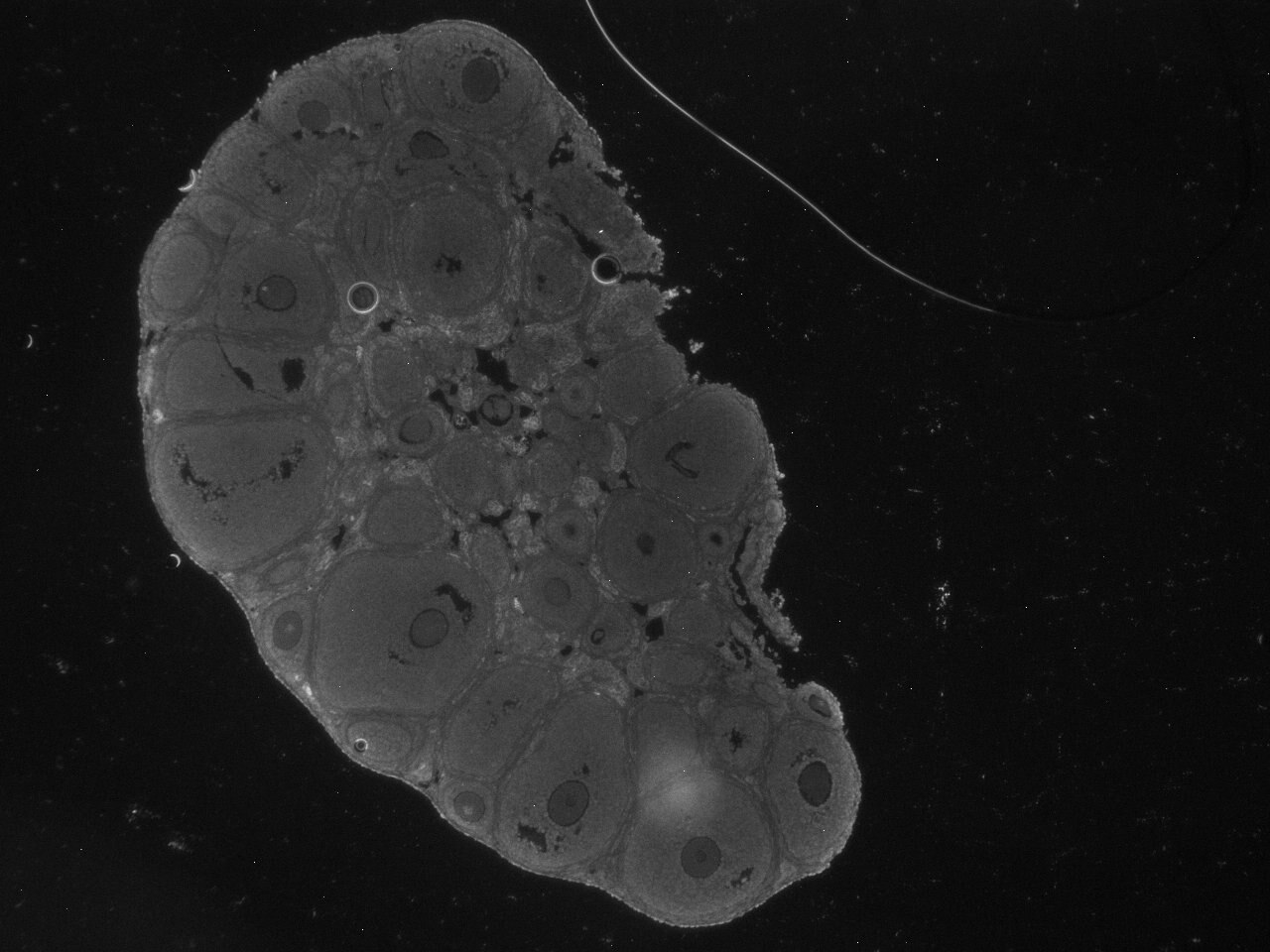
FH Kathryn (Verified Customer) (04-22-2022) | This CDK5 antibody works very well to stain the nuclei of developed mouse oocytes.
 |

