at dilution of 1:10000 incubated at room temperature for 1.5 hours. This data was developed using the same antibody clone with 60143-1-PBS in a different storage buffer formulation.")

 with human placenta tissue lysate 1520ug. This data was developed using the same antibody clone with 60143-1-PBS in a different storage buffer formulation.")
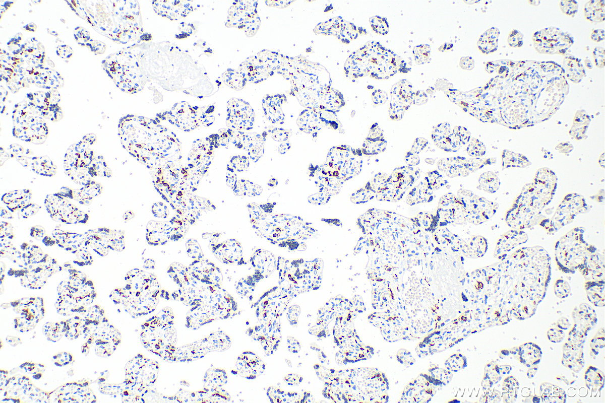
 at dilution of 1:20000 (under 10x lens). Heat mediated antigen retrieval with Tris-EDTA buffer (pH 9.0). This data was developed using the same antibody clone with 60143-1-PBS in a different storage buffer formulation.")
 at dilution of 1:20000 (under 40x lens). Heat mediated antigen retrieval with Tris-EDTA buffer (pH 9.0). This data was developed using the same antibody clone with 60143-1-PBS in a different storage buffer formulation.")
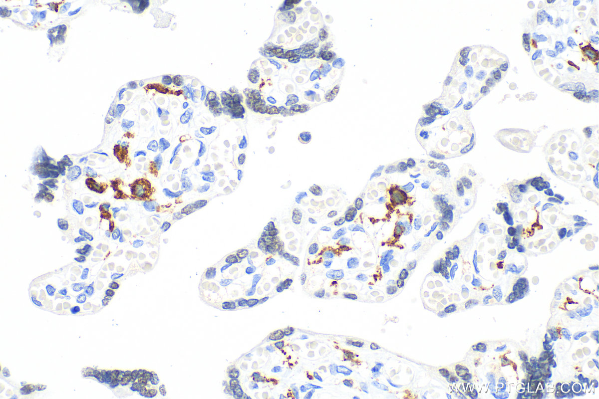
 at dilution of 1:80000 (under 10x lens). Heat mediated antigen retrieval with Tris-EDTA buffer (pH 9.0). This data was developed using the same antibody clone with 60143-1-PBS in a different storage buffer formulation.")
 at dilution of 1:80000 (under 40x lens). Heat mediated antigen retrieval with Tris-EDTA buffer (pH 9.0). This data was developed using the same antibody clone with 60143-1-PBS in a different storage buffer formulation.")
 at dilution of 1:20000 (under 10x lens). Heat mediated antigen retrieval with Tris-EDTA buffer (pH 9.0). This data was developed using the same antibody clone with 60143-1-PBS in a different storage buffer formulation.")
 at dilution of 1:20000 (under 40x lens). Heat mediated antigen retrieval with Tris-EDTA buffer (pH 9.0). This data was developed using the same antibody clone with 60143-1-PBS in a different storage buffer formulation.")
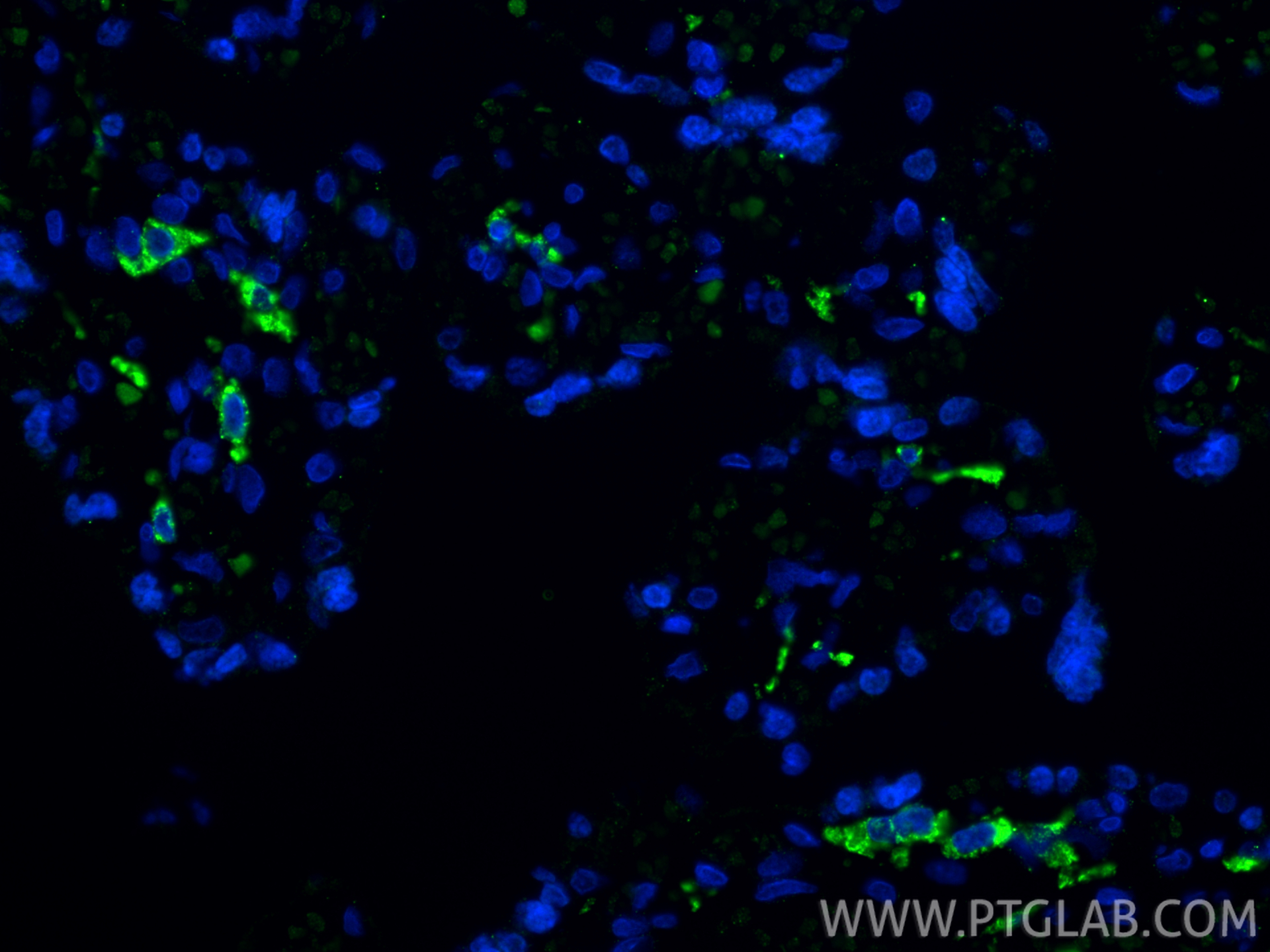
 fixed human lung cancer tissue using CD206 antibody (60143-1-Ig, Clone: 2A6A10 ) at dilution of 1:400 and CoraLite®488-Conjugated AffiniPure Goat Anti-Mouse IgG(H+L). This data was developed using the same antibody clone with 60143-1-PBS in a different storage buffer formulation.")
 fixed human lung cancer tissue using CD206 antibody (60143-1-Ig, Clone: 2A6A10 ) at dilution of 1:400 and CoraLite®488-Conjugated AffiniPure Goat Anti-Mouse IgG(H+L). This data was developed using the same antibody clone with 60143-1-PBS in a different storage buffer formulation.")
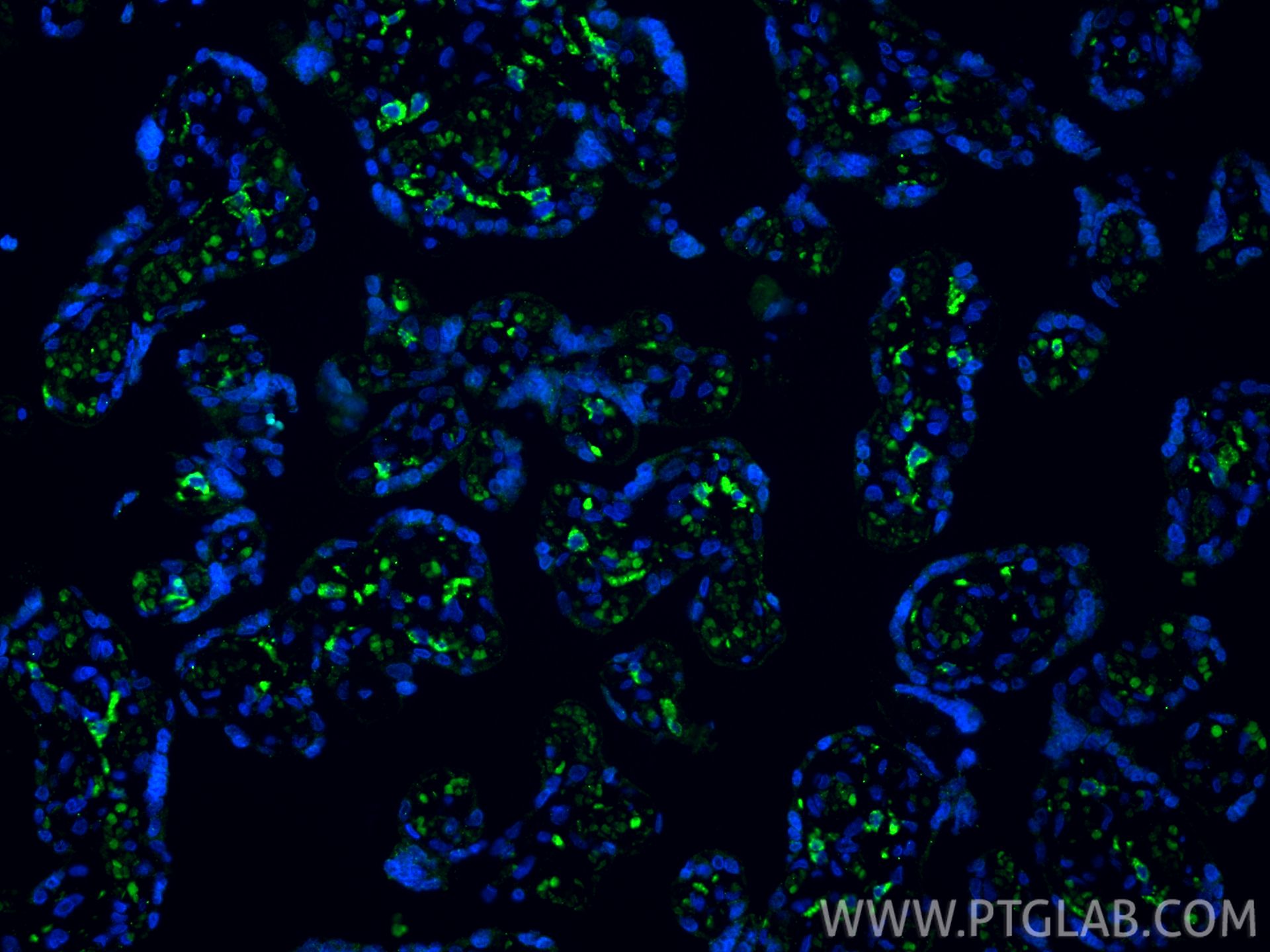
 fixed paraffin-embedded human placenta tissue using CD206 antibody (60143-1-Ig, Clone: 2A6A10 ) at dilution of 1:800 and CoraLite®488-Conjugated Goat Anti-Mouse IgG(H+L) (SA00013-1). Heat mediated antigen retrieval with Tris-EDTA buffer (pH 9.0). This data was developed using the same antibody clone with 60143-1-PBS in a different storage buffer formulation.")
 fixed paraffin-embedded human placenta tissue using CD206 antibody (60143-1-Ig, Clone: 2A6A10 ) at dilution of 1:800 and CoraLite®488-Conjugated Goat Anti-Mouse IgG(H+L) (SA00013-1). Heat mediated antigen retrieval with Tris-EDTA buffer (pH 9.0). This data was developed using the same antibody clone with 60143-1-PBS in a different storage buffer formulation.")
Product Information
60143-1-PBS targets CD206 in WB, IHC, IF-P, IP, ELISA applications and shows reactivity with human samples.
| Tested Reactivity | human |
| Host / Isotype | Mouse / IgG2a |
| Class | Monoclonal |
| Type | Antibody |
| Immunogen |
Peptide Predict reactive species |
| Full Name | mannose receptor, C type 1 |
| Calculated Molecular Weight | 166 kDa |
| Observed Molecular Weight | 170 kDa |
| GenBank Accession Number | NM_002438 |
| Gene Symbol | CD206 |
| Gene ID (NCBI) | 4360 |
| RRID | AB_2144924 |
| Conjugate | Unconjugated |
| Form | Liquid |
| Purification Method | Protein A purification |
| UNIPROT ID | P22897 |
| Storage Buffer | PBS only, pH 7.3. |
| Storage Conditions | Store at -80°C. |
Background Information
Background
CD206 (macrophage mannose receptor 1) is a lectin-type endocytic receptor expressed on selected macrophages, dendritic cells, and non-vascular endothelium and plays a role in antigen processing and presentation, phagocytosis, and intracellular signaling.
1. What is the molecular weight of CD206?
The molecular size of full-length CD206 is 170-180 kDa, depending on the exact tissue-specific glycosylation pattern (PMID: 19427834). Additionally, CD206 can be cleaved off and a soluble form (sMR) lacking the tail, with a slightly lower molecular weight, can be released to the cell medium (PMID: 9722572).
2. What is the subcellular localization of CD206?
CD206 is a type I membrane protein composed of a large extracellular multidomain, a transmembrane domain, and a short cytoplasmic tail. It is present at the plasma membrane and in endosomes, as CD206 undergoes constant recycling between the plasma membrane and endosomal compartment.
3. Is CD206 post-translationally modified?
CD206 undergoes quite extensive post-translational modifications, predominantly N-linked glycosylation that affects ligand binding recognition and affinity (PMID: 22966131).
4. Can CD206 marker be used as a marker of M2 macrophages?
The activation of macrophages with various stimuli leads to their polarization into classical (M1) or alternatively activated (M2) subtypes spectrums and both subtypes differ in their regulatory and effector functions (PMID: 24669294). Pathogens and IFN-γ promote M1 polarization, while IL-4 released during parasite infections and allergen response promotes M2 polarization. Classically, the markers of M2 macrophages include CD206, as well as arginase-1 (ARG1; https://www.ptglab.com/products/ARG1-Antibody-16001-1-AP.htm), CD163 (https://www.ptglab.com/products/CD163-Antibody-16646-1-AP.htm), and thrombospondin 1 (TSP1/ THBS1; https://www.ptglab.com/products/TSP1-Antibody-18304-1-AP.htm).
5. How can you polarize macrophages into M2 direction?
One of the most commonly used methods is stimulation by the addition of IL-4 cytokine. We recommend using our animal-free human IL-4 (https://www.ptglab.com/products/recombinant-human-il-4.htm).