Various lysates were subjected to SDS PAGE followed by western blot with 67366-1-Ig (CAPN3 antibody) at dilution of 1:3000 incubated at room temperature for 1.5 hours.
Various lysates were subjected to SDS PAGE followed by western blot with 67366-1-Ig (CAPN3 antibody) at dilution of 1:3000 incubated at room temperature for 1.5 hours.
IHC staining of mouse skeletal muscle using 67366-1-Ig
Immunohistochemical analysis of paraffin-embedded mouse skeletal muscle tissue slide using 67366-1-Ig (CAPN3 antibody) at dilution of 1:200 (under 10x lens). Heat mediated antigen retrieval with Tris-EDTA buffer (pH 9.0).
The Proteintech guarantee covers Proteintech antibodies in any species and any application, including those not listed on the datasheet. If the antibody doesn’t perform, you can receive a hassle-free refund or credit note.
human skeletal muscle tissue, pig skeletal muscle tissue, rat skeletal muscle tissue, mouse skeletal muscle tissue
Positive IHC detected in
mouse skeletal muscle tissue Note: suggested antigen retrieval with TE buffer pH 9.0; (*) Alternatively, antigen retrieval may be performed with citrate buffer pH 6.0
Positive IF/ICC detected in
U2OS cells
Recommended dilution
Application
Dilution
Western Blot (WB)
WB : 1:1000-1:6000
Immunohistochemistry (IHC)
IHC : 1:50-1:500
Immunofluorescence (IF)/ICC
IF/ICC : 1:1000-1:4000
It is recommended that this reagent should be titrated in each testing system to obtain optimal results.
Sample-dependent, Check data in validation data gallery.
PBS with 0.02% sodium azide and 50% glycerol , pH 7.3
Storage Conditions
Store at -20°C. Stable for one year after shipment. Aliquoting is unnecessary for -20oC storage. 20ul sizes contain 0.1% BSA.
Background Information
Calpain 3 (CAPN3), a major intracellular protease, is a muscle-specific member of the calpain large subunit family that specifically binds to titin. CAPN3 plays a role in the dysferlin protein complex and that disruption of CAPN3 function may affect muscle membrane repair and remodeling. CAPN3 has some isoforms with MW of 94, 84, 93, 36, 18 kDa. CAPN3 autolysis generates a small N-terminal fragment of 34 kDa and a large C-terminal fragment whose size ranges from 55 to 60 kDa during self-processing (PMID: 9794799, 14645524).
Various lysates were subjected to SDS PAGE followed by western blot with 67366-1-Ig (CAPN3 antibody) at dilution of 1:3000 incubated at room temperature for 1.5 hours.
IHC Figures
IHC staining of mouse skeletal muscle using 67366-1-Ig
Immunohistochemical analysis of paraffin-embedded mouse skeletal muscle tissue slide using 67366-1-Ig (CAPN3 antibody) at dilution of 1:200 (under 10x lens). Heat mediated antigen retrieval with Tris-EDTA buffer (pH 9.0).
IHC staining of mouse skeletal muscle using 67366-1-Ig
Immunohistochemical analysis of paraffin-embedded mouse skeletal muscle tissue slide using 67366-1-Ig (CAPN3 antibody) at dilution of 1:200 (under 40x lens). Heat mediated antigen retrieval with Tris-EDTA buffer (pH 9.0).
IF/ICC Figures
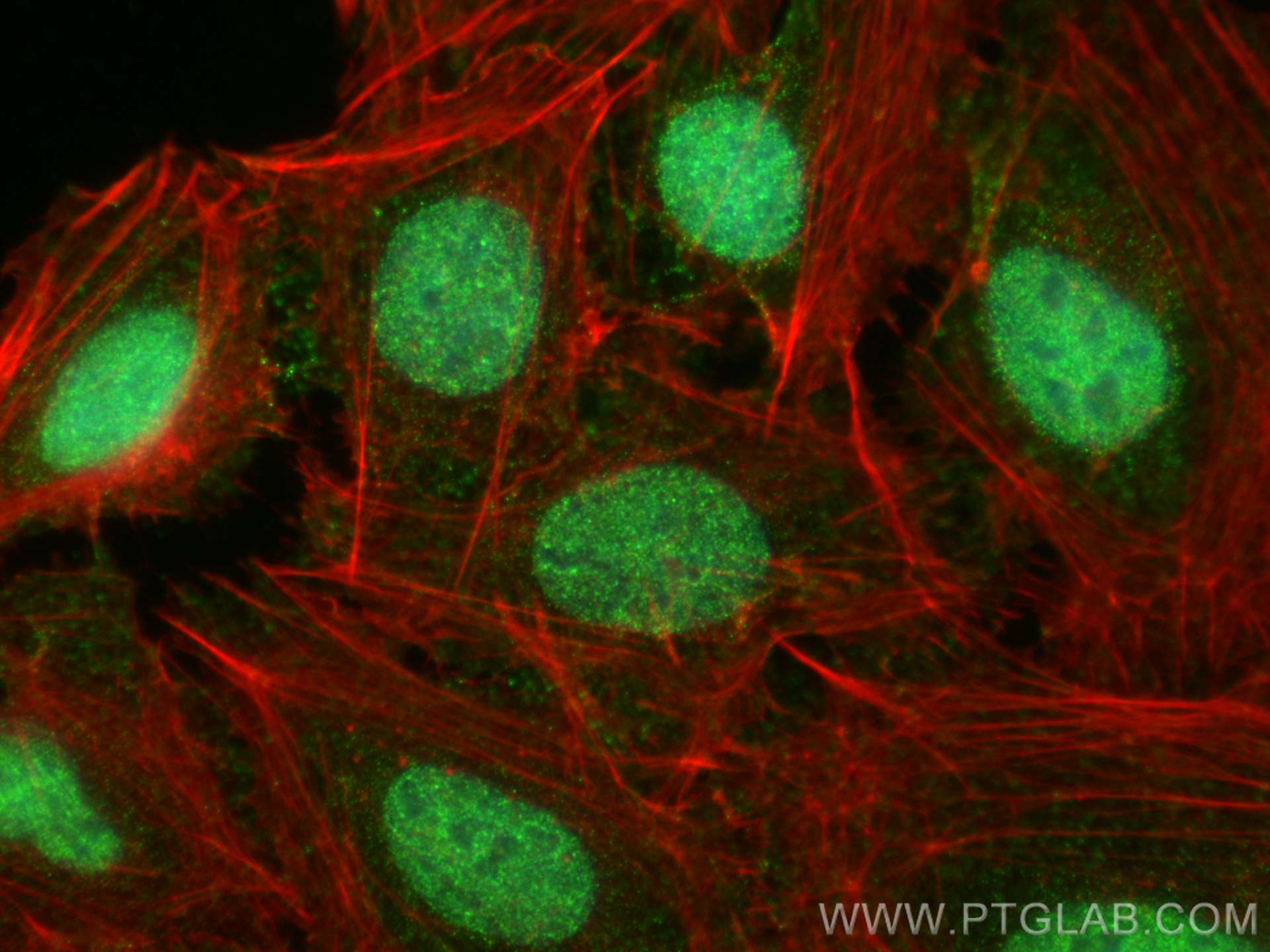
IF Staining of U2OS using 67366-1-Ig
Immunofluorescent analysis of (4% PFA) fixed U2OS cells using CAPN3 antibody (67366-1-Ig, Clone: 1G12A5 ) at dilution of 1:2000 and CoraLite®488-Conjugated AffiniPure Goat Anti-Mouse IgG(H+L) (SA00013-1), CL594-phalloidin (red).
The species listed in Tested Reactivity are in-house verified and applicable species. For unlisted species, please refer to the homology analysis of the immunogen sequence and related species. For rabbit polyclonal antibodies, homology >70% is recommended. For mouse monoclonal antibodies and rabbit recombinant antibodies, homology >90% is recommended. Generally, the higher the homology, the greater the applicability. However, there will be certain differences in protein expression in different species, tissues or cells. Therefore, the homology analysis results are for reference only and do not serve as a guarantee.
At Proteintech, we pride ourselves on our antibody quality, customer service and transparency. As such, we are comparing our antibodies with other vendors, enabling easy identification and comparisons of key data to help you choose the suitable antibody for your needs.
We have selected the top cited antibodies from these vendors for you to compare.
 at dilution of 1:3000 incubated at room temperature for 1.5 hours.")
 at dilution of 1:200 (under 10x lens). Heat mediated antigen retrieval with Tris-EDTA buffer (pH 9.0).")
 at dilution of 1:200 (under 40x lens). Heat mediated antigen retrieval with Tris-EDTA buffer (pH 9.0).")
 fixed U2OS cells using CAPN3 antibody (67366-1-Ig, Clone: 1G12A5 ) at dilution of 1:2000 and CoraLite®488-Conjugated AffiniPure Goat Anti-Mouse IgG(H+L) (SA00013-1), CL594-phalloidin (red).")