Tested Applications
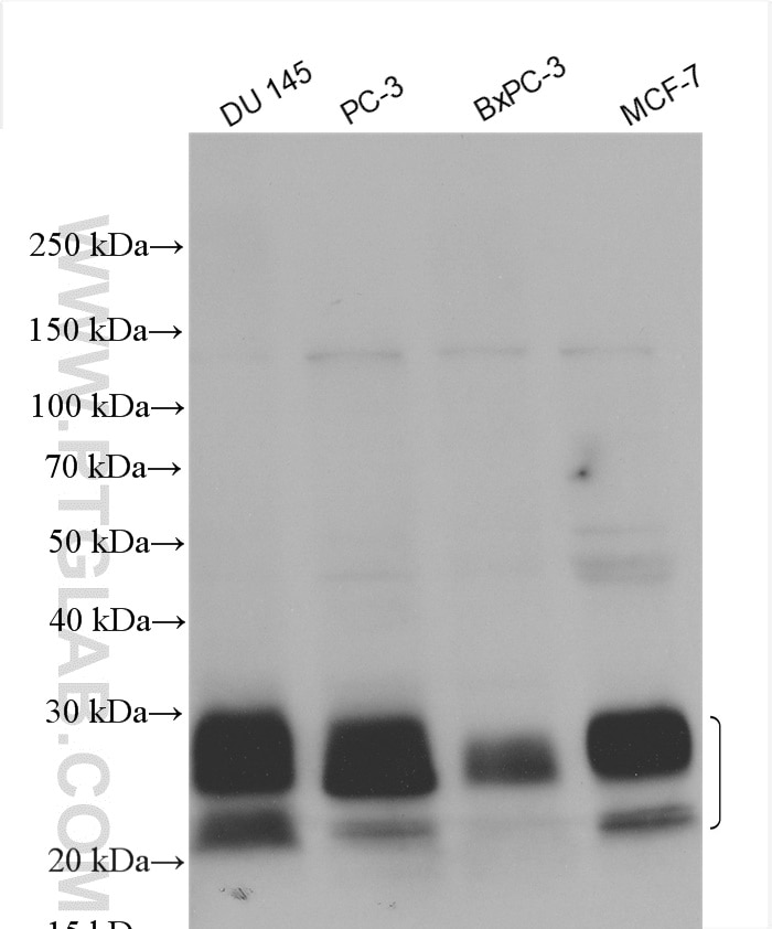
| Positive WB detected in | DU 145 cells, PC-3 cells, BxPC-3 cells, MCF-7 cells |
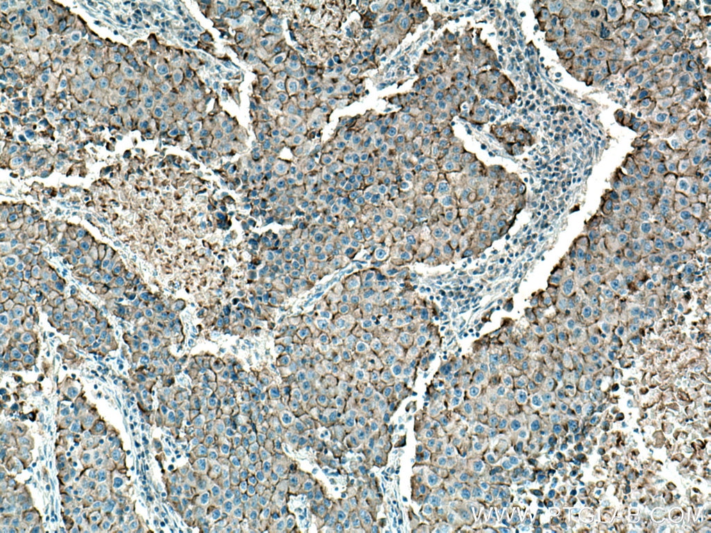
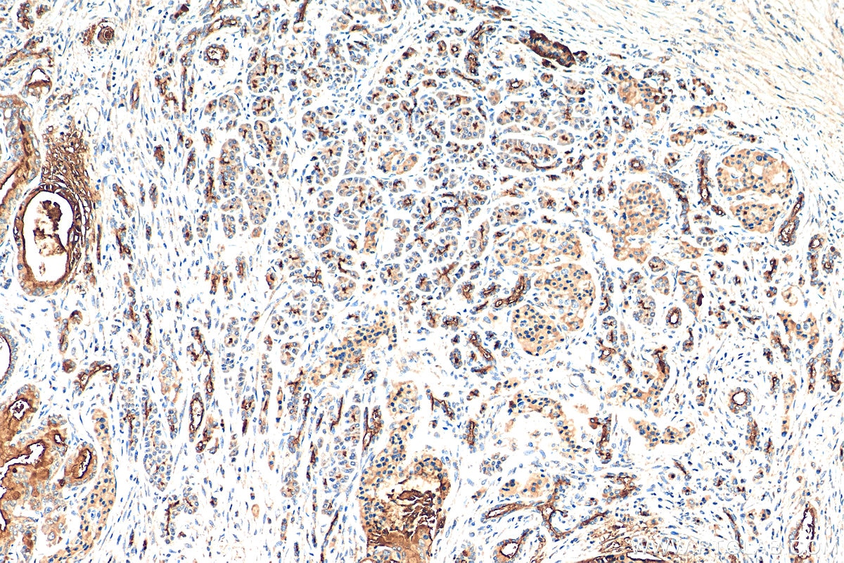
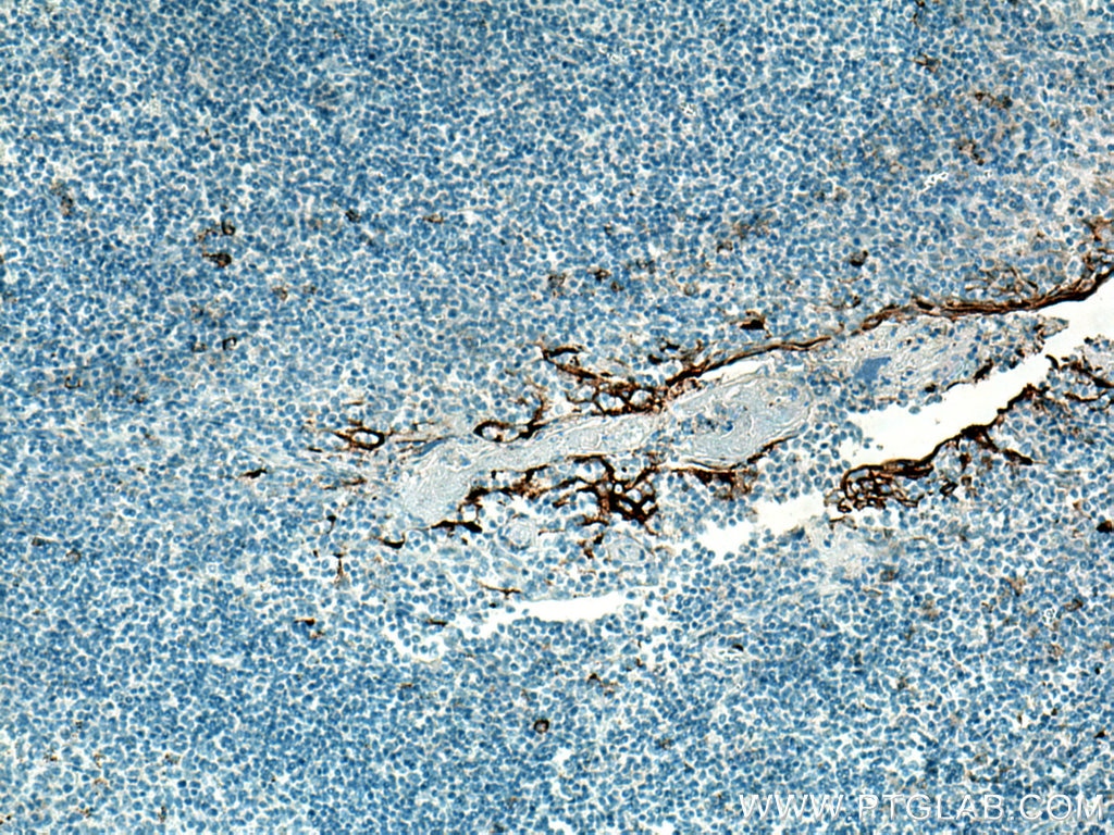
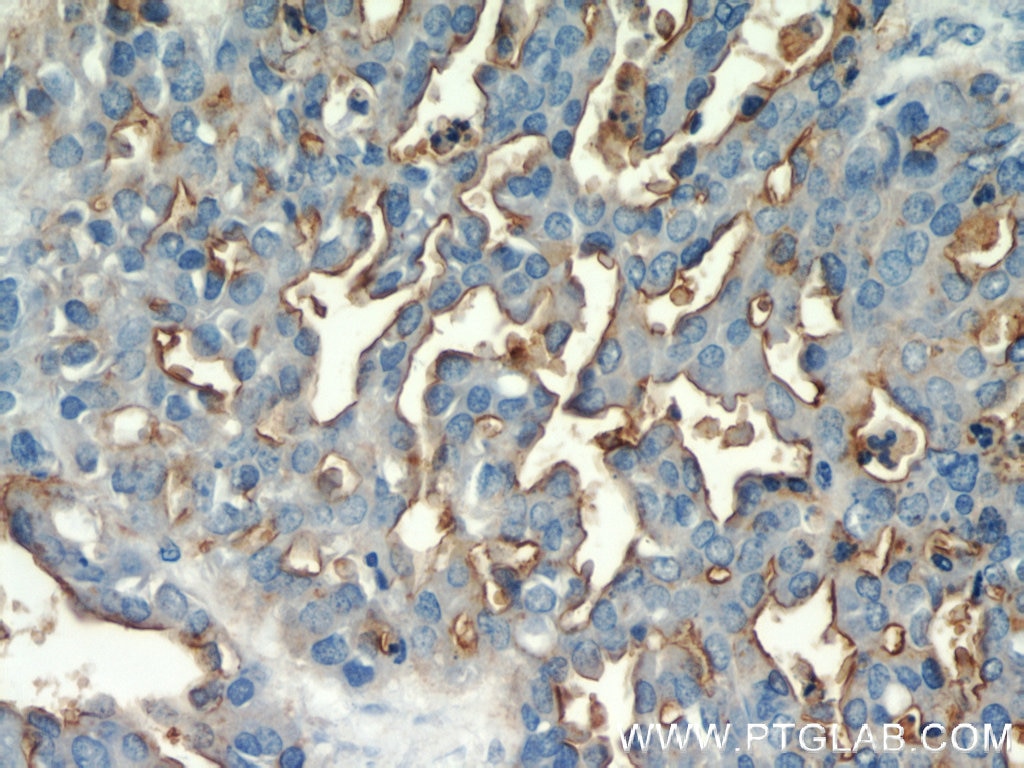
| Positive IHC detected in | human pancreas cancer tissue, human tonsillitis tissue, human breast cancer tissue, human ovary tumor tissue Note: suggested antigen retrieval with TE buffer pH 9.0; (*) Alternatively, antigen retrieval may be performed with citrate buffer pH 6.0 |
Recommended dilution
| Application | Dilution |
|---|---|
| Western Blot (WB) | WB : 1:500-1:2000 |
| Immunohistochemistry (IHC) | IHC : 1:50-1:500 |
| It is recommended that this reagent should be titrated in each testing system to obtain optimal results. | |
| Sample-dependent, Check data in validation data gallery. | |
Published Applications
| KD/KO | See 1 publications below |
| WB | See 11 publications below |
| IHC | See 4 publications below |
| IF | See 3 publications below |
| CoIP | See 1 publications below |
Product Information
23614-1-AP targets MUC1/CA15-3 C-terminal in WB, IHC, IF, CoIP, ELISA applications and shows reactivity with human samples.
| Tested Reactivity | human |
| Cited Reactivity | human, mouse |
| Host / Isotype | Rabbit / IgG |
| Class | Polyclonal |
| Type | Antibody |
| Immunogen |
CatNo: Ag20366 Product name: Recombinant human CA15-3,MUC1 protein Source: e coli.-derived, PET30a Tag: 6*His Domain: 1055-1255 aa of BC120975 Sequence: NSSLEDPSTDYYQELQRDISEMFLQIYKQGGFLGLSNIKFRPGSVVVQLTLAFREGTINVHDVETQFNQYKTEAASRYNLTISDVSVSDVPFPFSAQSGAGVPGWGIALLVLVCVLVALAIVYLIALAVCQCRRKNYGQLDIFPARDTYHPMSEYPTYHTHGRYVPPSSTDRSPYEKVSAGNGGSSLSYTNPAVAATSANL Predict reactive species |
| Full Name | mucin 1, cell surface associated |
| Calculated Molecular Weight | 1264 aa, 123 kDa |
| Observed Molecular Weight | 23-28 kDa |
| GenBank Accession Number | BC120975 |
| Gene Symbol | MUC1/CA15-3 |
| Gene ID (NCBI) | 4582 |
| RRID | AB_2879304 |
| Conjugate | Unconjugated |
| Form | Liquid |
| Purification Method | Antigen affinity purification |
| UNIPROT ID | P15941 |
| Storage Buffer | PBS with 0.02% sodium azide and 50% glycerol, pH 7.3. |
| Storage Conditions | Store at -20°C. Stable for one year after shipment. Aliquoting is unnecessary for -20oC storage. 20ul sizes contain 0.1% BSA. |
Background Information
MUC1 is a type I transmembrane glycoprotein expressed by various epithelial cells of the female reproductive tract, lung, breast, kidney, stomach, and pancreas. MUC1 is transcribed as a large precursor gene product, and upon translation, is cleaved in the endoplasmic reticulum, yielding two subunits: the large extracellular N-terminal subunit (MUC1-N, about 120-200 kDa) and the small cytoplasmic C-terminal subunit (MUC1-C, about 23-30 kDa). Among the known mucins, MUC1 is best studied and plays crucial roles in regulating many cellular properties, including cell proliferation, apoptosis, adhesion, and invasion. MUC1 is overexpressed in a wide range of human epithelial malignancies. This antibody was raised against the C-terminal region of human MUC1, thus it recognizes the 23-30 kDa cytoplasmic MUC1.
Protocols
| Product Specific Protocols | |
|---|---|
| IHC protocol for MUC1/CA15-3 C-terminal antibody 23614-1-AP | Download protocol |
| WB protocol for MUC1/CA15-3 C-terminal antibody 23614-1-AP | Download protocol |
| Standard Protocols | |
|---|---|
| Click here to view our Standard Protocols |
Publications
| Species | Application | Title |
|---|---|---|
ACS Sens Rolling Circle Amplification-Assisted Flow Cytometry Approach for Simultaneous Profiling of Exosomal Surface Proteins. | ||
Cell Cycle LINC02535/miR-30a-5p/GALNT3 axis contributes to lung adenocarcinoma progression via the NF- κ B signaling pathway. | ||
Sci Rep Long-term expansion of basal cells and the novel differentiation methods identify mechanisms for switching Claudin expression in normal epithelia | ||
Pathol Res Pract MUC1 promotes cancer stemness and predicts poor prognosis in osteosarcoma
| ||
Cell Death Discov Loss of QKI in macrophage aggravates inflammatory bowel disease through amplified ROS signaling and microbiota disproportion. | ||
Cell Death Discov Silencing of lncRNA MALAT1 facilitates erastin-induced ferroptosis in endometriosis through miR-145-5p/MUC1 signaling. |

