at dilution of 1:30000 incubated at room temperature for 1.5 hours.")
 at dilution of 1:1000 (under 10x lens). Heat mediated antigen retrieval with Tris-EDTA buffer (pH 9.0).")
 at dilution of 1:1000 (under 40x lens). Heat mediated antigen retrieval with Tris-EDTA buffer (pH 9.0).")
 at dilution of 1:1000 (under 10x lens). Heat mediated antigen retrieval with Tris-EDTA buffer (pH 9.0).")
 at dilution of 1:1000 (under 40x lens). Heat mediated antigen retrieval with Tris-EDTA buffer (pH 9.0).")
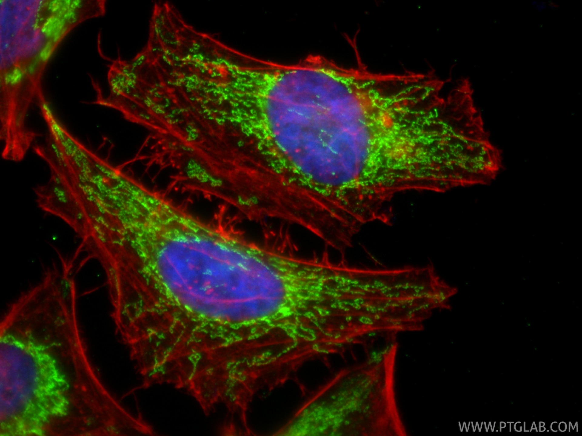
 fixed HepG2 cells using ATP5A1 antibody (82288-1-RR, Clone: 4O12 ) at dilution of 1:2000 and CoraLite®488-Conjugated AffiniPure Goat Anti-Rabbit IgG(H+L), CL594-Phalloidin (red).")
 fixed HeLa cells using ATP5A1 antibody (82288-1-RR, Clone: 4O12 ) at dilution of 1:1000 and CoraLite®594-Conjugated AffiniPure Goat Anti-Rabbit IgG(H+L).")
 fixed HeLa cells using ATP5A1 antibody (82288-1-RR, Clone: 4O12 ) at dilution of 1:1000 and CoraLite®488-Conjugated AffiniPure Goat Anti-Rabbit IgG(H+L), CL594-Phalloidin (red).")
 fixed HeLa cells using ATP5A1 antibody (82288-1-RR, Clone: 4O12 ) at dilution of 1:400 and CoraLite®488-Conjugated AffiniPure Goat Anti-Rabbit IgG(H+L), CL594-Phalloidin (red).")
 fixed HeLa cells using ATP5A1 antibody (82288-1-RR, Clone: 4O12 ) at dilution of 1:400 and CoraLite®488-Conjugated AffiniPure Goat Anti-Rabbit IgG(H+L).")
 fixed HeLa cells using ATP5A1 antibody (82288-1-RR, Clone: 4O12 ) at dilution of 1:400 and CoraLite®488-Conjugated Goat Anti-Rabbit IgG(H+L) (SA00013-2), CL594-phalloidin (red).")
 and CoraLite®488-Conjugated AffiniPure Goat Anti-Rabbit IgG(H+L) at dilution 1:1000 (red), or 0.4 ug Isotype Control. Cells were fixed with 4% PFA and permeabilized with Flow Cytometry Perm Buffer.")
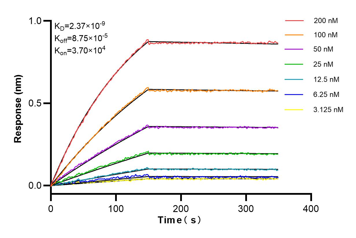
 kinetic assays of 82288-1-RR against Human ATP5A1 were performed. The affinity constant is 2.37 nM.")
Tested Applications
| Positive WB detected in | HEK-293 cells, HeLa cells, HepG2 cells, MCF-7 cells, Jurkat cells, NIH/3T3 cells, HSC-T6 cells |
| Positive IHC detected in | mouse brain tissue, human liver tissue Note: suggested antigen retrieval with TE buffer pH 9.0; (*) Alternatively, antigen retrieval may be performed with citrate buffer pH 6.0 |
| Positive IF/ICC detected in | HepG2 cells, HeLa cells |
| Positive FC (Intra) detected in | HeLa cells |
Recommended dilution
| Application | Dilution |
|---|---|
| Western Blot (WB) | WB : 1:5000-1:50000 |
| Immunohistochemistry (IHC) | IHC : 1:500-1:2000 |
| Immunofluorescence (IF)/ICC | IF/ICC : 1:1100-1:4400 |
| Flow Cytometry (FC) (INTRA) | FC (INTRA) : 0.40 ug per 10^6 cells in a 100 µl suspension |
| It is recommended that this reagent should be titrated in each testing system to obtain optimal results. | |
| Sample-dependent, Check data in validation data gallery. | |
Product Information
82288-1-RR targets ATP5A1 in WB, IHC, IF/ICC, FC (Intra), ELISA applications and shows reactivity with human, mouse, rat samples.
| Tested Reactivity | human, mouse, rat |
| Host / Isotype | Rabbit / IgG |
| Class | Recombinant |
| Type | Antibody |
| Immunogen |
CatNo: Ag6385 Product name: Recombinant human ATP5A1 protein Source: e coli.-derived, PGEX-4T Tag: GST Domain: 203-553 aa of BC064562 Sequence: GRGQRELIIGDRQTGKTSIAIDTIINQKRFNDGSDEKKKLYCIYVAIGQKRSTVAQLVKRLTDADAMKYTIVVSATASDAAPLQYLAPYSGCSMGEYFRDNGKHALIIYDDLSKQAVAYRQMSLLLRRPPGREAYPGDVFYLHSRLLERAAKMNDAFGGGSLTALPVIETQAGDVSAYIPTNVISITDGQIFLETELFYKGIRPAINVGLSVSRVGSAAQTRAMKQVAGTMKLELAQYREVAAFAQFGSDLDAATQQLLSRGVRLTELLKQGQYSPMAIEEQVAVIYAGVRGYLDKLEPSKITKFENAFLSHVVSQHQALLGTIRADGKISEQSDAKLKEIVTNFLAGFEA Predict reactive species |
| Full Name | ATP synthase, H+ transporting, mitochondrial F1 complex, alpha subunit 1, cardiac muscle |
| Calculated Molecular Weight | 60 kDa |
| Observed Molecular Weight | 50-55 kDa |
| GenBank Accession Number | BC064562 |
| Gene Symbol | ATP5A1 |
| Gene ID (NCBI) | 498 |
| RRID | AB_3086476 |
| Conjugate | Unconjugated |
| Form | Liquid |
| Purification Method | Protein A purification |
| UNIPROT ID | P25705 |
| Storage Buffer | PBS with 0.02% sodium azide and 50% glycerol, pH 7.3. |
| Storage Conditions | Store at -20°C. Stable for one year after shipment. Aliquoting is unnecessary for -20oC storage. 20ul sizes contain 0.1% BSA. |
Background Information
The ATP5A1 gene encodes the α subunit of mitochondrial ATP synthase which produces ATP from ADP in the presence of a proton gradient across the membrane. The mitochondrial ATP synthase, also known as Complex V or F1F0 ATP synthase, is a multi-subunit enzyme complex consisting of two functional domains, the F1-containing the catalytic core and the Fo-containing the membrane proton channel. F0 domain has 10 subunits: a,b, c, d, e, f, g, OSCP, A6L, and F6. F1 is composed of subunits α, β, γ, δ, ε, and a loosely attached inhibitor protein IF1. Recently defect in ATP5A1 has been linked to the fatal neonatal mitochondrial encephalopathy. ATP5A1 is localized in the mitochondria and anti-ATP5A1 can be used as the loading control for mitochondrial or Complex V proteins. This antibody recognizes the endogenous ATP5A1 protein in lysates from various cell lines and tissues. The predicted MW of ATP5A1 is 60 kDa, while it undergoes the transit peptide cleavage to become a mature form around 50-55 kDa. Several isoforms of ATP5A1 exist due to the alternative splicing.
Protocols
| Product Specific Protocols | |
|---|---|
| FC protocol for ATP5A1 antibody 82288-1-RR | Download protocol |
| IF protocol for ATP5A1 antibody 82288-1-RR | Download protocol |
| IHC protocol for ATP5A1 antibody 82288-1-RR | Download protocol |
| WB protocol for ATP5A1 antibody 82288-1-RR | Download protocol |
| Standard Protocols | |
|---|---|
| Click here to view our Standard Protocols |