Y79 cells were subjected to SDS PAGE followed by western blot with 20647-1-AP (ANO2 antibody) at dilution of 1:800 incubated at room temperature for 1.5 hours.
Y79 cells were subjected to SDS PAGE followed by western blot with 20647-1-AP (ANO2 antibody) at dilution of 1:800 incubated at room temperature for 1.5 hours.
IP experiment of mouse brain using 20647-1-AP
IP result of anti-ANO2 (IP:20647-1-AP, 4ug; Detection:20647-1-AP 1:300) with mouse brain tissue lysate 10000ug.
Immunohistochemical analysis of paraffin-embedded mouse eye tissue slide using 20647-1-AP (ANO2 antibody) at dilution of 1:200 (under 40x lens). Heat mediated antigen retrieval with Tris-EDTA buffer (pH 9.0).
IHC staining of rat eye using 20647-1-AP
Immunohistochemical analysis of paraffin-embedded rat eye tissue slide using 20647-1-AP (ANO2 antibody) at dilution of 1:200 (under 40x lens). Heat mediated antigen retrieval with Tris-EDTA buffer (pH 9.0).
Immunohistochemical analysis of paraffin-embedded rat eye tissue slide using 20647-1-AP (ANO2 antibody) at dilution of 1:200 (under 40x lens). Heat mediated antigen retrieval with Tris-EDTA buffer (pH 9.0).
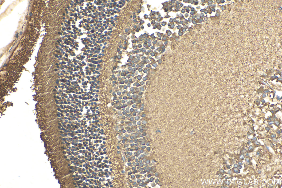
IHC staining of mouse eye using 20647-1-AP
Immunohistochemical analysis of paraffin-embedded mouse eye tissue slide using 20647-1-AP (ANO2 antibody) at dilution of 1:200 (under 40x lens). Heat mediated antigen retrieval with Tris-EDTA buffer (pH 9.0).
Immunohistochemical analysis of paraffin-embedded mouse eye tissue slide using 20647-1-AP (ANO2 antibody) at dilution of 1:200 (under 40x lens). Heat mediated antigen retrieval with Tris-EDTA buffer (pH 9.0).
IHC staining of human breast cancer using 20647-1-AP
Immunohistochemical analysis of paraffin-embedded human breast cancer tissue slide using 20647-1-AP (ANO2 Antibody) at dilution of 1:50 (under 10x lens).
Immunohistochemical analysis of paraffin-embedded human breast cancer tissue slide using 20647-1-AP (ANO2 Antibody) at dilution of 1:50 (under 10x lens).
IHC staining of human breast cancer using 20647-1-AP
Immunohistochemical analysis of paraffin-embedded human breast cancer tissue slide using 20647-1-AP (ANO2 Antibody) at dilution of 1:50 (under 40x lens).
Immunohistochemical analysis of paraffin-embedded human breast cancer tissue slide using 20647-1-AP (ANO2 Antibody) at dilution of 1:50 (under 40x lens).
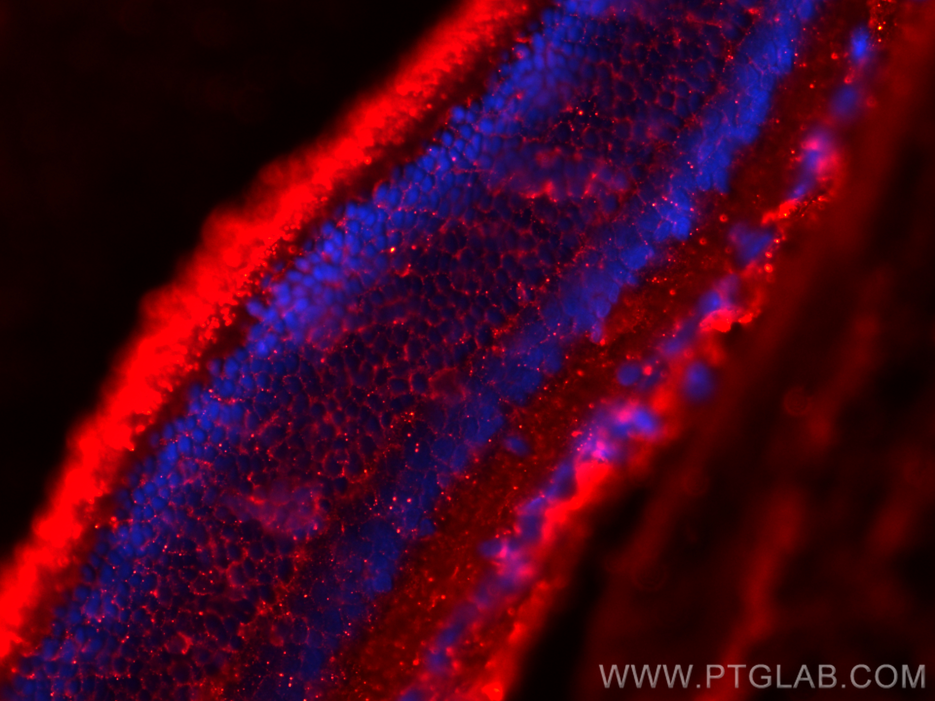
IF Staining of rat eye using 20647-1-AP
Immunofluorescent analysis of (4% PFA) fixed paraffin-embedded rat eye tissue using ANO2 antibody (20647-1-AP) at dilution of 1:400 and CoraLite®594-Conjugated Goat Anti-Rabbit IgG(H+L) (SA00013-4). Heat mediated antigen retrieval with Tris-EDTA buffer (pH 9.0).
Immunofluorescent analysis of (4% PFA) fixed paraffin-embedded rat eye tissue using ANO2 antibody (20647-1-AP) at dilution of 1:400 and CoraLite®594-Conjugated Goat Anti-Rabbit IgG(H+L) (SA00013-4). Heat mediated antigen retrieval with Tris-EDTA buffer (pH 9.0).
The Proteintech guarantee covers Proteintech antibodies in any species and any application, including those not listed on the datasheet. If the antibody doesn’t perform, you can receive a hassle-free refund or credit note.
mouse eye tissue, human breast cancer tissue, rat eye tissue Note: suggested antigen retrieval with TE buffer pH 9.0; (*) Alternatively, antigen retrieval may be performed with citrate buffer pH 6.0
Positive IF-P detected in
rat eye tissue
Recommended dilution
Application
Dilution
Western Blot (WB)
WB : 1:500-1:1000
Immunoprecipitation (IP)
IP : 0.5-4.0 ug for 1.0-3.0 mg of total protein lysate
Immunohistochemistry (IHC)
IHC : 1:50-1:500
Immunofluorescence (IF)-P
IF-P : 1:200-1:800
It is recommended that this reagent should be titrated in each testing system to obtain optimal results.
Sample-dependent, Check data in validation data gallery.
PBS with 0.02% sodium azide and 50% glycerol , pH 7.3
Storage Conditions
Store at -20°C. Stable for one year after shipment. Aliquoting is unnecessary for -20oC storage. 20ul sizes contain 0.1% BSA.
Background Information
ANO2, also named as C12orf3 and TMEM16B, belongs to the anoctamin family. It acts as a calcium-activated chloride channel (CaCC), mostly in photoreceptors. It may mediate olfactory amplification in olfactory sensory neurons (OSNs) and light perception amplification in retina.
Y79 cells were subjected to SDS PAGE followed by western blot with 20647-1-AP (ANO2 antibody) at dilution of 1:800 incubated at room temperature for 1.5 hours.
IHC Figures
IHC staining of mouse eye using 20647-1-AP
Immunohistochemical analysis of paraffin-embedded mouse eye tissue slide using 20647-1-AP (ANO2 antibody) at dilution of 1:200 (under 10x lens). Heat mediated antigen retrieval with Tris-EDTA buffer (pH 9.0).
IHC staining of mouse eye using 20647-1-AP
Immunohistochemical analysis of paraffin-embedded mouse eye tissue slide using 20647-1-AP (ANO2 antibody) at dilution of 1:200 (under 40x lens). Heat mediated antigen retrieval with Tris-EDTA buffer (pH 9.0).
IHC staining of rat eye using 20647-1-AP
Immunohistochemical analysis of paraffin-embedded rat eye tissue slide using 20647-1-AP (ANO2 antibody) at dilution of 1:200 (under 40x lens). Heat mediated antigen retrieval with Tris-EDTA buffer (pH 9.0).
IHC staining of mouse eye using 20647-1-AP
Immunohistochemical analysis of paraffin-embedded mouse eye tissue slide using 20647-1-AP (ANO2 antibody) at dilution of 1:200 (under 40x lens). Heat mediated antigen retrieval with Tris-EDTA buffer (pH 9.0).
IHC staining of human breast cancer using 20647-1-AP
Immunohistochemical analysis of paraffin-embedded human breast cancer tissue slide using 20647-1-AP (ANO2 Antibody) at dilution of 1:50 (under 10x lens).
IHC staining of human breast cancer using 20647-1-AP
Immunohistochemical analysis of paraffin-embedded human breast cancer tissue slide using 20647-1-AP (ANO2 Antibody) at dilution of 1:50 (under 40x lens).
IP Figures
IP experiment of mouse brain using 20647-1-AP
IP result of anti-ANO2 (IP:20647-1-AP, 4ug; Detection:20647-1-AP 1:300) with mouse brain tissue lysate 10000ug.
IF-P Figures
IF Staining of rat eye using 20647-1-AP
Immunofluorescent analysis of (4% PFA) fixed paraffin-embedded rat eye tissue using ANO2 antibody (20647-1-AP) at dilution of 1:400 and CoraLite®594-Conjugated Goat Anti-Rabbit IgG(H+L) (SA00013-4). Heat mediated antigen retrieval with Tris-EDTA buffer (pH 9.0).
The species listed in Tested Reactivity are in-house verified and applicable species. For unlisted species, please refer to the homology analysis of the immunogen sequence and related species. For rabbit polyclonal antibodies, homology >70% is recommended. For mouse monoclonal antibodies and rabbit recombinant antibodies, homology >90% is recommended. Generally, the higher the homology, the greater the applicability. However, there will be certain differences in protein expression in different species, tissues or cells. Therefore, the homology analysis results are for reference only and do not serve as a guarantee.
At Proteintech, we pride ourselves on our antibody quality, customer service and transparency. As such, we are comparing our antibodies with other vendors, enabling easy identification and comparisons of key data to help you choose the suitable antibody for your needs.
We have selected the top cited antibodies from these vendors for you to compare.
Proteintech
KD/KO VALIDATED
ANO2 Polyclonal antibody
Catalog Number
20647-1-AP
Citations
4
Dilutions
WB : 1:500-1:1000 IP : 0.5-4.0 ug for IP and 0.5-4.0 ug for 1.0-3.0 mg of total protein lysate for WB IHC : 1:50-1:500 IF-P : 1:200-1:800
Applications
WB, IHC, IF-P, IP, ELISA
Reactivity
human, mouse, rat
Product Guarantee
Covers any species including not listed on datasheet
Covers any applications including not listed on datasheet
 at dilution of 1:800 incubated at room temperature for 1.5 hours.")
 with mouse brain tissue lysate 10000ug.")
 at dilution of 1:200 (under 10x lens). Heat mediated antigen retrieval with Tris-EDTA buffer (pH 9.0).")
 at dilution of 1:200 (under 40x lens). Heat mediated antigen retrieval with Tris-EDTA buffer (pH 9.0).")
 at dilution of 1:200 (under 40x lens). Heat mediated antigen retrieval with Tris-EDTA buffer (pH 9.0).")
 at dilution of 1:200 (under 40x lens). Heat mediated antigen retrieval with Tris-EDTA buffer (pH 9.0).")
 at dilution of 1:50 (under 10x lens).")
 at dilution of 1:50 (under 40x lens).")
 fixed paraffin-embedded rat eye tissue using ANO2 antibody (20647-1-AP) at dilution of 1:400 and CoraLite®594-Conjugated Goat Anti-Rabbit IgG(H+L) (SA00013-4). Heat mediated antigen retrieval with Tris-EDTA buffer (pH 9.0).")