Tested Applications
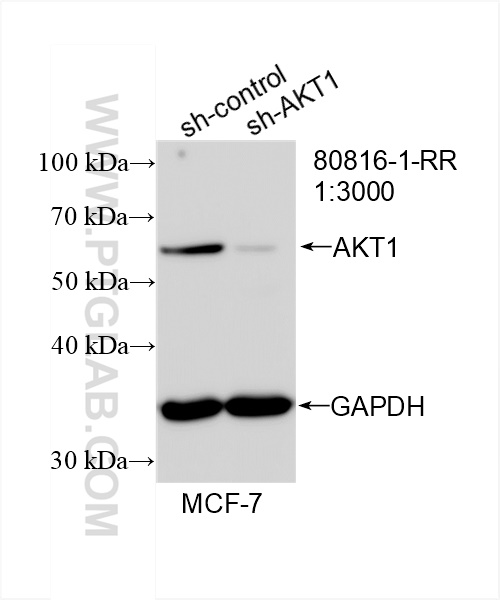
| Positive WB detected in | HEK-293 cells, MCF-7 cells, HeLa cells, A549 cells, Jurkat cells, K-562 cells, NIH/3T3 cells, RAW 264.7 cells, HSC-T6 cells, PC-12 cells |
| Positive IP detected in | HEK-293 cells |
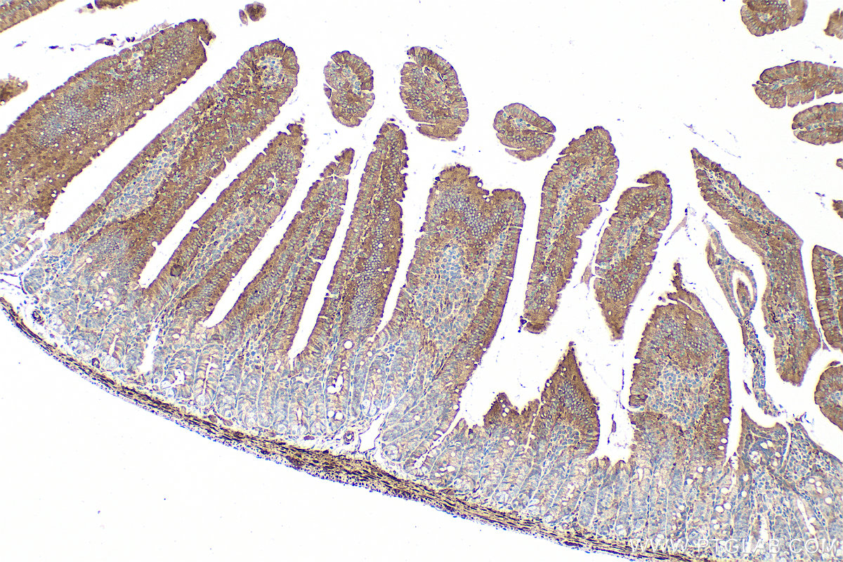
| Positive IHC detected in | human ovary tumor tissue, mouse small intestine tissue Note: suggested antigen retrieval with TE buffer pH 9.0; (*) Alternatively, antigen retrieval may be performed with citrate buffer pH 6.0 |
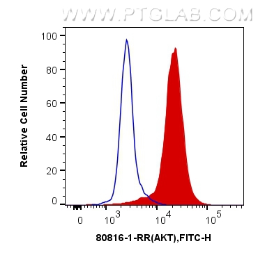
| Positive FC (Intra) detected in | Jurkat cells |
Recommended dilution
| Application | Dilution |
|---|---|
| Western Blot (WB) | WB : 1:5000-1:50000 |
| Immunoprecipitation (IP) | IP : 0.5-4.0 ug for 1.0-3.0 mg of total protein lysate |
| Immunohistochemistry (IHC) | IHC : 1:500-1:2000 |
| Flow Cytometry (FC) (INTRA) | FC (INTRA) : 0.40 ug per 10^6 cells in a 100 µl suspension |
| It is recommended that this reagent should be titrated in each testing system to obtain optimal results. | |
| Sample-dependent, Check data in validation data gallery. | |
Published Applications
| WB | See 10 publications below |
| IHC | See 1 publications below |
Product Information
80816-1-RR targets AKT1-Specific in WB, IHC, FC (Intra), IP, ELISA applications and shows reactivity with human, mouse, rat samples.
| Tested Reactivity | human, mouse, rat |
| Cited Reactivity | human, mouse, rat |
| Host / Isotype | Rabbit / IgG |
| Class | Recombinant |
| Type | Antibody |
| Immunogen |
CatNo: Ag0213 Product name: Recombinant human AKT protein Source: e coli.-derived, PGEX-4T Tag: GST Domain: 1-224 aa of BC000479 Sequence: MSDVAIVKEGWLHKRGEYIKTWRPRYFLLKNDGTFIGYKERPQDVDQREAPLNNFSVAQCQLMKTERPRPNTFIIRCLQWTTVIERTFHVETPEEREEWTTAIQTVADGLKKQEEEEMDFRSGSPSDNSGAEEMEVSLAKPKHRVTMNEFEYLKLLGKGTFGKVILVKEKATGRYYAMKILKKEVIVAKDEVAHTLTENRVLQNSRHPFLTALKYSFQTHDRLC Predict reactive species |
| Full Name | v-akt murine thymoma viral oncogene homolog 1 |
| Calculated Molecular Weight | 56 kDa |
| Observed Molecular Weight | 56-62 kDa |
| GenBank Accession Number | BC000479 |
| Gene Symbol | AKT1 |
| Gene ID (NCBI) | 207 |
| RRID | AB_2935546 |
| Conjugate | Unconjugated |
| Form | Liquid |
| Purification Method | Protein A purification |
| UNIPROT ID | P31749 |
| Storage Buffer | PBS with 0.02% sodium azide and 50% glycerol, pH 7.3. |
| Storage Conditions | Store at -20°C. Stable for one year after shipment. Aliquoting is unnecessary for -20oC storage. 20ul sizes contain 0.1% BSA. |
Background Information
The serine-threonine protein kinase AKT1 is catalytically inactive in serum-starved primary and immortalized fibroblasts. AKT1 and the related AKT2 are activated by platelet-derived growth factor. The activation is rapid and specific, and it is abrogated by mutations in the pleckstrin homology domain of AKT1. It was shown that the activation occurs through phosphatidylinositol 3-kinase. In the developing nervous system AKT is a critical mediator of growth factor-induced neuronal survival. Survival factors can suppress apoptosis in a transcription-independent manner by activating the serine/threonine kinase AKT1, which then phosphorylates and inactivates components of the apoptotic machinery. 80816-1-RR recognizes AKT1 specifically.
Protocols
| Product Specific Protocols | |
|---|---|
| FC protocol for AKT1-Specific antibody 80816-1-RR | Download protocol |
| IHC protocol for AKT1-Specific antibody 80816-1-RR | Download protocol |
| IP protocol for AKT1-Specific antibody 80816-1-RR | Download protocol |
| WB protocol for AKT1-Specific antibody 80816-1-RR | Download protocol |
| Standard Protocols | |
|---|---|
| Click here to view our Standard Protocols |
Publications
| Species | Application | Title |
|---|---|---|
Int J Mol Sci Rotenone Induces Parkinsonism with Constipation Symptoms in Mice by Disrupting the Gut Microecosystem, Inhibiting the PI3K-AKT Signaling Pathway and Gastrointestinal Motility | ||
Int Immunopharmacol Geniposide ameliorates psoriatic skin inflammation by inhibiting the TLR4/MyD88/NF-κB p65 signaling pathway and MMP9 | ||
Pathol Res Pract Identification of bioactive components in Wuji Wan and their protective effects against gastric ulcer via regulating PIK3CA-mediated PI3K/AKT pathway. | ||
Int J Biol Macromol An inulin-type polysaccharide from Atractylodis Macrocephalae Rhizoma can relieve psoriasis | ||
J Pharmacol Sci Astaxanthin suppress ferroptosis through the Akt1-FoxO3a signaling pathway to alleviates brain injury after intracerebral hemorrhage. | ||
Front Nutr Xianyu capsule ameliorates neuroinflammatory and glycerophospholipid metabolism in lithium-pilocarpine-induced acute epilepsy. |