Various lysates were subjected to SDS PAGE followed by western blot with 15264-1-AP (Adenylosuccinate lyase antibody) at dilution of 1:3000 incubated at room temperature for 1.5 hours.
Various lysates were subjected to SDS PAGE followed by western blot with 15264-1-AP (Adenylosuccinate lyase antibody) at dilution of 1:3000 incubated at room temperature for 1.5 hours.
WB analysis of HeLa using 15264-1-AP
HeLa cells were subjected to SDS PAGE followed by western blot with 15264-1-AP (Adenylosuccinate lyase antibody) at dilution of 1:400 incubated at room temperature for 1.5 hours.
HeLa cells were subjected to SDS PAGE followed by western blot with 15264-1-AP (Adenylosuccinate lyase antibody) at dilution of 1:400 incubated at room temperature for 1.5 hours.
WB analysis of HepG2 using 15264-1-AP
HepG2 cells were subjected to SDS PAGE followed by western blot with 15264-1-AP (Adenylosuccinate lyase antibody) at dilution of 1:400 incubated at room temperature for 1.5 hours.
HepG2 cells were subjected to SDS PAGE followed by western blot with 15264-1-AP (Adenylosuccinate lyase antibody) at dilution of 1:400 incubated at room temperature for 1.5 hours.
IHC staining of human liver cancer using 15264-1-AP
Immunohistochemical analysis of paraffin-embedded human liver cancer using 15264-1-AP (Adenylosuccinate lyase antibody) at dilution of 1:50 (under 10x lens).
Immunohistochemical analysis of paraffin-embedded human liver cancer using 15264-1-AP (Adenylosuccinate lyase antibody) at dilution of 1:50 (under 10x lens).
IHC staining of human liver cancer using 15264-1-AP
Immunohistochemical analysis of paraffin-embedded human liver cancer using 15264-1-AP (Adenylosuccinate lyase antibody) at dilution of 1:50 (under 40x lens).
Immunohistochemical analysis of paraffin-embedded human liver cancer using 15264-1-AP (Adenylosuccinate lyase antibody) at dilution of 1:50 (under 40x lens).
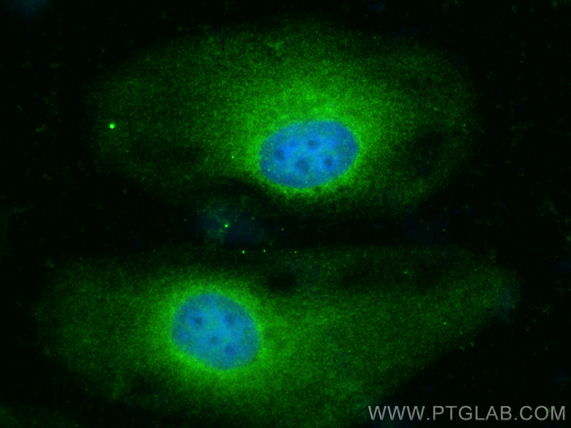
IF Staining of HeLa using 15264-1-AP
Immunofluorescent analysis of (-20°C Ethanol) fixed HeLa cells using Adenylosuccinate lyase antibody (15264-1-AP) at dilution of 1:400 and CoraLite®488-Conjugated Goat Anti-Rabbit IgG(H+L) (SA00013-2).
Immunofluorescent analysis of (-20°C Ethanol) fixed HeLa cells using Adenylosuccinate lyase antibody (15264-1-AP) at dilution of 1:400 and CoraLite®488-Conjugated Goat Anti-Rabbit IgG(H+L) (SA00013-2).
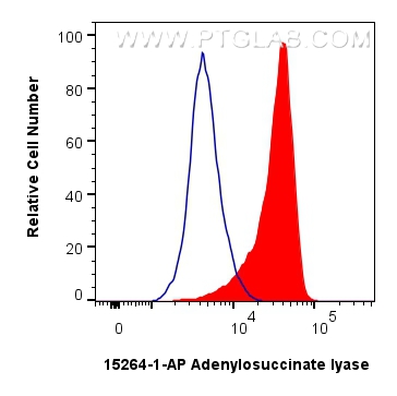
FC experiment of HeLa using 15264-1-AP
1x10^6 HeLa cells were intracellularly stained with 0.25 ug Adenylosuccinate lyase Polyclonal antibody (15264-1-AP) and CoraLite®488-Conjugated Goat Anti-Rabbit IgG(H+L) (SA00013-2)(red), or 0.25 ug Rabbit IgG control Rabbit PolyAb (30000-0-AP) (blue). Cells were fixed with 4% PFA and permeabilized with Flow Cytometry Perm Buffer (PF00011-C).
1x10^6 HeLa cells were intracellularly stained with 0.25 ug Adenylosuccinate lyase Polyclonal antibody (15264-1-AP) and CoraLite®488-Conjugated Goat Anti-Rabbit IgG(H+L) (SA00013-2)(red), or 0.25 ug Rabbit IgG control Rabbit PolyAb (30000-0-AP) (blue). Cells were fixed with 4% PFA and permeabilized with Flow Cytometry Perm Buffer (PF00011-C).
The Proteintech guarantee covers Proteintech antibodies in any species and any application, including those not listed on the datasheet. If the antibody doesn’t perform, you can receive a hassle-free refund or credit note.
HeLa cells, HepG2 cells, NIH/3T3 cells, RAW264.7 cells
Positive IHC detected in
human liver cancer tissue Note: suggested antigen retrieval with TE buffer pH 9.0; (*) Alternatively, antigen retrieval may be performed with citrate buffer pH 6.0
Positive IF/ICC detected in
HeLa cells
Positive FC (Intra) detected in
HeLa cells
Recommended dilution
Application
Dilution
Western Blot (WB)
WB : 1:1000-1:6000
Immunohistochemistry (IHC)
IHC : 1:20-1:200
Immunofluorescence (IF)/ICC
IF/ICC : 1:200-1:800
Flow Cytometry (FC) (INTRA)
FC (INTRA) : 0.25 ug per 10^6 cells in a 100 µl suspension
It is recommended that this reagent should be titrated in each testing system to obtain optimal results.
Sample-dependent, Check data in validation data gallery.
PBS with 0.02% sodium azide and 50% glycerol pH 7.3.
Storage Conditions
Store at -20°C. Stable for one year after shipment. Aliquoting is unnecessary for -20oC storage. 20ul sizes contain 0.1% BSA.
Background Information
ADSL(adenylosuccinate lyase) is also named as AMPS, ASase, ASL and belongs to the lyase 1 family. It is an enzyme involved in 2 pathways of purine nucleotide metabolism and catalyzes cleavage of succinyl groups to yield fumarate(PMID:18524658). Defects in ADSL are the cause of adenylosuccinase deficiency (ADSL deficiency). In humans, mutations in ADSL lead to an inborn error of metabolism originally characterized by developmental delay, often with autistic features(PMID:20884265)..The ADSL enzymatic activity is reduced in lymphocytes and red blood cells of the patient with severe psychomotor retardation(PMID:9545543). It has 2 isoforms produced by alternative splicing.
Various lysates were subjected to SDS PAGE followed by western blot with 15264-1-AP (Adenylosuccinate lyase antibody) at dilution of 1:3000 incubated at room temperature for 1.5 hours.
WB analysis of HeLa using 15264-1-AP
HeLa cells were subjected to SDS PAGE followed by western blot with 15264-1-AP (Adenylosuccinate lyase antibody) at dilution of 1:400 incubated at room temperature for 1.5 hours.
WB analysis of HepG2 using 15264-1-AP
HepG2 cells were subjected to SDS PAGE followed by western blot with 15264-1-AP (Adenylosuccinate lyase antibody) at dilution of 1:400 incubated at room temperature for 1.5 hours.
IHC Figures
IHC staining of human liver cancer using 15264-1-AP
Immunohistochemical analysis of paraffin-embedded human liver cancer using 15264-1-AP (Adenylosuccinate lyase antibody) at dilution of 1:50 (under 10x lens).
IHC staining of human liver cancer using 15264-1-AP
Immunohistochemical analysis of paraffin-embedded human liver cancer using 15264-1-AP (Adenylosuccinate lyase antibody) at dilution of 1:50 (under 40x lens).
IF/ICC Figures
IF Staining of HeLa using 15264-1-AP
Immunofluorescent analysis of (-20°C Ethanol) fixed HeLa cells using Adenylosuccinate lyase antibody (15264-1-AP) at dilution of 1:400 and CoraLite®488-Conjugated Goat Anti-Rabbit IgG(H+L) (SA00013-2).
FC (INTRA) Figures
FC experiment of HeLa using 15264-1-AP
1x10^6 HeLa cells were intracellularly stained with 0.25 ug Adenylosuccinate lyase Polyclonal antibody (15264-1-AP) and CoraLite®488-Conjugated Goat Anti-Rabbit IgG(H+L) (SA00013-2)(red), or 0.25 ug Rabbit IgG control Rabbit PolyAb (30000-0-AP) (blue). Cells were fixed with 4% PFA and permeabilized with Flow Cytometry Perm Buffer (PF00011-C).
The species listed in Tested Reactivity are in-house verified and applicable species. For unlisted species, please refer to the homology analysis of the immunogen sequence and related species. For rabbit polyclonal antibodies, homology >70% is recommended. For mouse monoclonal antibodies and rabbit recombinant antibodies, homology >90% is recommended. Generally, the higher the homology, the greater the applicability. However, there will be certain differences in protein expression in different species, tissues or cells. Therefore, the homology analysis results are for reference only and do not serve as a guarantee.
At Proteintech, we pride ourselves on our antibody quality, customer service and transparency. As such, we are comparing our antibodies with other vendors, enabling easy identification and comparisons of key data to help you choose the suitable antibody for your needs.
We have selected the top cited antibodies from these vendors for you to compare.
Proteintech
ADSL Polyclonal antibody
Catalog Number
15264-1-AP
Citations
4
Dilutions
WB : 1:1000-1:6000 IHC : 1:20-1:200 IF/ICC : 1:200-1:800 FC (INTRA) : 0.25 ug per 10^6 cells in a 100 µl suspension
Applications
WB, IHC, IF/ICC, FC (Intra), ELISA
Reactivity
human, mouse, rat
Product Guarantee
Covers any species including not listed on datasheet
Covers any applications including not listed on datasheet
 at dilution of 1:3000 incubated at room temperature for 1.5 hours.")
 at dilution of 1:400 incubated at room temperature for 1.5 hours.")
 at dilution of 1:400 incubated at room temperature for 1.5 hours.")
 at dilution of 1:50 (under 10x lens).")
 at dilution of 1:50 (under 40x lens).")
 fixed HeLa cells using Adenylosuccinate lyase antibody (15264-1-AP) at dilution of 1:400 and CoraLite®488-Conjugated Goat Anti-Rabbit IgG(H+L) (SA00013-2).")
 and CoraLite®488-Conjugated Goat Anti-Rabbit IgG(H+L) (SA00013-2)(red), or 0.25 ug Rabbit IgG control Rabbit PolyAb (30000-0-AP) (blue). Cells were fixed with 4% PFA and permeabilized with Flow Cytometry Perm Buffer (PF00011-C).")