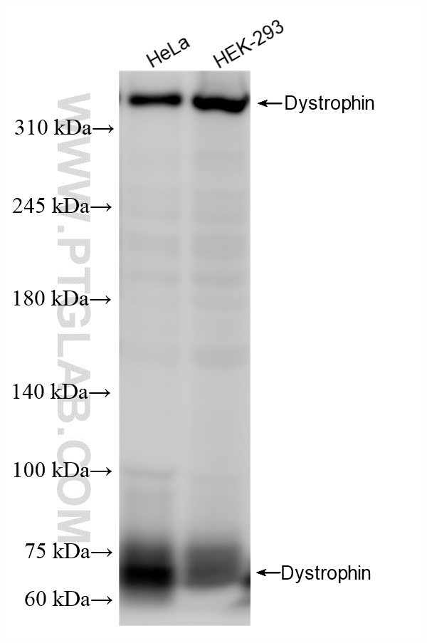
Various lysates were subjected to SDS PAGE followed by western blot with 83609-5-RR (DMD antibody) at dilution of 1:20000 incubated at room temperature for 1.5 hours.
Various lysates were subjected to SDS PAGE followed by western blot with 83609-5-RR (DMD antibody) at dilution of 1:20000 incubated at room temperature for 1.5 hours.
IHC staining of mouse skeletal muscle using 83609-5-RR
Immunohistochemical analysis of paraffin-embedded mouse skeletal muscle tissue slide using 83609-5-RR (Dystrophin antibody) at dilution of 1:200 (under 40x lens). Heat mediated antigen retrieval with Tris-EDTA buffer (pH 9.0).
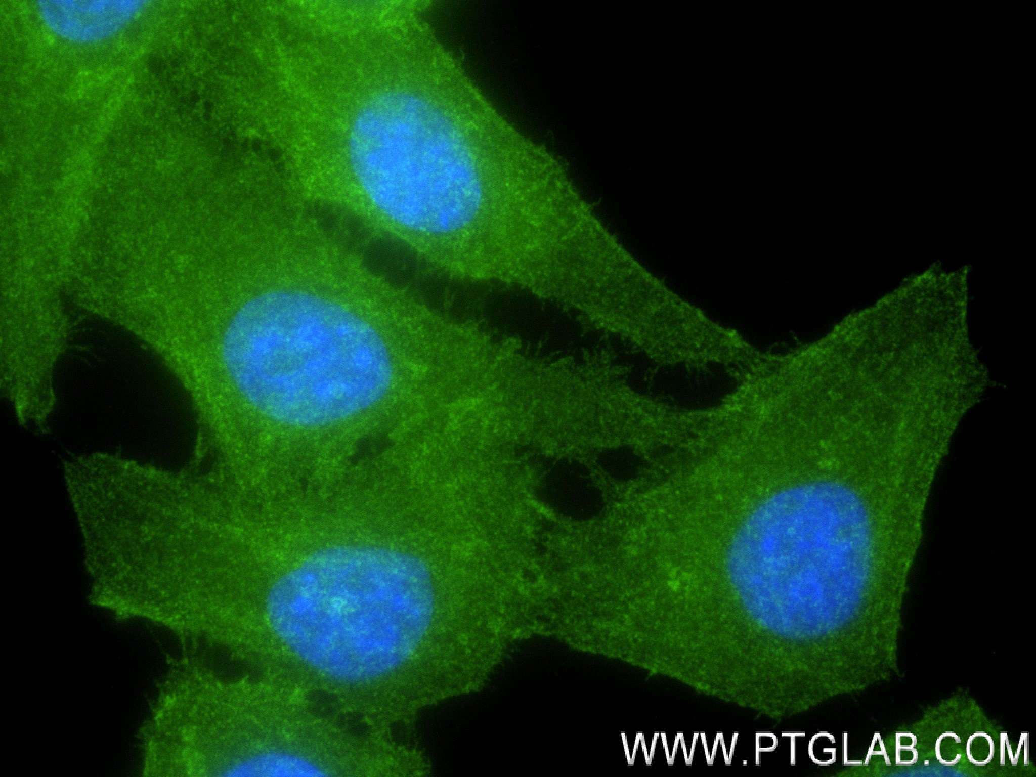
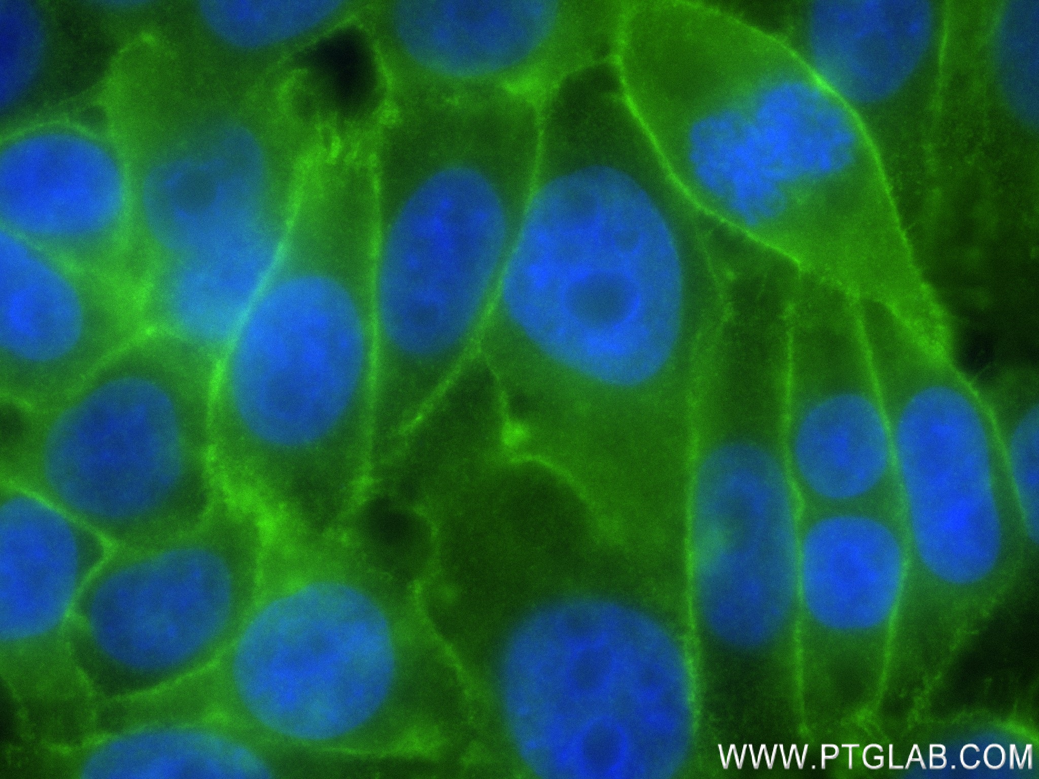
Immunofluorescent analysis of (4% PFA) fixed HepG2 cells using Dystrophin antibody (83609-5-RR, Clone: 240567A3 ) at dilution of 1:250 and CoraLite®488-Conjugated AffiniPure Goat Anti-Rabbit IgG(H+L) (SA00013-2).
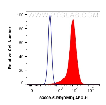
FC experiment of HepG2 using 83609-5-RR
1x10^6 HepG2 cells were intracellularly stained with 0.25 ug Dystrophin Recombinant antibody (83609-5-RR, Clone:240567A3) and APC-Conjugated AffiniPure Goat Anti-Rabbit IgG(H+L)(red), or 0.25 ug Isotype Control (blue). Cells were fixed with 4% PFA and permeabilized with Flow Cytometry Perm Buffer (PF00011-C).
1x10^6 HepG2 cells were intracellularly stained with 0.25 ug Dystrophin Recombinant antibody (83609-5-RR, Clone:240567A3) and APC-Conjugated AffiniPure Goat Anti-Rabbit IgG(H+L)(red), or 0.25 ug Isotype Control (blue). Cells were fixed with 4% PFA and permeabilized with Flow Cytometry Perm Buffer (PF00011-C).
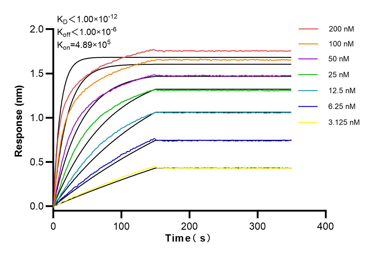
Affinity and Kinetic Characterization of 83609-5-RR
Biolayer interferometry (BLl) kinetic assays of 83609-5-RR
against Human Dystrophin were performed. The affinity constant is below 1 pM.
The Proteintech guarantee covers Proteintech antibodies in any species and any application, including those not listed on the datasheet. If the antibody doesn’t perform, you can receive a hassle-free refund or credit note.
mouse skeletal muscle tissue Note: suggested antigen retrieval with TE buffer pH 9.0; (*) Alternatively, antigen retrieval may be performed with citrate buffer pH 6.0
Positive IF-P detected in
rat brain tissue
Positive IF/ICC detected in
HepG2 cells
Positive FC (Intra) detected in
HepG2 cells
Recommended dilution
Application
Dilution
Western Blot (WB)
WB : 1:5000-1:50000
Immunohistochemistry (IHC)
IHC : 1:50-1:500
Immunofluorescence (IF)-P
IF-P : 1:200-1:800
Immunofluorescence (IF)/ICC
IF/ICC : 1:125-1:500
Flow Cytometry (FC) (INTRA)
FC (INTRA) : 0.25 ug per 10^6 cells in a 100 µl suspension
It is recommended that this reagent should be titrated in each testing system to obtain optimal results.
Sample-dependent, Check data in validation data gallery.
Product Information
83609-5-RR targets Dystrophin in WB, IHC, IF/ICC, IF-P, FC (Intra), ELISA applications and shows reactivity with human, mouse, rat samples.
PBS with 0.02% sodium azide and 50% glycerol , pH 7.3
Storage Conditions
Store at -20°C. Stable for one year after shipment. Aliquoting is unnecessary for -20oC storage. 20ul sizes contain 0.1% BSA.
Background Information
Dystrophin (DMD or BMD) is a large muscle protein whose mutations cause Duchenne muscular dystrophy (DMD) and Becker muscular dystrophy (BMD), the childhood neuromuscular disorders that result in progressive muscle weakness, respiratory difficulties and cardiovascular dysfunction. Dystrophin is a crucial component of the dystrophin-glycoprotein complex which is essential for muscle membrane integrity and stability. Dystrophin is located on the cytoplasmic face of the sarcolemma and connects the cytoskeletal network to the sarcolemma and extracellular matrix. Multiple isoforms of dystrophin exist due to the alternative splicing, with a wide range of MW (69-72, 110-143, 271, 426 kDa). Most tissues contain transcripts of several isoforms.
Various lysates were subjected to SDS PAGE followed by western blot with 83609-5-RR (DMD antibody) at dilution of 1:20000 incubated at room temperature for 1.5 hours.
IHC Figures
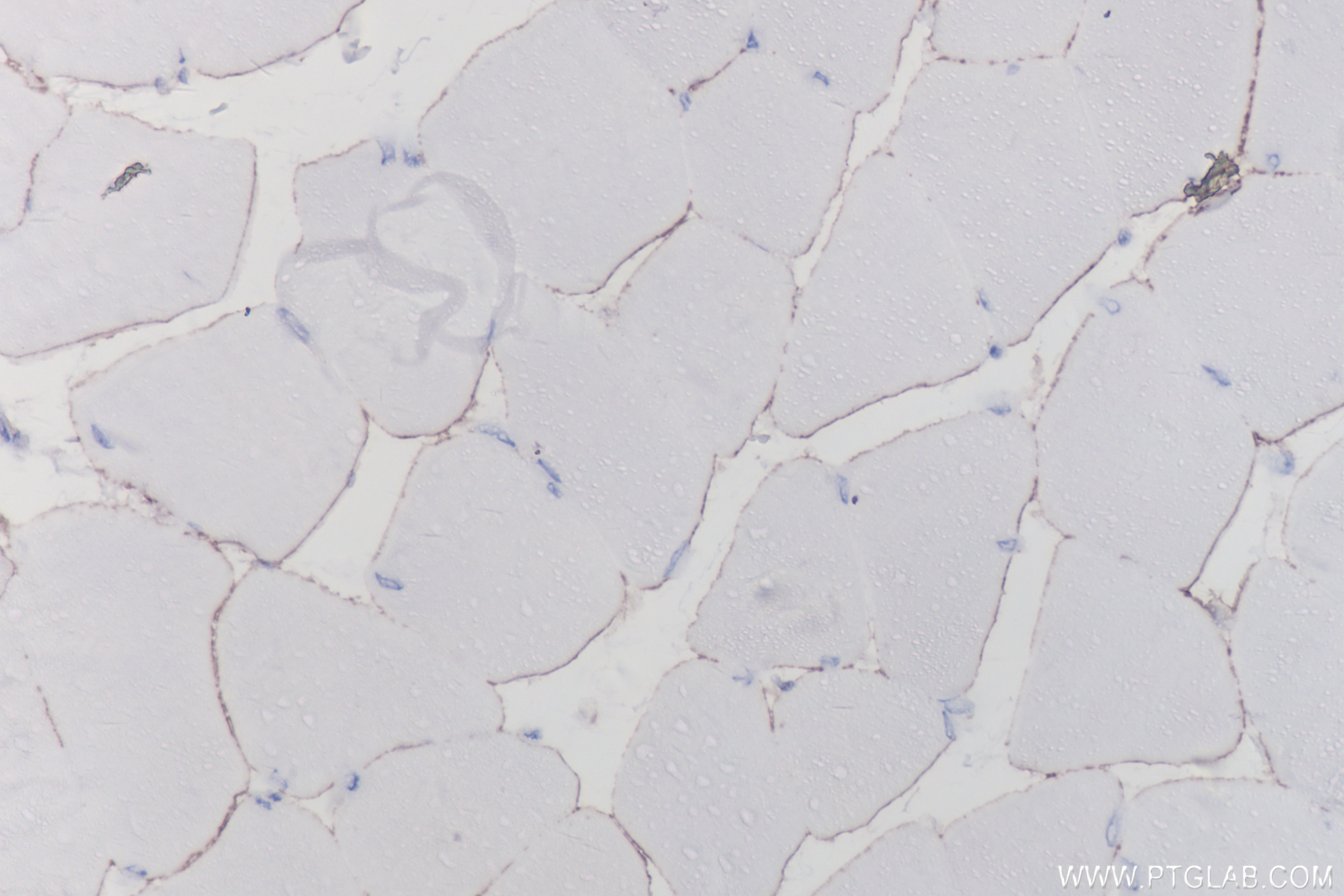
IHC staining of mouse skeletal muscle using 83609-5-RR
Immunohistochemical analysis of paraffin-embedded mouse skeletal muscle tissue slide using 83609-5-RR (Dystrophin antibody) at dilution of 1:200 (under 40x lens). Heat mediated antigen retrieval with Tris-EDTA buffer (pH 9.0).
IF-P Figures
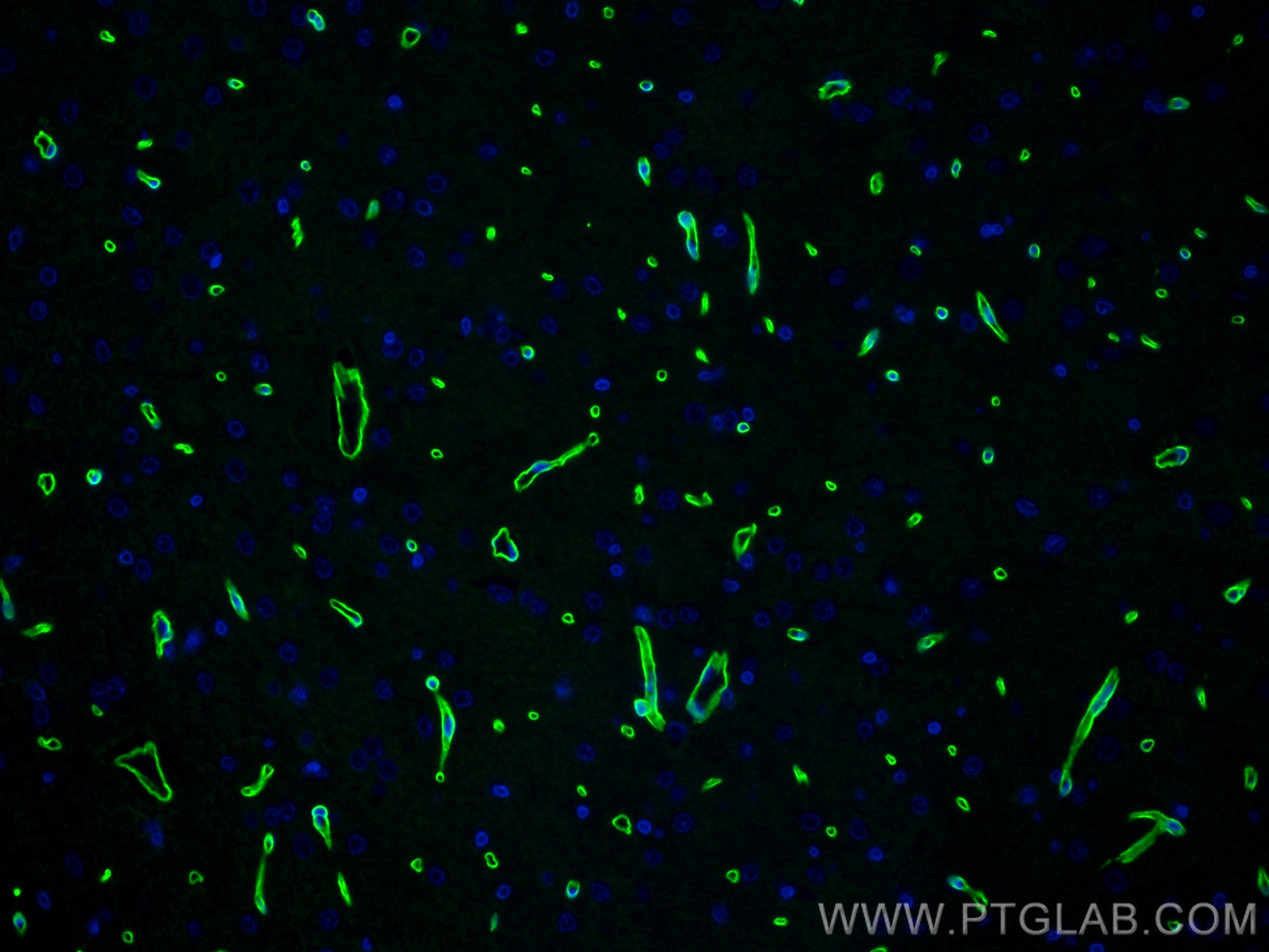
IF Staining of rat brain using 83609-5-RR
Immunofluorescent analysis of (4% PFA) fixed paraffin-embedded rat brain tissue using Dystrophin antibody (83609-5-RR, Clone: 240567A3 ) at dilution of 1:400 and Multi-rAb CoraLite ® Plus 488-Goat Anti-Rabbit Recombinant Secondary Antibody (H+L) (RGAR002). Heat mediated antigen retrieval with Tris-EDTA buffer (pH 9.0).
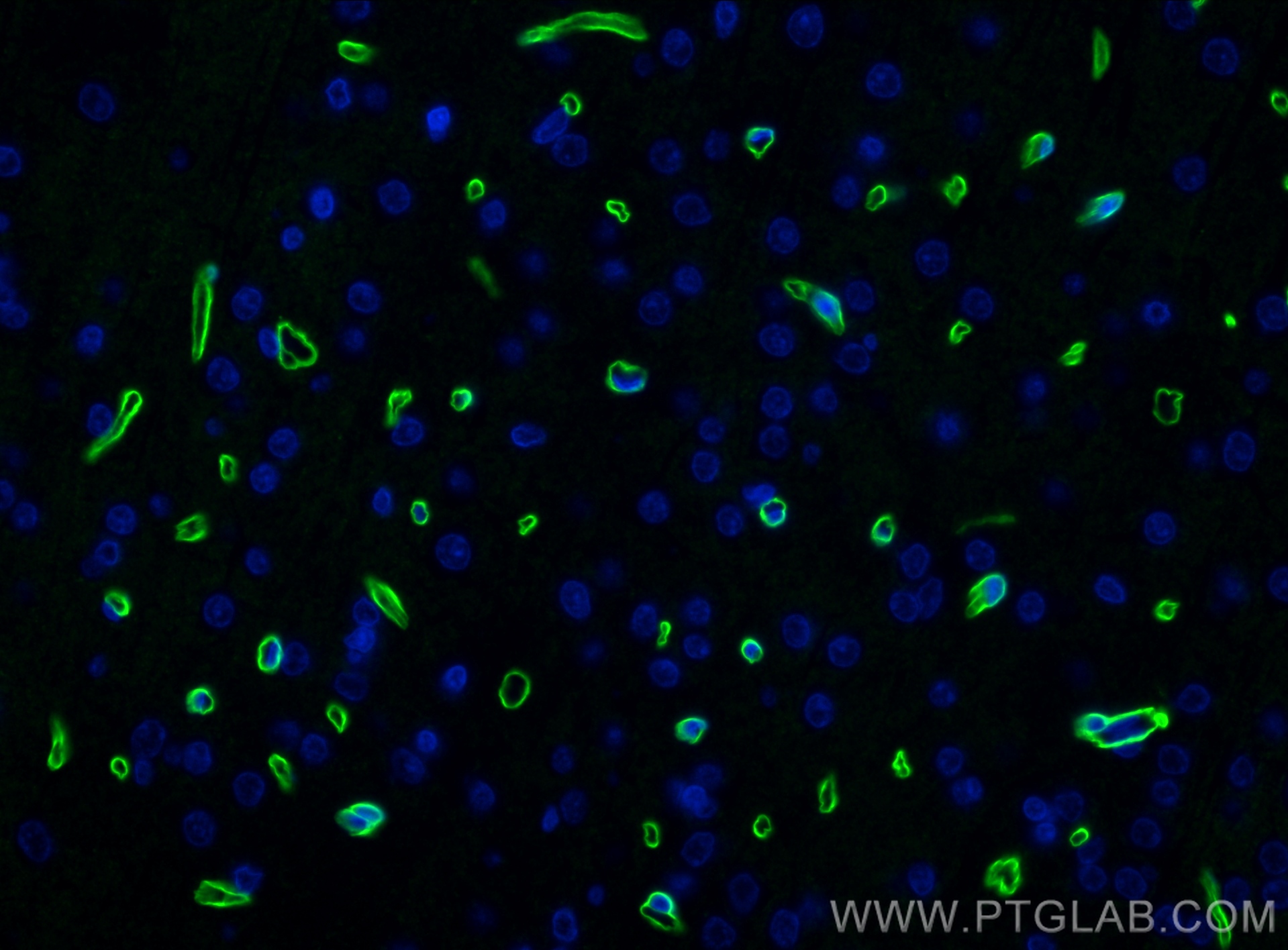
IF/ICC Figures
IF Staining of HepG2 using 83609-5-RR
Immunofluorescent analysis of (4% PFA) fixed HepG2 cells using Dystrophin antibody (83609-5-RR, Clone: 240567A3 ) at dilution of 1:250 and CoraLite®488-Conjugated AffiniPure Goat Anti-Rabbit IgG(H+L) (SA00013-2).
IF Staining of HepG2 using 83609-5-RR
Immunofluorescent analysis of (4% PFA) fixed HepG2 cells using Dystrophin antibody (83609-5-RR, Clone: 240567A3 ) at dilution of 1:250 and CoraLite®488-Conjugated AffiniPure Goat Anti-Rabbit IgG(H+L) (SA00013-2).
FC (INTRA) Figures
FC experiment of HepG2 using 83609-5-RR
1x10^6 HepG2 cells were intracellularly stained with 0.25 ug Dystrophin Recombinant antibody (83609-5-RR, Clone:240567A3) and APC-Conjugated AffiniPure Goat Anti-Rabbit IgG(H+L)(red), or 0.25 ug Isotype Control (blue). Cells were fixed with 4% PFA and permeabilized with Flow Cytometry Perm Buffer (PF00011-C).
AFFINITY Figures
Affinity and Kinetic Characterization of 83609-5-RR
Biolayer interferometry (BLl) kinetic assays of 83609-5-RR
against Human Dystrophin were performed. The affinity constant is below 1 pM.
The species listed in Tested Reactivity are in-house verified and applicable species. For unlisted species, please refer to the homology analysis of the immunogen sequence and related species. For rabbit polyclonal antibodies, homology >70% is recommended. For mouse monoclonal antibodies and rabbit recombinant antibodies, homology >90% is recommended. Generally, the higher the homology, the greater the applicability. However, there will be certain differences in protein expression in different species, tissues or cells. Therefore, the homology analysis results are for reference only and do not serve as a guarantee.
At Proteintech, we pride ourselves on our antibody quality, customer service and transparency. As such, we are comparing our antibodies with other vendors, enabling easy identification and comparisons of key data to help you choose the suitable antibody for your needs.
We have selected the top cited antibodies from these vendors for you to compare.
Proteintech
Dystrophin Recombinant antibody
Catalog Number
83609-5-RR
Citations
-
Dilutions
WB : 1:5000-1:50000 IHC : 1:50-1:500 IF-P : 1:200-1:800 IF/ICC : 1:125-1:500 FC (INTRA) : 0.25 ug per 10^6 cells in a 100 µl suspension
Applications
WB, IHC, IF/ICC, IF-P, FC (Intra), ELISA
Reactivity
human, mouse, rat
Product Guarantee
Covers any species including not listed on datasheet
Covers any applications including not listed on datasheet
 at dilution of 1:20000 incubated at room temperature for 1.5 hours.")
 at dilution of 1:200 (under 40x lens). Heat mediated antigen retrieval with Tris-EDTA buffer (pH 9.0).")
 fixed paraffin-embedded rat brain tissue using Dystrophin antibody (83609-5-RR, Clone: 240567A3 ) at dilution of 1:400 and Multi-rAb CoraLite ® Plus 488-Goat Anti-Rabbit Recombinant Secondary Antibody (H+L) (RGAR002). Heat mediated antigen retrieval with Tris-EDTA buffer (pH 9.0).")
 fixed paraffin-embedded rat brain tissue using Dystrophin antibody (83609-5-RR, Clone: 240567A3 ) at dilution of 1:400 and Multi-rAb CoraLite ® Plus 488-Goat Anti-Rabbit Recombinant Secondary Antibody (H+L) (RGAR002). Heat mediated antigen retrieval with Tris-EDTA buffer (pH 9.0).")
 fixed HepG2 cells using Dystrophin antibody (83609-5-RR, Clone: 240567A3 ) at dilution of 1:250 and CoraLite®488-Conjugated AffiniPure Goat Anti-Rabbit IgG(H+L) (SA00013-2).")
 fixed HepG2 cells using Dystrophin antibody (83609-5-RR, Clone: 240567A3 ) at dilution of 1:250 and CoraLite®488-Conjugated AffiniPure Goat Anti-Rabbit IgG(H+L) (SA00013-2).")
 and APC-Conjugated AffiniPure Goat Anti-Rabbit IgG(H+L)(red), or 0.25 ug Isotype Control (blue). Cells were fixed with 4% PFA and permeabilized with Flow Cytometry Perm Buffer (PF00011-C).")
 kinetic assays of 83609-5-RR
against Human Dystrophin were performed. The affinity constant is below 1 pM.")