at dilution of 1:20000 incubated at room temperature for 1.5 hours.")
 at dilution of 1:20000 incubated at room temperature for 1.5 hours.")
 at dilution of 1:20000 incubated at room temperature for 1.5 hours.")
 at dilution of 1:10000 incubated at room temperature for 1.5 hours.")
 at dilution of 1:20000 incubated at room temperature for 1.5 hours.")
 at dilution of 1:20000 incubated at room temperature for 1.5 hours.")
 at dilution of 1:20000 incubated at room temperature for 1.5 hours.")
 at dilution of 1:20000 incubated at room temperature for 1.5 hours.")
 fixed mouse testis tissue using STAR antibody (67130-1-Ig, Clone: 1G6E6 ) at dilution of 1:400 and CoraLite®488-Conjugated AffiniPure Goat Anti-Mouse IgG(H+L).")
 fixed mouse testis tissue using STAR antibody (67130-1-Ig, Clone: 1G6E6 ) at dilution of 1:400 and CoraLite®488-Conjugated AffiniPure Goat Anti-Mouse IgG(H+L).")
 fixed frozen OCT-embedded mouse testis tissue using 67130-1-Ig (STAR antibody) at dilution of 1:400, CoraLite®555 CL555-17178 (TNP1 antibody, orange) and CoraLite® Plus 488 CL488-13720 (BOULE antibody, green).")
Tested Applications
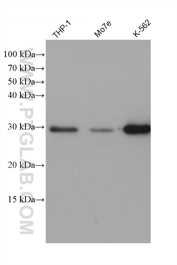
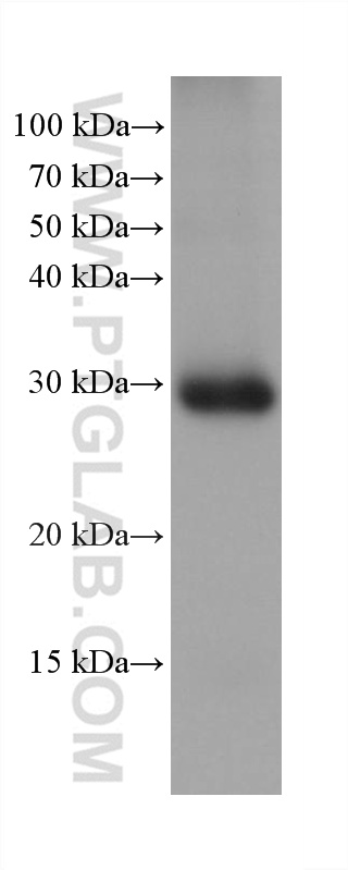
| Positive WB detected in | pig adrenal gland tissue, human adrenal gland tissue, human testis tissue, K-562 cells, rat adrenal gland tissue, rat testis tissue, THP-1 cells, Mo7e cells |
| Positive IF-P detected in | mouse testis tissue |
| Positive IF-Fro detected in | mouse testis tissue |
Recommended dilution
| Application | Dilution |
|---|---|
| Western Blot (WB) | WB : 1:5000-1:50000 |
| Immunofluorescence (IF)-P | IF-P : 1:200-1:800 |
| Immunofluorescence (IF)-FRO | IF-FRO : 1:200-1:800 |
| It is recommended that this reagent should be titrated in each testing system to obtain optimal results. | |
| Sample-dependent, Check data in validation data gallery. | |
Published Applications
| WB | See 3 publications below |
| IHC | See 1 publications below |
| IF | See 4 publications below |
Product Information
67130-1-Ig targets STAR in WB, IHC, IF-P, IF-Fro, ELISA applications and shows reactivity with human, mouse, rat, pig samples.
| Tested Reactivity | human, mouse, rat, pig |
| Cited Reactivity | mouse, pig |
| Host / Isotype | Mouse / IgG1 |
| Class | Monoclonal |
| Type | Antibody |
| Immunogen |
CatNo: Ag28698 Product name: Recombinant human STAR protein Source: e coli.-derived, PET28a Tag: 6*His Domain: 1-285 aa of BC010550 Sequence: MLLATFKLCAGSSYRHMRNMKGLRQQAVMAISQELNRRALGGPTPSTWINQVRRRSSLLGSRLEETLYSDQELAYLQQGEEAMQKALGILSNQEGWKKESQQDNGDKVMSKVVPDVGKVFRLEVVVDQPMERLYEELVERMEAMGEWNPNVKEIKVLQKIGKDTFITHELAAEAAGNLVGPRDFVSVRCAKRRGSTCVLAGMATDFGNMPEQKGVIRAEHGPTCMVLHPLAGSPSKTKLTWLLSIDLKGWLPKSIINQVLSQTQVDFANHLRKRLESHPASEARC Predict reactive species |
| Full Name | steroidogenic acute regulatory protein |
| Calculated Molecular Weight | 285 aa, 32 kDa |
| Observed Molecular Weight | 28-32 kDa |
| GenBank Accession Number | BC010550 |
| Gene Symbol | STAR |
| Gene ID (NCBI) | 6770 |
| RRID | AB_2882429 |
| Conjugate | Unconjugated |
| Form | Liquid |
| Purification Method | Protein G purification |
| UNIPROT ID | P49675 |
| Storage Buffer | PBS with 0.02% sodium azide and 50% glycerol, pH 7.3. |
| Storage Conditions | Store at -20°C. Stable for one year after shipment. Aliquoting is unnecessary for -20oC storage. 20ul sizes contain 0.1% BSA. |
Background Information
Steroidogenic acute regulatory protein (StAR) is a key player in acute regulation of hormone-induced steroidogenesis, by facilitating cholesterol transport from cellular stores to inner mitochondria membrane where the cholesterol was conversed to pregnenolone. StAR is prevalently expressed in mitochondria of steroid-producing adrenal and gonadal tissue
Protocols
| Product Specific Protocols | |
|---|---|
| IF protocol for STAR antibody 67130-1-Ig | Download protocol |
| WB protocol for STAR antibody 67130-1-Ig | Download protocol |
| Standard Protocols | |
|---|---|
| Click here to view our Standard Protocols |
Publications
| Species | Application | Title |
|---|---|---|
Mol Cell Endocrinol HE4 overexpression in mice leads to leydig cell hyperplasia and spermatogensis impairment: Pathological implications for oligospermia | ||
J Ethnopharmacol Saffron extract alleviates D-gal-induced late-onset hypogonadism by activating the PI3K-Akt-Nrf2 signaling pathway | ||
Drug Des Devel Ther Semaglutide Alleviates Ovary Inflammation via the AMPK/SIRT1/NF‑κB Signaling Pathway in Polycystic Ovary Syndrome Mice | ||
Cell Rep High matrix stiffness triggers testosterone decline in aging males by disrupting stem Leydig cell pool homeostasis | ||
Theriogenology Impacts of elevated temperature on morphology, oxidative stress levels, and testosterone synthesis in ex vivo cultured porcine testicular tissue | ||
Toxicol Appl Pharmacol Reproductive effects of pubertal exposure to neonicotinoid thiacloprid in immature male mice |