Various lysates were subjected to SDS PAGE followed by western blot with 66759-1-Ig (PSMB8 antibody) at dilution of 1:10000 incubated at room temperature for 1.5 hours.
Various lysates were subjected to SDS PAGE followed by western blot with 66759-1-Ig (PSMB8 antibody) at dilution of 1:10000 incubated at room temperature for 1.5 hours.
WB analysis of HSC-T6 using 66759-1-Ig
HSC-T6 cells were subjected to SDS PAGE followed by western blot with 66759-1-Ig (PSMB8 antibody) at dilution of 1:10000 incubated at room temperature for 1.5 hours.
HSC-T6 cells were subjected to SDS PAGE followed by western blot with 66759-1-Ig (PSMB8 antibody) at dilution of 1:10000 incubated at room temperature for 1.5 hours.
IHC staining of human lung cancer using 66759-1-Ig
Immunohistochemical analysis of paraffin-embedded human lung cancer tissue slide using 66759-1-Ig (PSMB8 antibody) at dilution of 1:1000 (under 10x lens. Heat mediated antigen retrieval with Tris-EDTA buffer (pH 9.0).
Immunohistochemical analysis of paraffin-embedded human lung cancer tissue slide using 66759-1-Ig (PSMB8 antibody) at dilution of 1:1000 (under 10x lens. Heat mediated antigen retrieval with Tris-EDTA buffer (pH 9.0).
IHC staining of human lung cancer using 66759-1-Ig
Immunohistochemical analysis of paraffin-embedded human lung cancer tissue slide using 66759-1-Ig (PSMB8 antibody) at dilution of 1:1000 (under 40x lens. Heat mediated antigen retrieval with Tris-EDTA buffer (pH 9.0).
Immunohistochemical analysis of paraffin-embedded human lung cancer tissue slide using 66759-1-Ig (PSMB8 antibody) at dilution of 1:1000 (under 40x lens. Heat mediated antigen retrieval with Tris-EDTA buffer (pH 9.0).
IHC staining of mouse colon using 66759-1-Ig
Immunohistochemical analysis of paraffin-embedded mouse colon tissue slide using 66759-1-Ig (PSMB8 antibody) at dilution of 1:8000 (under 10x lens). Heat mediated antigen retrieval with Tris-EDTA buffer (pH 9.0).
Immunohistochemical analysis of paraffin-embedded mouse stomach tissue slide using 66759-1-Ig (PSMB8 antibody) at dilution of 1:8000 (under 10x lens). Heat mediated antigen retrieval with Tris-EDTA buffer (pH 9.0).
IHC staining of rat colon using 66759-1-Ig
Immunohistochemical analysis of paraffin-embedded rat small intestine tissue slide using 66759-1-Ig (PSMB8 antibody) at dilution of 1:8000 (under 10x lens). Heat mediated antigen retrieval with Tris-EDTA buffer (pH 9.0).
Immunohistochemical analysis of paraffin-embedded rat small intestine tissue slide using 66759-1-Ig (PSMB8 antibody) at dilution of 1:8000 (under 10x lens). Heat mediated antigen retrieval with Tris-EDTA buffer (pH 9.0).
IHC staining of human oesophagus cancer using 66759-1-Ig
Immunohistochemical analysis of paraffin-embedded human oesophagus cancer tissue slide using 66759-1-Ig (PSMB8 antibody) at dilution of 1:8000 (under 10x lens). Heat mediated antigen retrieval with Tris-EDTA buffer (pH 9.0).
Immunohistochemical analysis of paraffin-embedded human oesophagus cancer tissue slide using 66759-1-Ig (PSMB8 antibody) at dilution of 1:8000 (under 10x lens). Heat mediated antigen retrieval with Tris-EDTA buffer (pH 9.0).
IHC staining of rat stomach using 66759-1-Ig
Immunohistochemical analysis of paraffin-embedded rat stomach tissue slide using 66759-1-Ig (PSMB8 antibody) at dilution of 1:8000 (under 10x lens). Heat mediated antigen retrieval with Tris-EDTA buffer (pH 9.0).
Immunohistochemical analysis of paraffin-embedded rat stomach tissue slide using 66759-1-Ig (PSMB8 antibody) at dilution of 1:8000 (under 10x lens). Heat mediated antigen retrieval with Tris-EDTA buffer (pH 9.0).
IHC staining of human liver cancer using 66759-1-Ig
Immunohistochemical analysis of paraffin-embedded human liver cancer tissue slide using 66759-1-Ig (PSMB8 antibody) at dilution of 1:1000 (under 10x lens. Heat mediated antigen retrieval with Tris-EDTA buffer (pH 9.0).
Immunohistochemical analysis of paraffin-embedded human liver cancer tissue slide using 66759-1-Ig (PSMB8 antibody) at dilution of 1:1000 (under 10x lens. Heat mediated antigen retrieval with Tris-EDTA buffer (pH 9.0).
IHC staining of human liver cancer using 66759-1-Ig
Immunohistochemical analysis of paraffin-embedded human liver cancer tissue slide using 66759-1-Ig (PSMB8 antibody) at dilution of 1:1000 (under 40x lens. Heat mediated antigen retrieval with Tris-EDTA buffer (pH 9.0).
Immunohistochemical analysis of paraffin-embedded human liver cancer tissue slide using 66759-1-Ig (PSMB8 antibody) at dilution of 1:1000 (under 40x lens. Heat mediated antigen retrieval with Tris-EDTA buffer (pH 9.0).
IF Staining of human lung cancer using 66759-1-Ig
Immunofluorescent analysis of (4% PFA) fixed human lung cancer tissue using PSMB8 antibody (66759-1-Ig, Clone: 2A5B6 ) at dilution of 1:400 and CoraLite®488-Conjugated Goat Anti-Mouse IgG(H+L).
Immunofluorescent analysis of (4% PFA) fixed human lung cancer tissue using PSMB8 antibody (66759-1-Ig, Clone: 2A5B6 ) at dilution of 1:400 and CoraLite®488-Conjugated Goat Anti-Mouse IgG(H+L).
IF Staining of human liver cancer using 66759-1-Ig
Immunofluorescent analysis of (4% PFA) fixed human liver cancer tissue using PSMB8 antibody (66759-1-Ig, Clone: 2A5B6 ) at dilution of 1:400 and CoraLite®488-Conjugated Goat Anti-Mouse IgG(H+L).
Immunofluorescent analysis of (4% PFA) fixed human liver cancer tissue using PSMB8 antibody (66759-1-Ig, Clone: 2A5B6 ) at dilution of 1:400 and CoraLite®488-Conjugated Goat Anti-Mouse IgG(H+L).
IF Staining of HeLa using 66759-1-Ig
Immunofluorescent analysis of (4% PFA) fixed HeLa cells using PSMB8 antibody (66759-1-Ig, Clone: 2A5B6 ) at dilution of 1:400 and CoraLite®488-Conjugated Goat Anti-Mouse IgG(H+L), CL594-Phalloidin (red).
Immunofluorescent analysis of (4% PFA) fixed HeLa cells using PSMB8 antibody (66759-1-Ig, Clone: 2A5B6 ) at dilution of 1:400 and CoraLite®488-Conjugated Goat Anti-Mouse IgG(H+L), CL594-Phalloidin (red).
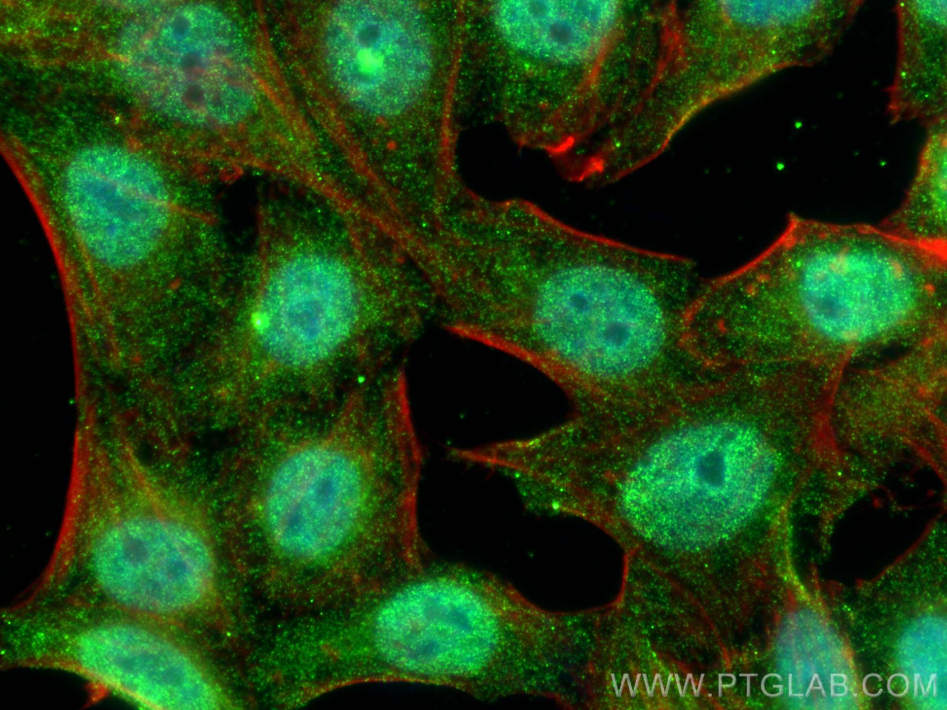
IF Staining of HepG2 using 66759-1-Ig
Immunofluorescent analysis of (4% PFA) fixed HepG2 cells using PSMB8 antibody (66759-1-Ig, Clone: 2A5B6 ) at dilution of 1:800 and Multi-rAb CoraLite ® Plus 488-Goat Anti-Mouse Recombinant Secondary Antibody (H+L) (RGAM002), CL594-phalloidin (red).
The Proteintech guarantee covers Proteintech antibodies in any species and any application, including those not listed on the datasheet. If the antibody doesn’t perform, you can receive a hassle-free refund or credit note.
human lung cancer tissue, human liver cancer tissue, human oesophagus cancer tissue, mouse colon tissue, mouse stomach tissue, rat colon tissue, rat stomach tissue Note: suggested antigen retrieval with TE buffer pH 9.0; (*) Alternatively, antigen retrieval may be performed with citrate buffer pH 6.0
Positive IF-P detected in
human lung cancer tissue, human liver cancer tissue
Positive IF/ICC detected in
HepG2 cells, HeLa cells
Recommended dilution
Application
Dilution
Western Blot (WB)
WB : 1:5000-1:50000
Immunohistochemistry (IHC)
IHC : 1:500-1:2000
Immunofluorescence (IF)-P
IF-P : 1:200-1:800
Immunofluorescence (IF)/ICC
IF/ICC : 1:400-1:1600
It is recommended that this reagent should be titrated in each testing system to obtain optimal results.
Sample-dependent, Check data in validation data gallery.
Product Information
66759-1-Ig targets PSMB8 in WB, IHC, IF/ICC, IF-P, ELISA applications and shows reactivity with human, mouse, rat samples.
PBS with 0.02% sodium azide and 50% glycerol , pH 7.3
Storage Conditions
Store at -20°C. Stable for one year after shipment. Aliquoting is unnecessary for -20oC storage. 20ul sizes contain 0.1% BSA.
Background Information
PSMB8(Proteasome subunit beta type-8) is also named as LMP7, PSMB5i, RING10, Y2 and belongs to the peptidase T1B family. The gene encodes the chymotrypsin-like catalytic subunit of the immunoproteasome(PMID: 19525961). PSMB8 has a role in controlling pathogenic immune responses and may be a target in autoimmune disorders. Its prosequence is not essential for incorporation of PSMB8 into the maturing proteasome, but it increased the efficiency of PSMB8 incorporation and proteasome maturation(PMID: 10926487). The pro-PSMB8 is a 276aa protein with the molecular mass of 30 kDa, and the mature form is about 23kDa due to the 72aa propeptide cleaved. Defects in PSMB8 are the cause of Nakajo syndrome (NKJO).
Various lysates were subjected to SDS PAGE followed by western blot with 66759-1-Ig (PSMB8 antibody) at dilution of 1:10000 incubated at room temperature for 1.5 hours.
WB analysis of HSC-T6 using 66759-1-Ig
HSC-T6 cells were subjected to SDS PAGE followed by western blot with 66759-1-Ig (PSMB8 antibody) at dilution of 1:10000 incubated at room temperature for 1.5 hours.
IHC Figures
IHC staining of human lung cancer using 66759-1-Ig
Immunohistochemical analysis of paraffin-embedded human lung cancer tissue slide using 66759-1-Ig (PSMB8 antibody) at dilution of 1:1000 (under 10x lens. Heat mediated antigen retrieval with Tris-EDTA buffer (pH 9.0).
IHC staining of human lung cancer using 66759-1-Ig
Immunohistochemical analysis of paraffin-embedded human lung cancer tissue slide using 66759-1-Ig (PSMB8 antibody) at dilution of 1:1000 (under 40x lens. Heat mediated antigen retrieval with Tris-EDTA buffer (pH 9.0).
IHC staining of mouse colon using 66759-1-Ig
Immunohistochemical analysis of paraffin-embedded mouse colon tissue slide using 66759-1-Ig (PSMB8 antibody) at dilution of 1:8000 (under 10x lens). Heat mediated antigen retrieval with Tris-EDTA buffer (pH 9.0).
IHC staining of mouse stomach using 66759-1-Ig
Immunohistochemical analysis of paraffin-embedded mouse stomach tissue slide using 66759-1-Ig (PSMB8 antibody) at dilution of 1:8000 (under 10x lens). Heat mediated antigen retrieval with Tris-EDTA buffer (pH 9.0).
IHC staining of rat colon using 66759-1-Ig
Immunohistochemical analysis of paraffin-embedded rat small intestine tissue slide using 66759-1-Ig (PSMB8 antibody) at dilution of 1:8000 (under 10x lens). Heat mediated antigen retrieval with Tris-EDTA buffer (pH 9.0).
IHC staining of human oesophagus cancer using 66759-1-Ig
Immunohistochemical analysis of paraffin-embedded human oesophagus cancer tissue slide using 66759-1-Ig (PSMB8 antibody) at dilution of 1:8000 (under 10x lens). Heat mediated antigen retrieval with Tris-EDTA buffer (pH 9.0).
IHC staining of rat stomach using 66759-1-Ig
Immunohistochemical analysis of paraffin-embedded rat stomach tissue slide using 66759-1-Ig (PSMB8 antibody) at dilution of 1:8000 (under 10x lens). Heat mediated antigen retrieval with Tris-EDTA buffer (pH 9.0).
IHC staining of human liver cancer using 66759-1-Ig
Immunohistochemical analysis of paraffin-embedded human liver cancer tissue slide using 66759-1-Ig (PSMB8 antibody) at dilution of 1:1000 (under 10x lens. Heat mediated antigen retrieval with Tris-EDTA buffer (pH 9.0).
IHC staining of human liver cancer using 66759-1-Ig
Immunohistochemical analysis of paraffin-embedded human liver cancer tissue slide using 66759-1-Ig (PSMB8 antibody) at dilution of 1:1000 (under 40x lens. Heat mediated antigen retrieval with Tris-EDTA buffer (pH 9.0).
IF-P Figures
IF Staining of human lung cancer using 66759-1-Ig
Immunofluorescent analysis of (4% PFA) fixed human lung cancer tissue using PSMB8 antibody (66759-1-Ig, Clone: 2A5B6 ) at dilution of 1:400 and CoraLite®488-Conjugated Goat Anti-Mouse IgG(H+L).
IF Staining of human liver cancer using 66759-1-Ig
Immunofluorescent analysis of (4% PFA) fixed human liver cancer tissue using PSMB8 antibody (66759-1-Ig, Clone: 2A5B6 ) at dilution of 1:400 and CoraLite®488-Conjugated Goat Anti-Mouse IgG(H+L).
IF/ICC Figures
IF Staining of HeLa using 66759-1-Ig
Immunofluorescent analysis of (4% PFA) fixed HeLa cells using PSMB8 antibody (66759-1-Ig, Clone: 2A5B6 ) at dilution of 1:400 and CoraLite®488-Conjugated Goat Anti-Mouse IgG(H+L), CL594-Phalloidin (red).
IF Staining of HepG2 using 66759-1-Ig
Immunofluorescent analysis of (4% PFA) fixed HepG2 cells using PSMB8 antibody (66759-1-Ig, Clone: 2A5B6 ) at dilution of 1:800 and Multi-rAb CoraLite ® Plus 488-Goat Anti-Mouse Recombinant Secondary Antibody (H+L) (RGAM002), CL594-phalloidin (red).
The species listed in Tested Reactivity are in-house verified and applicable species. For unlisted species, please refer to the homology analysis of the immunogen sequence and related species. For rabbit polyclonal antibodies, homology >70% is recommended. For mouse monoclonal antibodies and rabbit recombinant antibodies, homology >90% is recommended. Generally, the higher the homology, the greater the applicability. However, there will be certain differences in protein expression in different species, tissues or cells. Therefore, the homology analysis results are for reference only and do not serve as a guarantee.
At Proteintech, we pride ourselves on our antibody quality, customer service and transparency. As such, we are comparing our antibodies with other vendors, enabling easy identification and comparisons of key data to help you choose the suitable antibody for your needs.
We have selected the top cited antibodies from these vendors for you to compare.
 at dilution of 1:10000 incubated at room temperature for 1.5 hours.")
 at dilution of 1:10000 incubated at room temperature for 1.5 hours.")
 at dilution of 1:1000 (under 10x lens. Heat mediated antigen retrieval with Tris-EDTA buffer (pH 9.0).")
 at dilution of 1:1000 (under 40x lens. Heat mediated antigen retrieval with Tris-EDTA buffer (pH 9.0).")
 at dilution of 1:8000 (under 10x lens). Heat mediated antigen retrieval with Tris-EDTA buffer (pH 9.0).")
 at dilution of 1:8000 (under 10x lens). Heat mediated antigen retrieval with Tris-EDTA buffer (pH 9.0).")
 at dilution of 1:8000 (under 10x lens). Heat mediated antigen retrieval with Tris-EDTA buffer (pH 9.0).")
 at dilution of 1:8000 (under 10x lens). Heat mediated antigen retrieval with Tris-EDTA buffer (pH 9.0).")
 at dilution of 1:8000 (under 10x lens). Heat mediated antigen retrieval with Tris-EDTA buffer (pH 9.0).")
 at dilution of 1:1000 (under 10x lens. Heat mediated antigen retrieval with Tris-EDTA buffer (pH 9.0).")
 at dilution of 1:1000 (under 40x lens. Heat mediated antigen retrieval with Tris-EDTA buffer (pH 9.0).")
 fixed human lung cancer tissue using PSMB8 antibody (66759-1-Ig, Clone: 2A5B6 ) at dilution of 1:400 and CoraLite®488-Conjugated Goat Anti-Mouse IgG(H+L).")
 fixed human liver cancer tissue using PSMB8 antibody (66759-1-Ig, Clone: 2A5B6 ) at dilution of 1:400 and CoraLite®488-Conjugated Goat Anti-Mouse IgG(H+L).")
 fixed HeLa cells using PSMB8 antibody (66759-1-Ig, Clone: 2A5B6 ) at dilution of 1:400 and CoraLite®488-Conjugated Goat Anti-Mouse IgG(H+L), CL594-Phalloidin (red).")
 fixed HepG2 cells using PSMB8 antibody (66759-1-Ig, Clone: 2A5B6 ) at dilution of 1:800 and Multi-rAb CoraLite ® Plus 488-Goat Anti-Mouse Recombinant Secondary Antibody (H+L) (RGAM002), CL594-phalloidin (red).")