Tested Applications
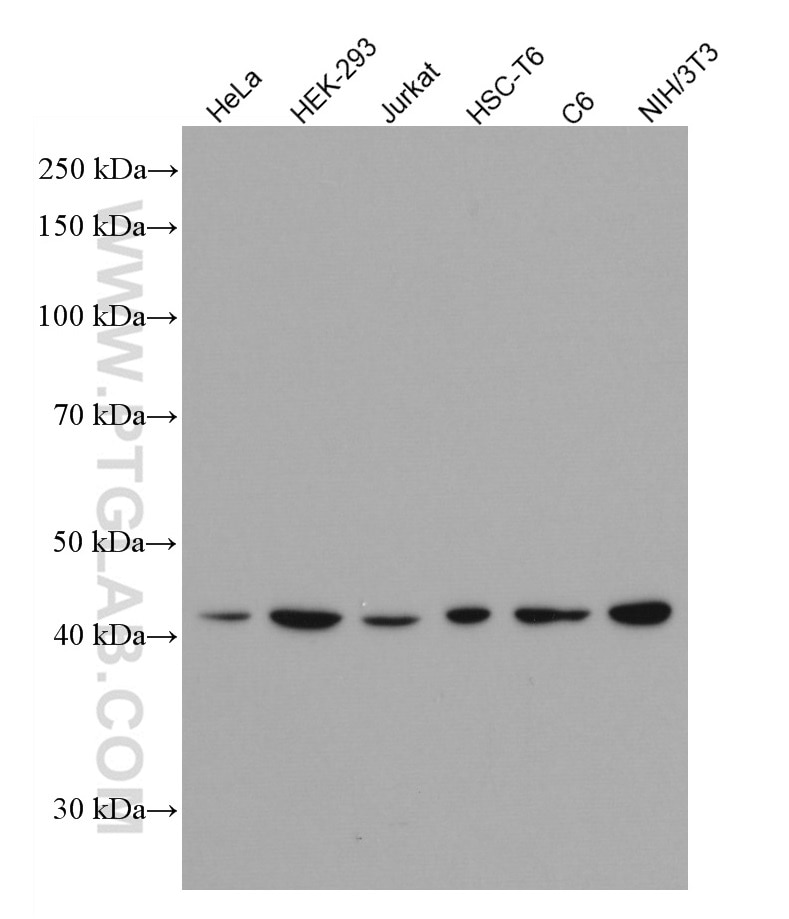
| Positive WB detected in | HeLa cells, HEK-293 cells, Jurkat cells, HSC-T6 cells, C6 cells, NIH/3T3 cells |
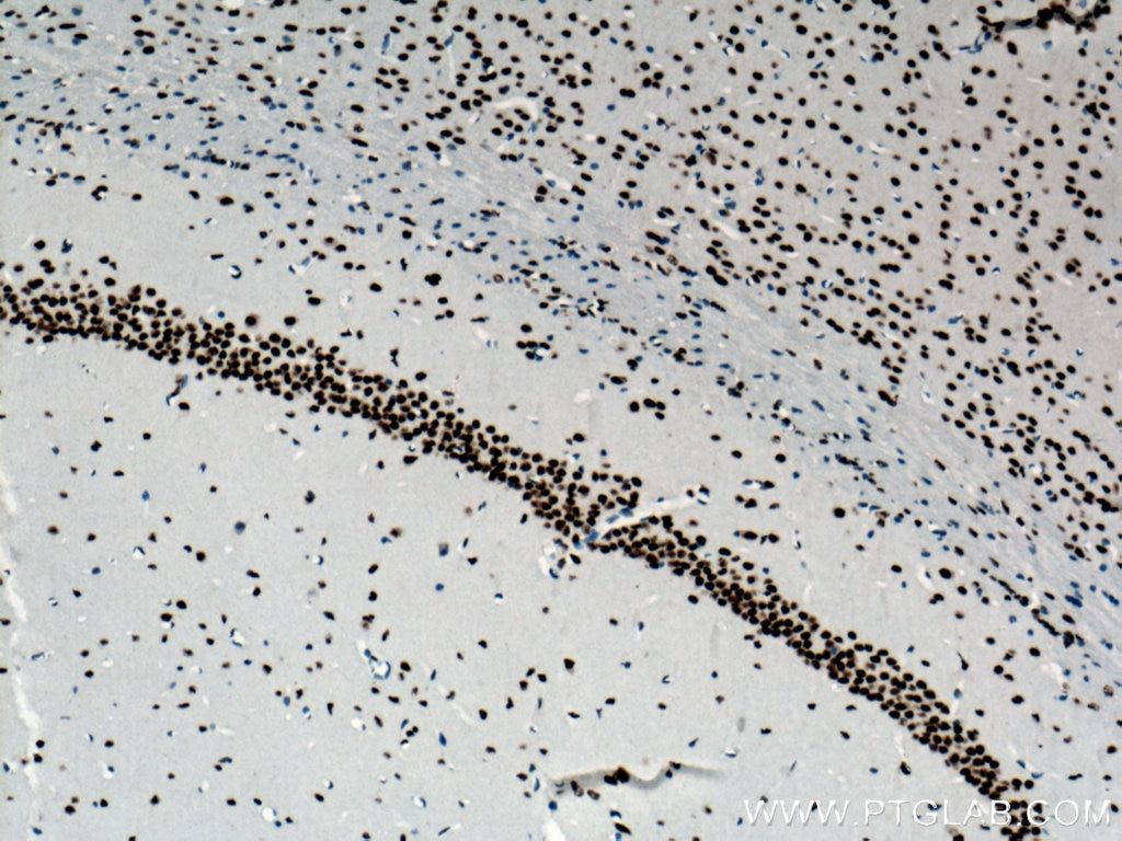
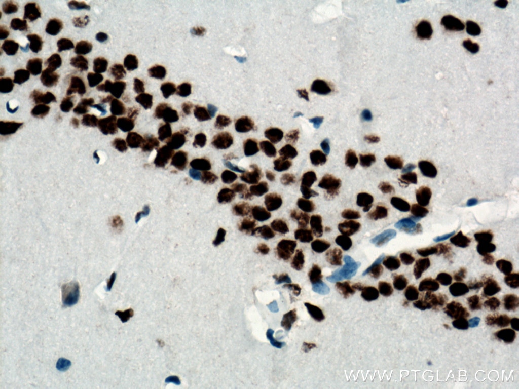
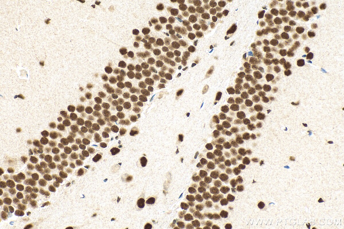
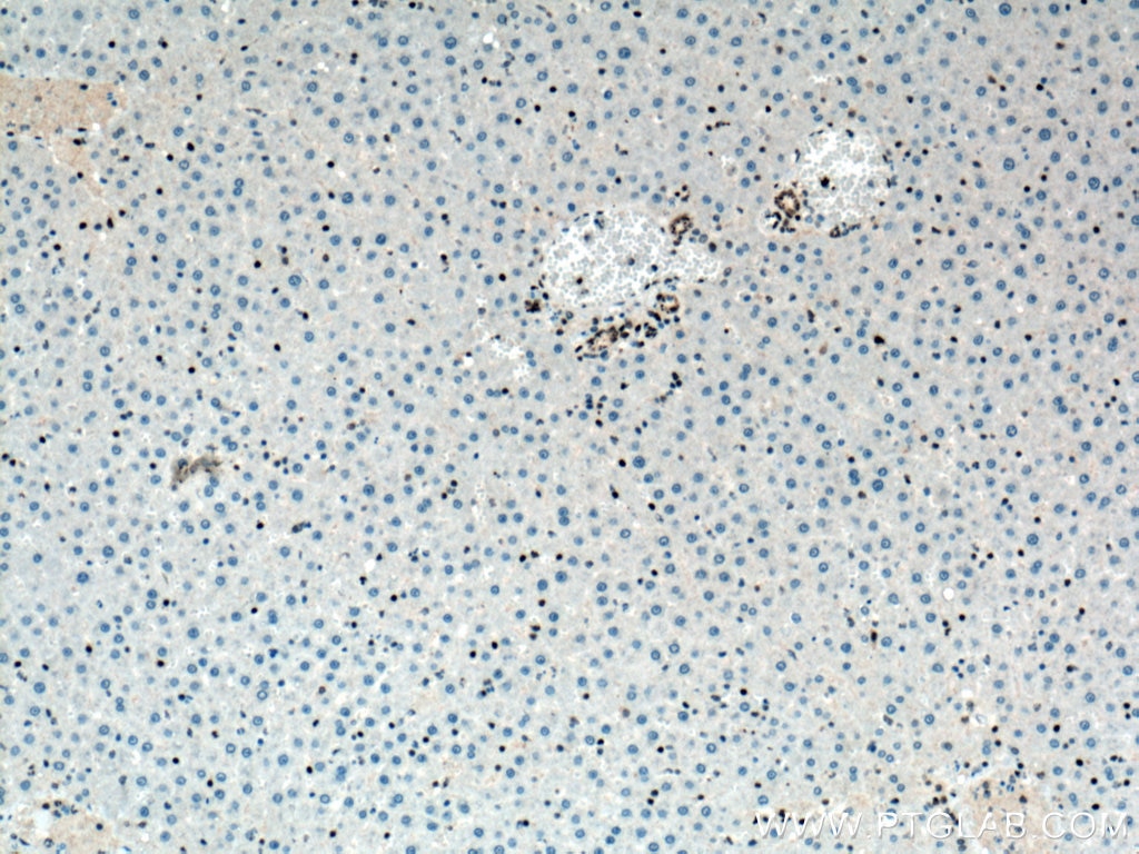
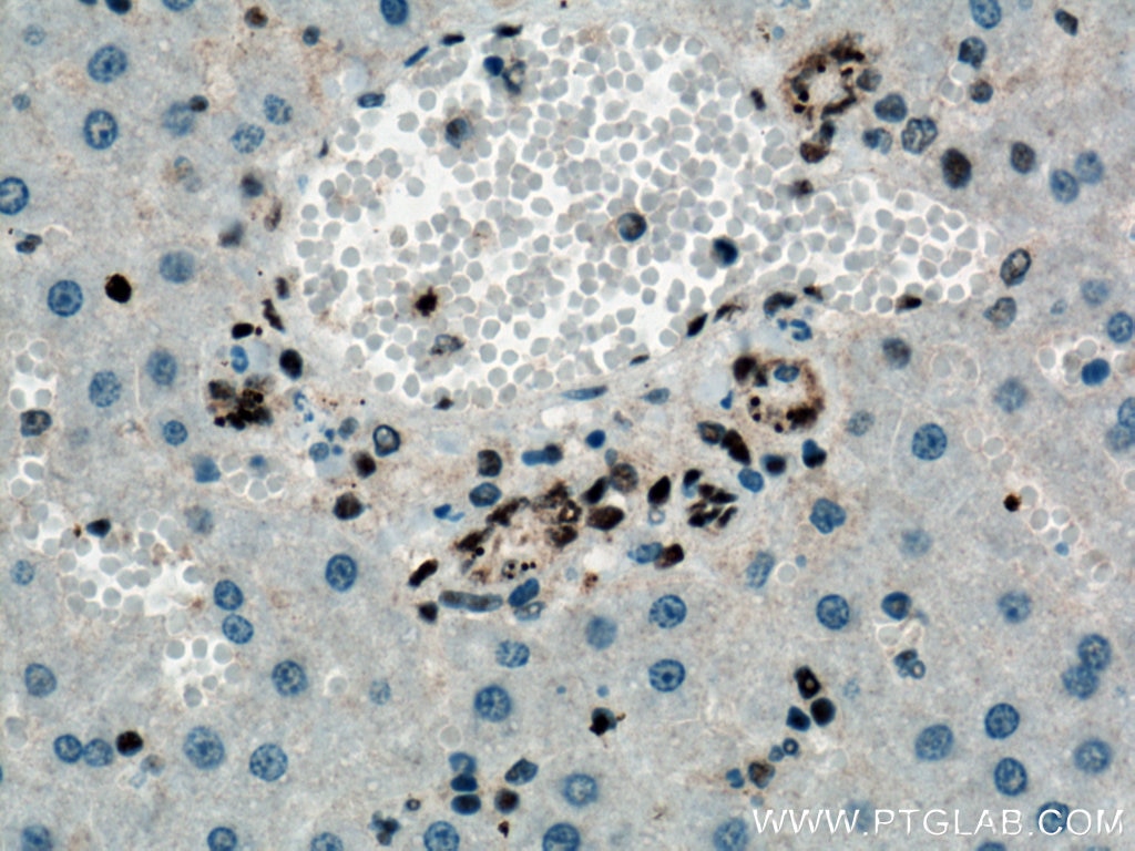
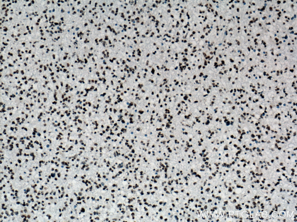
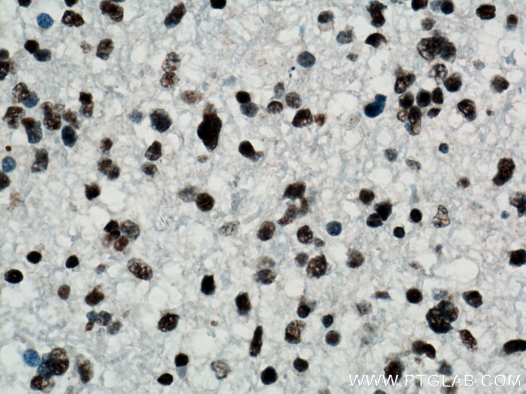
| Positive IHC detected in | mouse brain tissue, human gliomas tissue, rat liver tissue Note: suggested antigen retrieval with TE buffer pH 9.0; (*) Alternatively, antigen retrieval may be performed with citrate buffer pH 6.0 |
Recommended dilution
| Application | Dilution |
|---|---|
| Western Blot (WB) | WB : 1:5000-1:50000 |
| Immunohistochemistry (IHC) | IHC : 1:1000-1:4000 |
| It is recommended that this reagent should be titrated in each testing system to obtain optimal results. | |
| Sample-dependent, Check data in validation data gallery. | |
Published Applications
| WB | See 4 publications below |
| IHC | See 3 publications below |
Product Information
66734-1-Ig targets TDP-43 in WB, IHC, ELISA applications and shows reactivity with human, mouse, rat samples.
| Tested Reactivity | human, mouse, rat |
| Cited Reactivity | human, mouse |
| Host / Isotype | Mouse / IgG1 |
| Class | Monoclonal |
| Type | Antibody |
| Immunogen |
Recombinant protein Predict reactive species |
| Full Name | TAR DNA binding protein |
| Calculated Molecular Weight | 43 kDa |
| Observed Molecular Weight | 43 kDa |
| GenBank Accession Number | BC001487 |
| Gene Symbol | TDP-43 |
| Gene ID (NCBI) | 23435 |
| RRID | AB_2882084 |
| Conjugate | Unconjugated |
| Form | Liquid |
| Purification Method | Protein G purification |
| UNIPROT ID | Q13148 |
| Storage Buffer | PBS with 0.02% sodium azide and 50% glycerol, pH 7.3. |
| Storage Conditions | Store at -20°C. Stable for one year after shipment. Aliquoting is unnecessary for -20oC storage. 20ul sizes contain 0.1% BSA. |
Background Information
Transactivation response (TAR), DNA-binding protein of 43 kDa (also known as TARDBP or TDP-43), was first isolated as a transcriptional inactivator binding to the TAR DNA element of the HIV-1 virus. Neumann et al. (2006) found that a hyperphosphorylated, ubiquitinated, and cleaved form of TARDBP, known as pathologic TDP-43, is the major component of the tau-negative and ubiquitin-positive inclusions that characterize amyotrophic lateral sclerosis (ALS) and the most common pathological subtype of frontotemporal lobar degeneration (FTLD-U). 66734-1-Ig is a mouse monoclonal antibody recognizing the intact protein of TDP-43.
Protocols
| Product Specific Protocols | |
|---|---|
| IHC protocol for TDP-43 antibody 66734-1-Ig | Download protocol |
| WB protocol for TDP-43 antibody 66734-1-Ig | Download protocol |
| Standard Protocols | |
|---|---|
| Click here to view our Standard Protocols |
Publications
| Species | Application | Title |
|---|---|---|
Acta Neuropathol Neurons selectively targeted in frontotemporal dementia reveal early stage TDP-43 pathobiology. | ||
Int J Biol Sci Spreading of pathological TDP-43 along corticospinal tract axons induces ALS-like phenotypes in Atg5+/- mice. | ||
Int Immunopharmacol Paraquat exposure triggers amyloid-β and α-synuclein aggregation in the prefrontal cortex of mice: Suppression of microglial phagocytosis via IL-17A | ||
Exp Neurol Muscle-dominant wild-type TDP-43 expression induces myopathological changes featuring tubular aggregates and TDP-43-positive inclusions. | ||
Nat Commun TDP-43 proteinopathy in ALS is triggered by loss of ASRGL1 and associated with HML-2 expression |

