Tested Applications
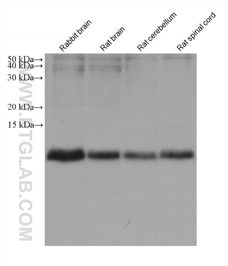
| Positive WB detected in | rabbit brain tissue, rat brain tissue, rat cerebellum tissue, rat spinal cord tissue |
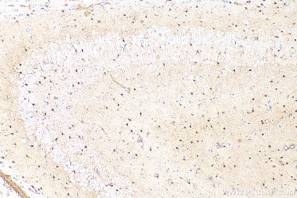
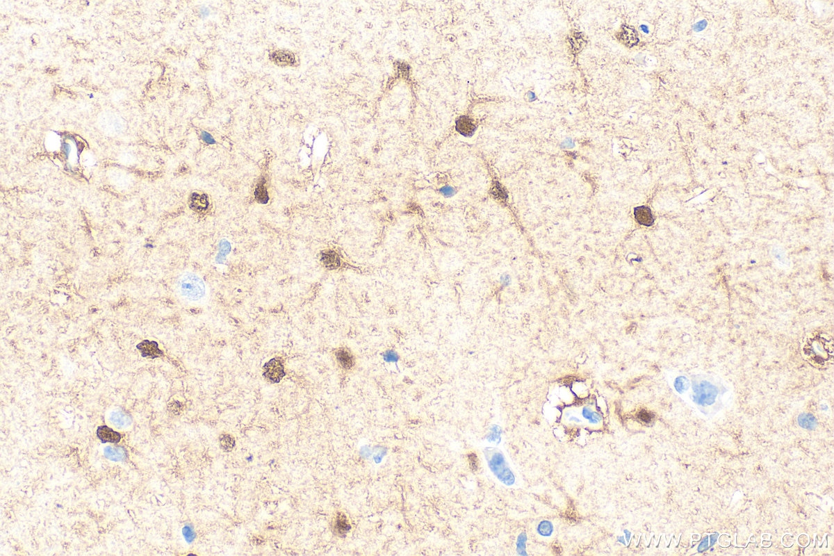
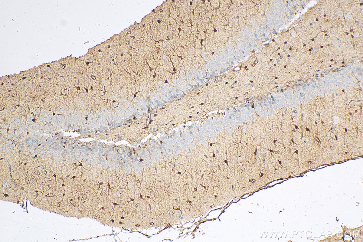
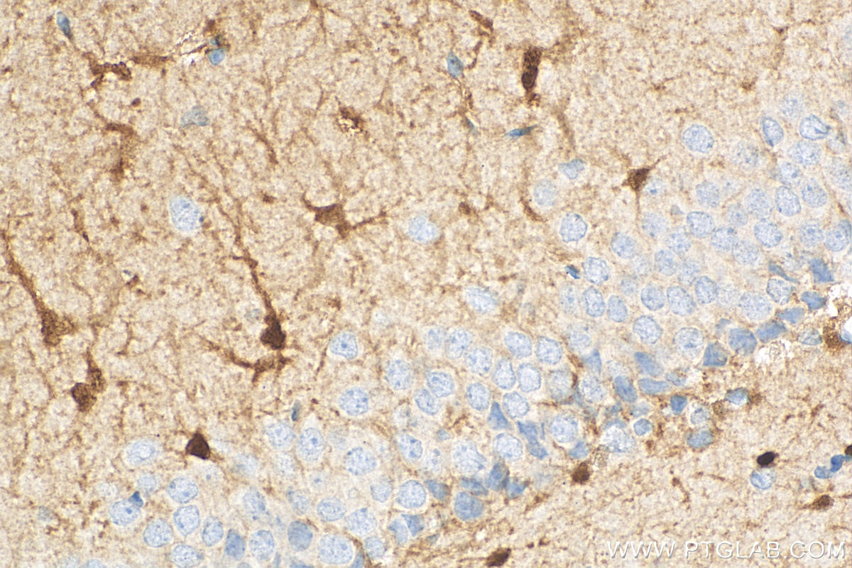
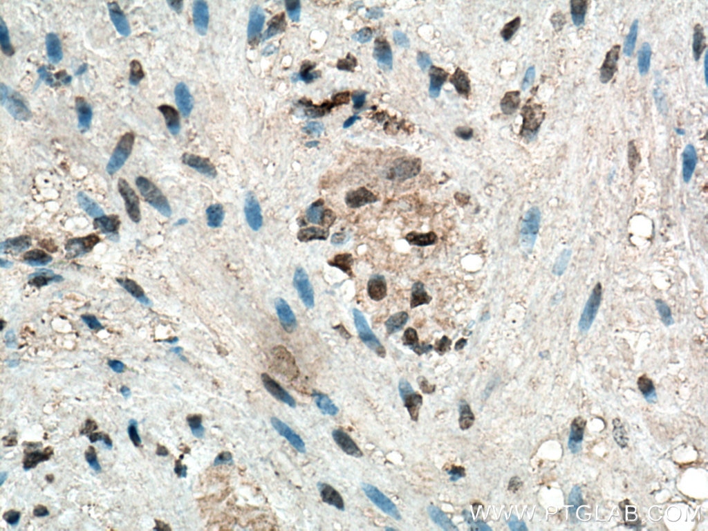
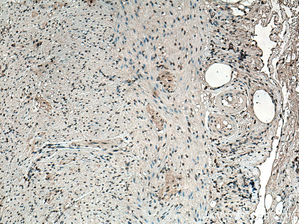
| Positive IHC detected in | rat brain tissue, human appendicitis tissue Note: suggested antigen retrieval with TE buffer pH 9.0; (*) Alternatively, antigen retrieval may be performed with citrate buffer pH 6.0 |
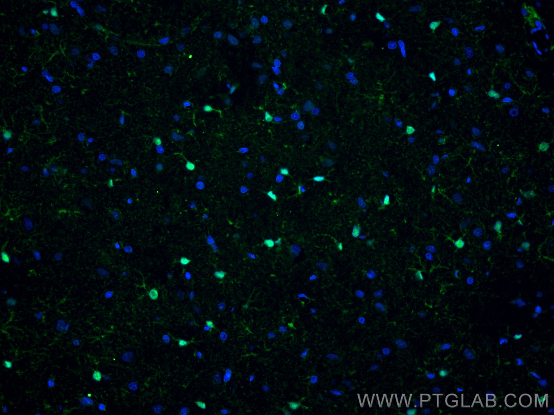
| Positive IF-P detected in | rat brain tissue |
Recommended dilution
| Application | Dilution |
|---|---|
| Western Blot (WB) | WB : 1:1000-1:2000 |
| Immunohistochemistry (IHC) | IHC : 1:1000-1:8000 |
| Immunofluorescence (IF)-P | IF-P : 1:200-1:800 |
| It is recommended that this reagent should be titrated in each testing system to obtain optimal results. | |
| Sample-dependent, Check data in validation data gallery. | |
Published Applications
| WB | See 5 publications below |
| IHC | See 3 publications below |
| IF | See 14 publications below |
Product Information
66616-1-Ig targets S100B in WB, IHC, IF-P, ELISA applications and shows reactivity with human, rat samples.
| Tested Reactivity | human, rat |
| Cited Reactivity | human, rat |
| Host / Isotype | Mouse / IgG1 |
| Class | Monoclonal |
| Type | Antibody |
| Immunogen |
CatNo: Ag7440 Product name: Recombinant human S100B protein Source: e coli.-derived, PGEX-4T Tag: GST Domain: 1-92 aa of BC001766 Sequence: MSELEKAMVALIDVFHQYSGREGDKHKLKKSELKELINNELSHFLEEIKEQEVVDKVMETLDNDGDGECDFQEFMAFVAMVTTACHEFFEHE Predict reactive species |
| Full Name | S100 calcium binding protein B |
| Calculated Molecular Weight | 11 kDa |
| Observed Molecular Weight | 11 kDa |
| GenBank Accession Number | BC001766 |
| Gene Symbol | S100 Beta |
| Gene ID (NCBI) | 6285 |
| RRID | AB_2881976 |
| Conjugate | Unconjugated |
| Form | Liquid |
| Purification Method | Protein G purification |
| UNIPROT ID | P04271 |
| Storage Buffer | PBS with 0.02% sodium azide and 50% glycerol, pH 7.3. |
| Storage Conditions | Store at -20°C. Stable for one year after shipment. Aliquoting is unnecessary for -20oC storage. 20ul sizes contain 0.1% BSA. |
Background Information
S100B is a member of the S100 family of proteins containing 2 EF-hand calcium-binding motifs and has been implicated in the regulation of cellular activities such as metabolism, motility and proliferation. In the nervous system, S100B is constitutively released by astrocytes into the extracellular space and at nanomolar concentrations, it can promote neurite outgrowth and protect neurons against oxidative stress. Within the central nervous system (CNS), S100B is thought to be a marker for astroglial activation, linking astrocyte dysfunction to schizophrenia. In addition to astrocytes, S100B is released from many cell types, such as, adipocytes, chondrocytes, cardiomyocytes and lymphocytes. Serum S100B represents a new biomarker for mood disorders.
Protocols
| Product Specific Protocols | |
|---|---|
| IF protocol for S100B antibody 66616-1-Ig | Download protocol |
| IHC protocol for S100B antibody 66616-1-Ig | Download protocol |
| WB protocol for S100B antibody 66616-1-Ig | Download protocol |
| Standard Protocols | |
|---|---|
| Click here to view our Standard Protocols |
Publications
| Species | Application | Title |
|---|---|---|
Acta Neuropathol Commun Repetitive mild TBI causes pTau aggregation in nigra without altering preexisting fibril induced Parkinson’s-like pathology burden | ||
ACS Omega Platelet-Rich Plasma Gel-Loaded Collagen/Chitosan Composite Film Accelerated Rat Sciatic Nerve Injury Repair | ||
Front Neurosci Nogo-C Inhibits Peripheral Nerve Regeneration by Regulating Schwann Cell Apoptosis and Dedifferentiation. | ||
Transl Oncol High throughput proteomic and metabolic profiling identified target correction of metabolic abnormalities as a novel therapeutic approach in head and neck paraganglioma. | ||
Bioact Mater Advanced nerve regeneration enabled by neural conformal electronic stimulators enhancing mitochondrial transport | ||
Cell Rep Astrocytic modulation of GABA transmission in the VTA limits progression of anxiety- and depression-like behaviors in adult mice |
Reviews
The reviews below have been submitted by verified Proteintech customers who received an incentive for providing their feedback.
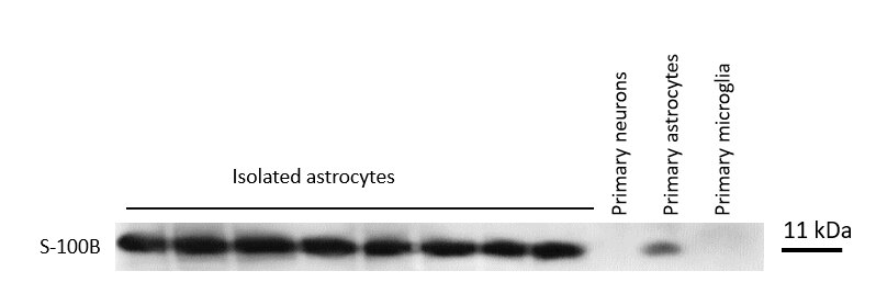
FH Kateryna (Verified Customer) (01-16-2025) | The S100B antibody was utilized to assess the identity of the immunomagnetically isolated astrocytic fraction by Western blot. As a positive control, mouse primary cortical astrocytes were used. As negative controls, mouse primary cortical neurons and microglial cells were used. The samples were processed by SDS-PAGE, and the separated proteins were transferred onto a nitrocellulose membrane. Blocking was performed with 5% non-fat dry milk, and the membrane was incubated overnight at 4°C with the S100B antibody. The antibody is specific and performs consistently.
 |
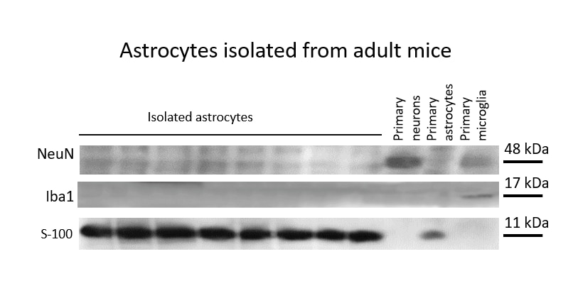
FH Kateryna (Verified Customer) (11-21-2024) | I use the S100B monoclonal antibody (66616-1-Ig) to validate the identity of immunomagnetically isolated astrocytes from mouse brain via Western blot and as a housekeeping protein for other protein measurements via Western blot. The Western blot is run under reducing conditions using SDS-PAGE, a nitrocellulose membrane, and 5% non-fat dry milk as a blocking solution. The antibody is highly sensitive, providing consistent bands at 11 kDa.
 |
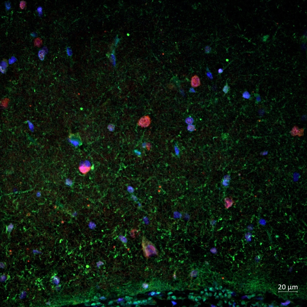
FH Reyes (Verified Customer) (07-02-2024) | s100b (in green) works in human brain FFPE tissue, but it does not yield the best results. (It colocalizes in a small way with NeuN in red).
 |
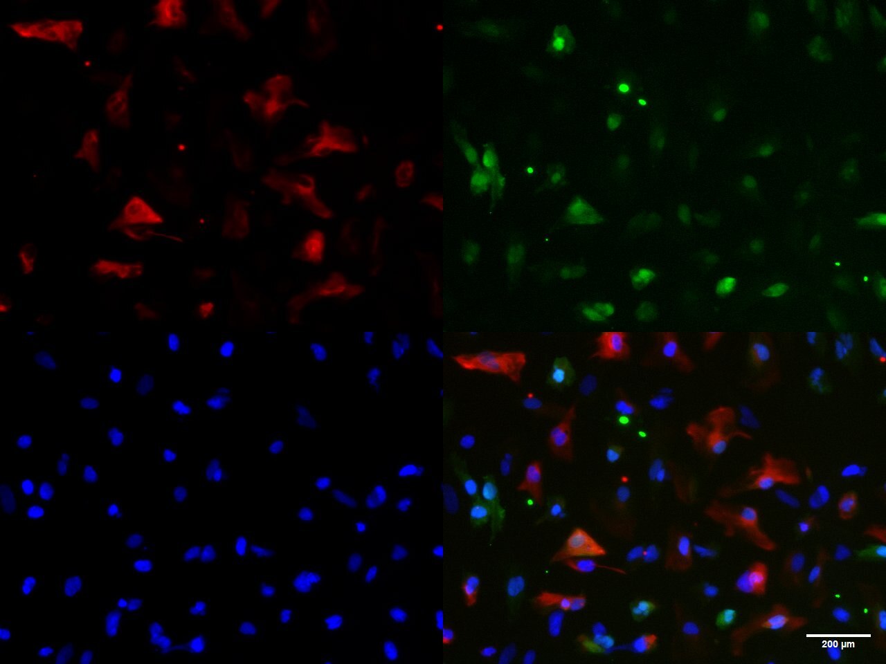
FH Gabriele (Verified Customer) (06-06-2024) | The antibody works well, staining positively the hiPSC-derived astrocytes in green (1:200). It can colocalize with GFAP (in red). Nuclei are stained with hoechst (in blue)
 |
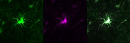
FH Clarisse (Verified Customer) (08-08-2022) | This antibody works very well as an astrocyte marker. In the image: in green mouse anti-S100B (1:500 - 66616-1-Ig) and in magenta rabbit anti-S100B (1:500 - 15146-1-AP). The rabbit antibody has a better performance.
 |

