mouse brain tissue were subjected to SDS PAGE followed by western blot with 66589-1-Ig (EGLN1 antibody) at dilution of 1:3000 incubated at room temperature for 1.5 hours.
mouse brain tissue were subjected to SDS PAGE followed by western blot with 66589-1-Ig (EGLN1 antibody) at dilution of 1:3000 incubated at room temperature for 1.5 hours.
WB analysis of HEK-293 using 66589-1-Ig
HEK-293 cells were subjected to SDS PAGE followed by western blot with 66589-1-Ig (EGLN1 antibody) at dilution of 1:3000 incubated at room temperature for 1.5 hours.
HEK-293 cells were subjected to SDS PAGE followed by western blot with 66589-1-Ig (EGLN1 antibody) at dilution of 1:3000 incubated at room temperature for 1.5 hours.
WB analysis of SH-SY5Y using 66589-1-Ig
SH-SY5Y cells were subjected to SDS PAGE followed by western blot with 66589-1-Ig (EGLN1 antibody) at dilution of 1:3000 incubated at room temperature for 1.5 hours.
SH-SY5Y cells were subjected to SDS PAGE followed by western blot with 66589-1-Ig (EGLN1 antibody) at dilution of 1:3000 incubated at room temperature for 1.5 hours.
WB analysis of pig brain using 66589-1-Ig
pig brain tissue were subjected to SDS PAGE followed by western blot with 66589-1-Ig (EGLN1 antibody) at dilution of 1:3000 incubated at room temperature for 1.5 hours.
pig brain tissue were subjected to SDS PAGE followed by western blot with 66589-1-Ig (EGLN1 antibody) at dilution of 1:3000 incubated at room temperature for 1.5 hours.
IHC staining of human testis using 66589-1-Ig
Immunohistochemical analysis of paraffin-embedded human testis tissue slide using 66589-1-Ig (EGLN1 antibody) at dilution of 1:300 (under 10x lens. Heat mediated antigen retrieval with Tris-EDTA buffer (pH 9.0).
Immunohistochemical analysis of paraffin-embedded human testis tissue slide using 66589-1-Ig (EGLN1 antibody) at dilution of 1:300 (under 10x lens. Heat mediated antigen retrieval with Tris-EDTA buffer (pH 9.0).
IHC staining of human testis using 66589-1-Ig
Immunohistochemical analysis of paraffin-embedded human testis tissue slide using 66589-1-Ig (EGLN1 antibody) at dilution of 1:300 (under 40x lens. Heat mediated antigen retrieval with Tris-EDTA buffer (pH 9.0).
Immunohistochemical analysis of paraffin-embedded human testis tissue slide using 66589-1-Ig (EGLN1 antibody) at dilution of 1:300 (under 40x lens. Heat mediated antigen retrieval with Tris-EDTA buffer (pH 9.0).
IHC staining of human kidney using 66589-1-Ig
Immunohistochemical analysis of paraffin-embedded human kidney tissue slide using 66589-1-Ig (EGLN1 antibody) at dilution of 1:300 (under 10x lens. Heat mediated antigen retrieval with Tris-EDTA buffer (pH 9.0).
Immunohistochemical analysis of paraffin-embedded human kidney tissue slide using 66589-1-Ig (EGLN1 antibody) at dilution of 1:300 (under 10x lens. Heat mediated antigen retrieval with Tris-EDTA buffer (pH 9.0).
IHC staining of human kidney using 66589-1-Ig
Immunohistochemical analysis of paraffin-embedded human kidney tissue slide using 66589-1-Ig (EGLN1 antibody) at dilution of 1:300 (under 40x lens. Heat mediated antigen retrieval with Tris-EDTA buffer (pH 9.0).
Immunohistochemical analysis of paraffin-embedded human kidney tissue slide using 66589-1-Ig (EGLN1 antibody) at dilution of 1:300 (under 40x lens. Heat mediated antigen retrieval with Tris-EDTA buffer (pH 9.0).
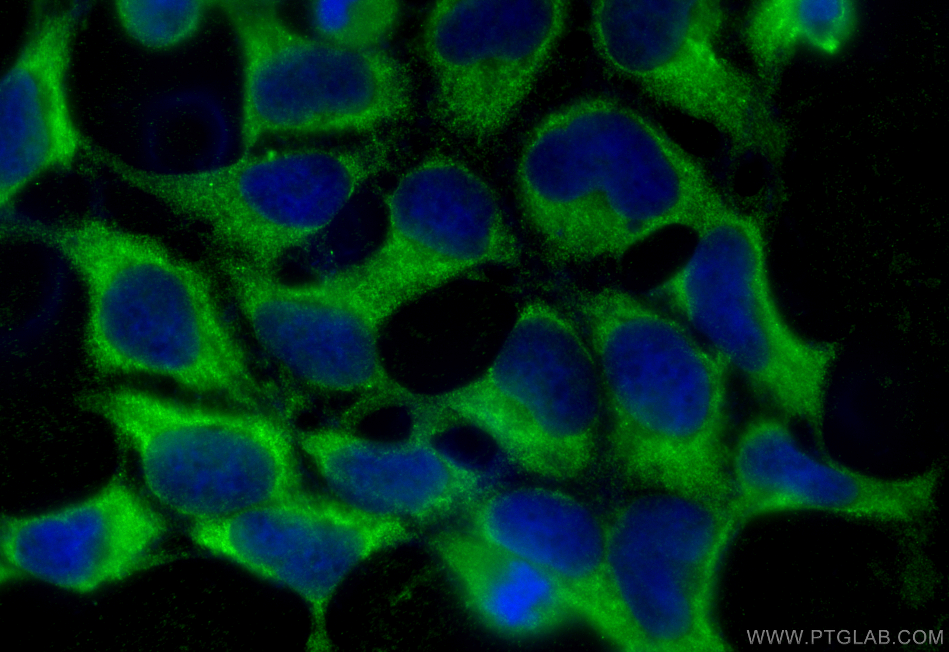
IF Staining of HEK-293 using 66589-1-Ig
Immunofluorescent analysis of (-20°C Ethanol) fixed HEK-293 cells using PHD2/EGLN1 antibody (66589-1-Ig, Clone: 1A2F1 ) at dilution of 1:800 and CoraLite®488-Conjugated Goat Anti-Mouse IgG(H+L) (SA00013-1).
Immunofluorescent analysis of (-20°C Ethanol) fixed HEK-293 cells using PHD2/EGLN1 antibody (66589-1-Ig, Clone: 1A2F1 ) at dilution of 1:800 and CoraLite®488-Conjugated Goat Anti-Mouse IgG(H+L) (SA00013-1).
The Proteintech guarantee covers Proteintech antibodies in any species and any application, including those not listed on the datasheet. If the antibody doesn’t perform, you can receive a hassle-free refund or credit note.
human testis tissue, human kidney tissue Note: suggested antigen retrieval with TE buffer pH 9.0; (*) Alternatively, antigen retrieval may be performed with citrate buffer pH 6.0
Positive IF/ICC detected in
HEK-293 cells
Recommended dilution
Application
Dilution
Western Blot (WB)
WB : 1:1000-1:6000
Immunohistochemistry (IHC)
IHC : 1:150-1:600
Immunofluorescence (IF)/ICC
IF/ICC : 1:400-1:1600
It is recommended that this reagent should be titrated in each testing system to obtain optimal results.
Sample-dependent, Check data in validation data gallery.
PBS with 0.02% sodium azide and 50% glycerol , pH 7.3
Storage Conditions
Store at -20°C. Stable for one year after shipment. Aliquoting is unnecessary for -20oC storage. 20ul sizes contain 0.1% BSA.
Background Information
EGLN1, also named as PHD2, SM-20, HPH-2 and HIF-PH2, catalyzes the post-translational formation of 4-hydroxyproline in hypoxia-inducible factor (HIF) alpha proteins. It hydroxylates HIF-1 alpha at 'Pro-402' and 'Pro-564', and HIF-2 alpha. EGLN1 functions as a cellular oxygen sensor and, under normoxic conditions, targets HIF through the hydroxylation for proteasomal degradation via the von Hippel-Lindau ubiquitination complex. Defects in EGLN1 are the cause of erythrocytosis familial type 3 (ECYT3). EGLN1 has 3 isoforms with MW of 46 kDa, 44 kDa and 36 kDa produced by alternative splicing. It mainly localizes in cytoplasm and can shuttle between the nucleus and cytoplasm (PubMed:19631610). The antibody is specific to EGLN1.
mouse brain tissue were subjected to SDS PAGE followed by western blot with 66589-1-Ig (EGLN1 antibody) at dilution of 1:3000 incubated at room temperature for 1.5 hours.
WB analysis of HEK-293 using 66589-1-Ig
HEK-293 cells were subjected to SDS PAGE followed by western blot with 66589-1-Ig (EGLN1 antibody) at dilution of 1:3000 incubated at room temperature for 1.5 hours.
WB analysis of SH-SY5Y using 66589-1-Ig
SH-SY5Y cells were subjected to SDS PAGE followed by western blot with 66589-1-Ig (EGLN1 antibody) at dilution of 1:3000 incubated at room temperature for 1.5 hours.
WB analysis of pig brain using 66589-1-Ig
pig brain tissue were subjected to SDS PAGE followed by western blot with 66589-1-Ig (EGLN1 antibody) at dilution of 1:3000 incubated at room temperature for 1.5 hours.
IHC Figures
IHC staining of human testis using 66589-1-Ig
Immunohistochemical analysis of paraffin-embedded human testis tissue slide using 66589-1-Ig (EGLN1 antibody) at dilution of 1:300 (under 10x lens. Heat mediated antigen retrieval with Tris-EDTA buffer (pH 9.0).
IHC staining of human testis using 66589-1-Ig
Immunohistochemical analysis of paraffin-embedded human testis tissue slide using 66589-1-Ig (EGLN1 antibody) at dilution of 1:300 (under 40x lens. Heat mediated antigen retrieval with Tris-EDTA buffer (pH 9.0).
IHC staining of human kidney using 66589-1-Ig
Immunohistochemical analysis of paraffin-embedded human kidney tissue slide using 66589-1-Ig (EGLN1 antibody) at dilution of 1:300 (under 10x lens. Heat mediated antigen retrieval with Tris-EDTA buffer (pH 9.0).
IHC staining of human kidney using 66589-1-Ig
Immunohistochemical analysis of paraffin-embedded human kidney tissue slide using 66589-1-Ig (EGLN1 antibody) at dilution of 1:300 (under 40x lens. Heat mediated antigen retrieval with Tris-EDTA buffer (pH 9.0).
IF/ICC Figures
IF Staining of HEK-293 using 66589-1-Ig
Immunofluorescent analysis of (-20°C Ethanol) fixed HEK-293 cells using PHD2/EGLN1 antibody (66589-1-Ig, Clone: 1A2F1 ) at dilution of 1:800 and CoraLite®488-Conjugated Goat Anti-Mouse IgG(H+L) (SA00013-1).
The species listed in Tested Reactivity are in-house verified and applicable species. For unlisted species, please refer to the homology analysis of the immunogen sequence and related species. For rabbit polyclonal antibodies, homology >70% is recommended. For mouse monoclonal antibodies and rabbit recombinant antibodies, homology >90% is recommended. Generally, the higher the homology, the greater the applicability. However, there will be certain differences in protein expression in different species, tissues or cells. Therefore, the homology analysis results are for reference only and do not serve as a guarantee.
At Proteintech, we pride ourselves on our antibody quality, customer service and transparency. As such, we are comparing our antibodies with other vendors, enabling easy identification and comparisons of key data to help you choose the suitable antibody for your needs.
We have selected the top cited antibodies from these vendors for you to compare.
 at dilution of 1:3000 incubated at room temperature for 1.5 hours.")
 at dilution of 1:3000 incubated at room temperature for 1.5 hours.")
 at dilution of 1:3000 incubated at room temperature for 1.5 hours.")
 at dilution of 1:3000 incubated at room temperature for 1.5 hours.")
 at dilution of 1:300 (under 10x lens. Heat mediated antigen retrieval with Tris-EDTA buffer (pH 9.0).")
 at dilution of 1:300 (under 40x lens. Heat mediated antigen retrieval with Tris-EDTA buffer (pH 9.0).")
 at dilution of 1:300 (under 10x lens. Heat mediated antigen retrieval with Tris-EDTA buffer (pH 9.0).")
 at dilution of 1:300 (under 40x lens. Heat mediated antigen retrieval with Tris-EDTA buffer (pH 9.0).")
 fixed HEK-293 cells using PHD2/EGLN1 antibody (66589-1-Ig, Clone: 1A2F1 ) at dilution of 1:800 and CoraLite®488-Conjugated Goat Anti-Mouse IgG(H+L) (SA00013-1).")