
Immunofluorescence Imaging of Live and Fixed Pancreatic Tissue Slices Using FlexAble 2.0 Antibody Labeling
Written by Vijay Sai Josyula, Ph.D. Candidate at Uppsala University
Key Benefits of FlexAble 2.0 Antibody Labeling for Imaging of Pancreatic Tissue Slices:
-
Saves up to 3 hours of bench time by eliminating the need for secondary antibody incubation
-
FlexAble 2.0 labeled antibodies demonstrated effective penetration in agarose-stabilized pancreatic tissue slices
-
Allows labeling of the exact amount needed, saving antibody amount and cost
-
Easy-to-follow protocol with minimal hands-on time
Introduction
If you work in pancreatic islet research, you have probably spent time with two familiar ex vivo systems: isolated islets and 3D organoids. Each has clear value, yet each comes with tradeoffs that can shape the data you collect. Islet isolation by differential centrifugation exposes fragile endocrine clusters to chemical and mechanical stress that can distort native architecture and compromise beta cell viability. Organoids restore a degree of 3D structure, but even the best models often fall short of reproducing the full spectrum of paracrine and juxtacrine signaling that occurs inside the intact pancreas.
Live pancreatic tissue slices offer a compelling alternative. By preserving the native microenvironment — including extracellular matrix contacts, local vasculature remnants, resident immune cells, and neighboring exocrine tissue — slices maintain physiological cell-to-cell connectivity that isolated systems frequently lose. Within this preserved context you can monitor insulin secretion at high resolution, capture real-time functional readouts such as calcium signaling, and quantify glucose- and hormone-stimulated responses across all endocrine cell types while still seeing what is happening in the surrounding tissue.
Because tissue slices retain structure without requiring whole animal experimentation beyond the initial preparation, they help reduce reliance on extended in vivo studies and the ethical and logistical constraints that go with them. The result is a robust, data-rich platform for diabetes research, beta cell regeneration work, and high-throughput pharmacological screening that keeps biology as close to native as you can get outside the body.
By maintaining native extracellular matrix architecture and intercellular contacts, live pancreatic tissue slices allow beta cell physiology to be studied within a preserved structural context that is not achievable with isolated islets or organoids.
The same live tissue slices, and even the same islets, can be fixed and used to extract even more information from each preparation while preserving spatial relationships. This application note outlines a method combining imaging of live pancreatic tissue slices with using Proteintech’s FlexAble 2.0 Antibody Labeling Kit.
Experimental Overview
In this approach, CX3CR1⁺/GFP reporter mice were used, in which islet macrophages are endogenously fluorescent. Macrophage behavior was first imaged in living slices following drug exposure. Subsequently, without discarding the tissue, the same slices were fixed and immunostained, allowing local nerve architecture to be mapped.
To visualize innervation, the norepinephrine transporter was targeted using a primary antibody that was directly conjugated to CoraLite® Plus 647 via the FlexAble 2.0 Antibody Labeling Kit from Proteintech. This kit allows you to stably label any unconjugated antibody with fluorescent dyes in just two steps and ten minutes without any buffer exchange. Reagent volumes for the conjugation were calculated using the FlexAble 2.0 calculator on the Proteintech website. By using FlexAble 2.0, unconjugated primary antibodies were converted into ready-to-use fluorescent reagents, eliminating the need for secondary antibody incubation. As a result, approximately three hours of bench time were saved. Since the kit works with as little as 0.5 µg of primary antibody and allows easy scale-up, it is possible to label exactly the amount of antibody needed — ultimately saving both antibody and cost. Notably, agarose present in the slices did not hinder penetration of the labeled antibody. Uniform penetration and consistent, high-contrast labeling were observed throughout the tissue, demonstrating the compatibility of this workflow with agarose-stabilized slices and its potential for extension to multiplex immunostaining applications.
Materials & Methods
Step-by-step protocol for live pancreatic tissue-slice technique
The live pancreatic tissue‑slice technique, originally established in the laboratory of Dr. Stephan Speier (1) , proceeds as follows:
-
Agarose Infusion: Under anesthesia, the common bile duct is cannulated and infused with 1.5% low‑melting‑point agarose solution to uniformly fill the pancreatic ducts and parenchyma.
-
Tissue Preparation: Once gelation is complete, the pancreas is excised, trimmed of surrounding connective tissue, ducts, and large vessels, and sectioned into ~5 mm³ blocks.
-
Vibratome Sectioning: Blocks are mounted on a vibratome and precision‑cut into 150 μm‑thick slices, preserving intact islet–exocrine interfaces.
-
Imaging and Functional Assays: Slices are transferred to a perfusion chamber and imaged on a confocal microscope, enabling high‑resolution calcium imaging, insulin granule tracking, and real‑time assessment of stimulus‑secretion coupling within an undisturbed microenvironment.
Step-by-step protocol for staining fixed pancreatic tissue slices
After the live imaging of CX3CR1+/GFP macrophages in drug-treated slices as described above, proceed as follows:
-
Fix the same slices in 4% paraformaldehyde (PFA) for 1 hour at 37°C.
-
Wash 2x in PBST for 15 minutes each at 37°C.
-
Conjugate the anti-norepinephrine transporter antibody with CoraLite® Plus 647 using FlexAble 2.0 according to the kit instructions (use the FlexCalc FlexAble 2.0 calculator to determine the antibody amount and buffer needed).
-
Stain slices with the conjugated antibody for 2 hours at 37°C.
-
Wash 3x in PBST for 15 minutes each at 37°C.
-
Add nuclear stain (Hoechst).
-
Apply coverslip and let cure overnight at 4°C
-
Image the slices to capture nerve architecture.
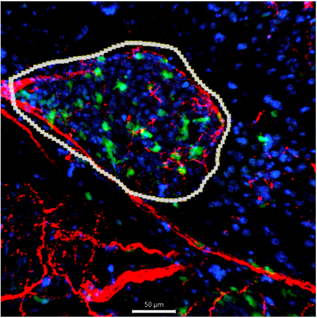 |
Pancreatic tissue section showing an islet (outlined in white) with resident macrophages (green) distributed both inside and around the islet. Nerve fibers (red), stained using the FlexAble 2.0 conjugation system, are seen in close association with the islet and surrounding exocrine tissue. Nuclei are counterstained in blue. This image highlights the structural proximity of macrophages and nerves to the endocrine islet environment. Green: Macrophages (GFP) Red: Nerves stained with anti-SLC6A2 (norepinephrine transporter) antibody conjugated with CoraLite® Plus 647 Antibody Labeling Kit (Cat. Nr.: KFA503) Blue: Nuclei (Hoechst) Encircled: Islet |
Conclusion
FlexAble 2.0 Antibody Labeling Kits offer a fast and reliable solution for researchers for almost any antibody application using fluorescent or enzymatic detection with HRP or biotin, such as standard and cyclic IF, chromogenic IHC, Western Blot, Flow Cytometry, and Tyramide Signal Amplification (TSA) multiplex experiments, to name a view. By enabling direct antibody labeling, they eliminate the need for secondary staining, significantly reducing preparation time while ensuring high-quality results for applications such as confocal microscopy.
References
-
Marciniak A, Cohrs CM, Tsata V, Chouinard JA, Selck C, Stertmann J, et al. Using pancreas tissue slices for in situ studies of islet of Langerhans and acinar cell biology. Nat Protoc. 2014 Dec;9(12):2809–22.

Related Content
FlexAble Antibody Labeling Kits | Proteintech
The FlexAble way to improve your immunofluorescence with IBEX | Proteintech Group
Imaging the Rainbow: Multiplexing with Same-Species Antibodies for Immunofluorescence | Proteintech…
Support
Newsletter Signup
Stay up-to-date with our latest news and events. New to Proteintech? Get 10% off your first order when you sign up.