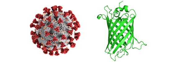
How fluorescent proteins can be applied in SARS-CoV-2 research
GFP or RFP and their derivatives are commonly used for virus research
Fluorescent proteins (FPs) like GFP or RFP and their derivatives are commonly used for virus research. Frequently, they are applied as fluorescent markers in microscopy or cell sorting experiments and as protein tags for protein purification, immunoprecipitation or protein-protein interaction assays. Here, we provide a short outline that describes how FPs have been used in the current SARS-CoV-2 research. Please note that this summary does not provide a complete overview; also, many referenced papers are pre-prints without peer reviews.

Application examples of fluorescent proteins in SARS-CoV-2 research
Virus protein purification
Although fluorescent protein tags are rarely applied in protein purification, the yellow fluorescent protein (YFP) has been used for purification of the receptor-binding domain (RBD) of the spike glycoprotein S [DOI: 10.1101/2020.04.16.045419]. Here, RBD-vYFP was expressed in mammalian cells and isolated from cell extract with an anti-GFP Nanobody resin (such as GFP-Trap). The RBD-vYFP fusion protein was eluted from the resin with an acidic buffer. Efforts to cleave off YFP failed and the full-length RBD-vYFP fusion was used for additional assays.
ChromoTek’s Nano-Traps such as GFP-Trap is well suited for the purification of FP-tagged virus proteins like RBD. Besides protein purification, the Nano-Traps can be used for immunoprecipitation of FP-tagged virus proteins and to find potential viral and host cell binding partners.
Fluorescence microscopy
FPs are ideal tools for visualization of virus proteins in microscopy and used as reporters. Two approaches are reported:
- The virus is labeled with the FP by introducing the FP reporter gene into the viral genome. When the virus infects cells and viral proteins are produced, also the FP is expressed. The resulting fluorescent signal labels the infected cells.
- Viral proteins are tagged with the FP and recombinantly expressed in cells. The FP signal can be used to analyze the location of the virus protein, for characterization and to find binding partners.
The downsides of FPs are their photobleaching and relative weak signal intensity in super-resolution microscopy. ChromoTek’s Nano-Boosters, e.g. GFP-Booster, which is a GFP Nanobody conjugated to fluorescent dyes, overcome these issues by locating a fluorescent dye to the FPs, which stabilizes and increases signal intensity.
Flow cytometry/ FACS
FP tags can also be used in flow cytometry/ FACS because cells are detected based on the fluorescent signal resulting from intracellular or cell surface FPs:
- Cells are infected by the virus coding for FP tagged viral protein(s). Cells, which express the FP, are counted or sorted based on the fluorescent signal.
- Cells expressing FP tagged virus proteins can be recorded or sorted.
- Cells are incubated with FP tagged viral proteins. Cells, which have a fluorescent signal based from the FP tagged viral protein binding to molecules on the cell surface are counted.
If the fluorescent signal of the FP is not strong enough or another fluorescent channel shall be used ChromoTek’s Nano-Booster can be applied. For example, if GFP shall be analyzed in the red channel, the GFP-Booster Alexa Fluor® 647 can be used to locate a red fluorescent dye to the GFP fusion protein via the GFP Nanobody.
Overview of publications using FPs
Related Content
Scientists unravel why Omicron spreads faster
Neuropilin-1 joins ACE2 as SARS-CoV-2 gateway
How Nanobodies could advance Sars-CoV diagnostics
Neutralizing antibodies and their role in COVID-19 research
Serious about Serology: Understanding COVID19 Antibody Tests
Characterization of COVID-19 antibodies by Bio-Layer Interferometry using Nano-CaptureLigands

Support
Newsletter Signup
Stay up-to-date with our latest news and events. New to Proteintech? Get 10% off your first order when you sign up.
