Get the Most from Your iPSCs: Improved Cardiomyocyte Differentiation
Written by Jamuna Karanankattil Sukumaran, Senior Scientist at Proteintech
Induced pluripotent stem cells (iPSCs) have become a powerful tool for generating cardiomyocytes, with exciting applications in disease modeling, drug screening, and regenerative medicine. However, getting reliable and high-yield differentiation results isn’t always straightforward—it takes careful planning and fine-tuning at every step. In this article, we’re sharing some practical, lab-tested tips and tricks to help researchers boost both the consistency and efficiency of cardiomyocyte generation from iPSCs.
-
The starting material—High-quality iPSCs
The key to a successful cardiomyocyte differentiation protocol starts with high-quality iPSCs. Before jumping into differentiation, make sure your iPSCs are in great shape. Here’s what to watch for:
-
The cells should look healthy, with dense, compact colonies and little to no signs of spontaneous differentiation.
-
Check that pluripotency markers like Nanog and OCT-4 are strongly expressed in more than 95% of the cells—this is crucial.
-
Timing is everything when it comes to passaging. Aim to passage the cells when they’re around 85–90% confluent to avoid triggering unwanted differentiation.
-
Use ROCK Inhibitor During Initial Seeding
When you're passaging iPSCs for differentiation, don’t skip the ROCK inhibitor—it’s a must for helping the cells survive the stress of replating.
-
Add ROCK inhibitor during the first 18–24 hours after seeding. It really helps with cell attachment and overall viability.
-
Don’t worry if the cells temporarily take on a spindle-like shape while the RI is in the media—that’s totally normal. They’ll usually bounce back to their usual colony morphology after you change the media and remove the RI.
-
Optimize iPSC Seeding Density
Getting the starting cell density right is crucial for successful cardiomyocyte differentiation—it can truly make or break your results.
-
A good starting point is around 15,000 cells/cm², but think of this as a baseline rather than a strict rule.
-
Keep in mind that different iPSC lines grow at different rates, and this variability can affect how well they differentiate.
-
It’s worth taking the time to optimize the seeding density for each line you work with to get the best possible outcome.
-
Use High-Quality, Consistent Growth Factors
The quality of your growth factors can have a bigger impact on differentiation than you might expect—and it’s often overlooked.
-
Whenever possible, go for animal component–free growth factors made in a human expression system. These are more likely to have proper folding and glycosylation, which can really improve the efficiency and consistency of differentiation.
-
Plus, they tend to offer better batch-to-batch consistency, which means fewer surprises in your experiments and higher cardiomyocyte purity down the line.

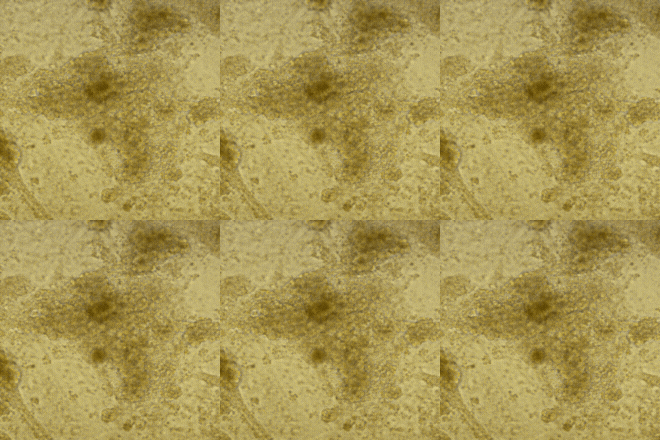
Beating Cardiomyocytes developed from Human iPSCs using Proteintech reagents
-
Fine-Tune CHIR Concentrations
CHIR99021, a Wnt pathway activator, is widely used to kick-start mesoderm differentiation—but it needs to be handled with care.
-
If you’re seeing a lot of cell death during or after CHIR treatment, it’s a sign to double-check your seeding density and how long you're exposing the cells.
-
Sometimes, simply lowering the CHIR concentration or shortening the treatment window can reduce toxicity without compromising mesoderm induction. A little fine-tuning here can go a long way in improving outcomes.
-
Precision is Key During the Mesoderm-to-Cardiac Transition
Getting the timing right for Wnt inhibition—typically using IWP2 or Wnt-C59—is crucial for guiding cells from the mesoderm stage into the cardiac lineage.
-
If the timing’s off, you might end up with low yields or unwanted cell types instead of cardiomyocytes.
-
To stay on track, it’s a good idea to monitor the cells during this transition—using flow cytometry or gene expression analysis—to make sure they’re following the right path toward becoming heart cells.
-
Minimize Physical and Environmental Stress
iPSCs and differentiating cells are quite sensitive to their surroundings, so gentle handling is key.
-
Try to avoid shaking, bumping, or moving the culture plates around too much—any sudden disturbance can stress the cells.
-
It’s also important to keep the temperature and CO₂ levels stable, especially during media changes and throughout the entire differentiation process. A little extra care here can make a big difference in your results.
A quick overview of the protocol we use in-house:
|
Day 0 |
Plate iPSC |
Stemflex media + ROCK Inhibitor |
|
Day 1 |
Media change |
Stemflex media + CHIR99021 (1uM) |
|
Day 2 |
Media change |
Stemflex media + CHIR99021 (1uM) |
|
Day 3 |
Media change |
Stemflex media + CHIR99021 (1uM) |
|
Day 4 |
Media change |
RPMI/B27 media + 100 ng/mL Activin A, 10 ng/mL bFGF, 1% KOSR |
|
Day 5 |
Media change |
RPMI/B27 media (no insulin) + 5 ng/mL BMP4, 5 ng/mL bFGF |
|
Day 9 |
Media change |
RPMI/B27 media (no insulin) |
|
Day 11 |
Media change |
RPMI/B27 media (with insulin) |
|
Day 12 |
- |
Beating cardiomyocytes |
 |
 |
|
Cardiomyocyte derived from iPSCs characterized using Cardiac Troponin T Polyclonal Antibody (15513-1-AP) |
Cardiomyocyte derived from iPSCs characterized using Alpha Actinin Polyclonal Antibody (11313-2-AP) |
To Sum Up
Cardiomyocyte differentiation is as much an art as it is a science. Success starts with healthy, high-quality iPSCs and is supported by reliable reagents—like HumanKine® growth factors—that ensure consistency at every step. From dialing in the right seeding density to fine-tuning Wnt signaling, careful attention to each stage of the process can go a long way in improving both efficiency and reproducibility. With a little patience and some line-specific tweaks, generating high-purity cardiomyocytes is surely achievable.
Growth factors for differentiation
|
Growth Factor |
Catalog Number |
|
bFGF/FGF-2 |
|
|
BMP4 |
|
|
Activin A |
|
|
VEGF |
Antibodies for Characterization
|
Characterization Marker |
Catalog Number |
|
cTnl |
|
|
NKX2.5 |
|
|
CtnT |
|
|
MYL2 |
|
|
ACTN2 |
Related Content
Immune Checkpoint Inhibitor Therapy: Revolutionizing Cancer Treatment | Proteintech Group
Cuproptosis in Health and Disease: Copper’s Double-Edged Sword | Proteintech Group
m5C Modifications in RNA and Cancer | Proteintech Group
The Role of Autophagy in Cancer
Support
Newsletter Signup
Stay up-to-date with our latest news and events. New to Proteintech? Get 10% off your first order when you sign up.

