- Phare
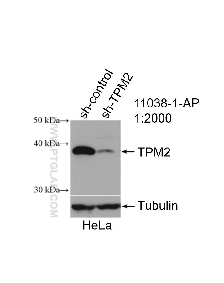
- Validé par KD/KO
Anticorps Polyclonal de lapin anti-TPM2
TPM2 Polyclonal Antibody for WB, IHC, IF/ICC, IP, ELISA
Hôte / Isotype
Lapin / IgG
Réactivité testée
Humain, rat, souris
Applications
WB, IHC, IF/ICC, IP, ELISA
Conjugaison
Non conjugué
N° de cat : 11038-1-AP
Synonymes
Galerie de données de validation
Applications testées
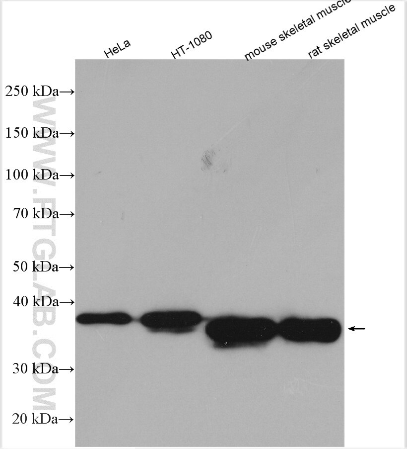
| Résultats positifs en WB | cellules HT-1080, cellules HeLa, muscle squelettique de rat, muscle squelettique de souris |
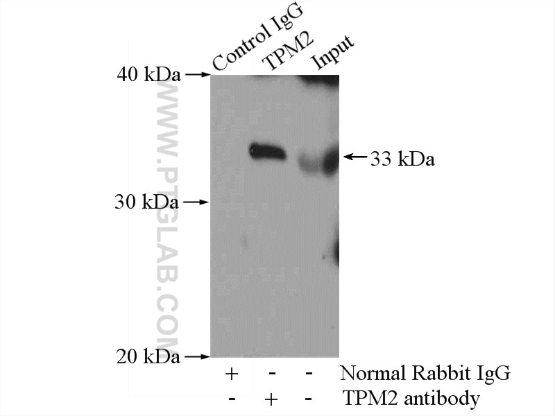
| Résultats positifs en IP | tissu de muscle squelettique de souris |
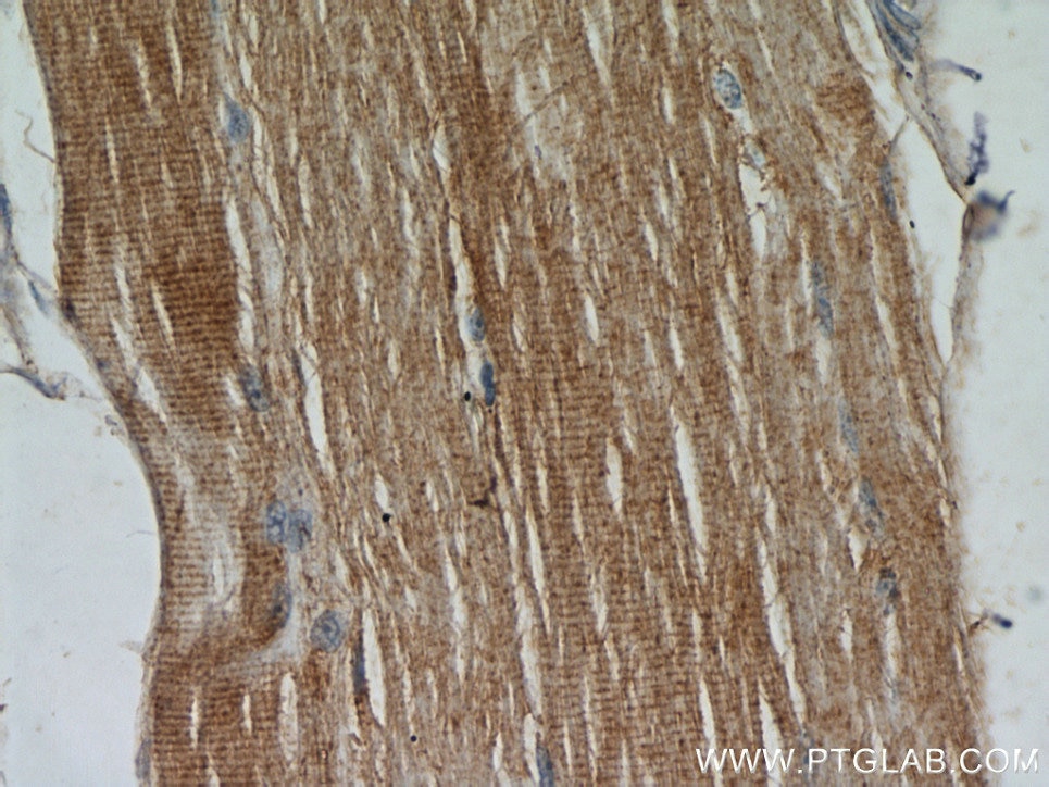
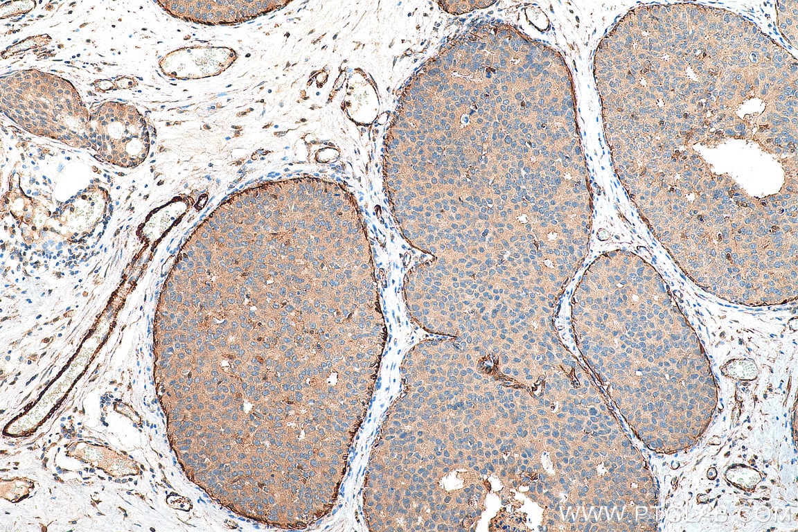
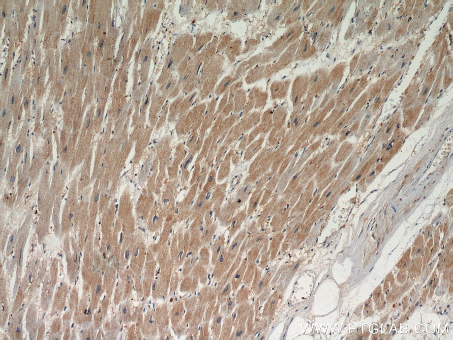
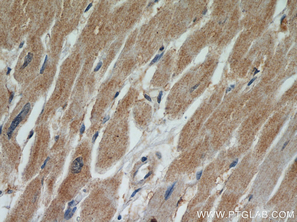
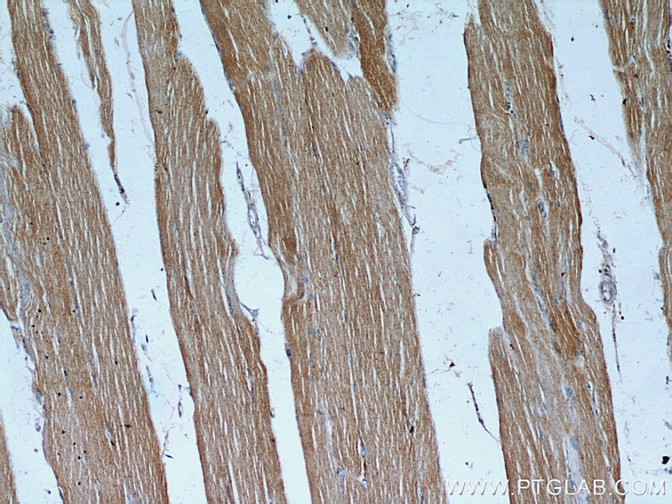
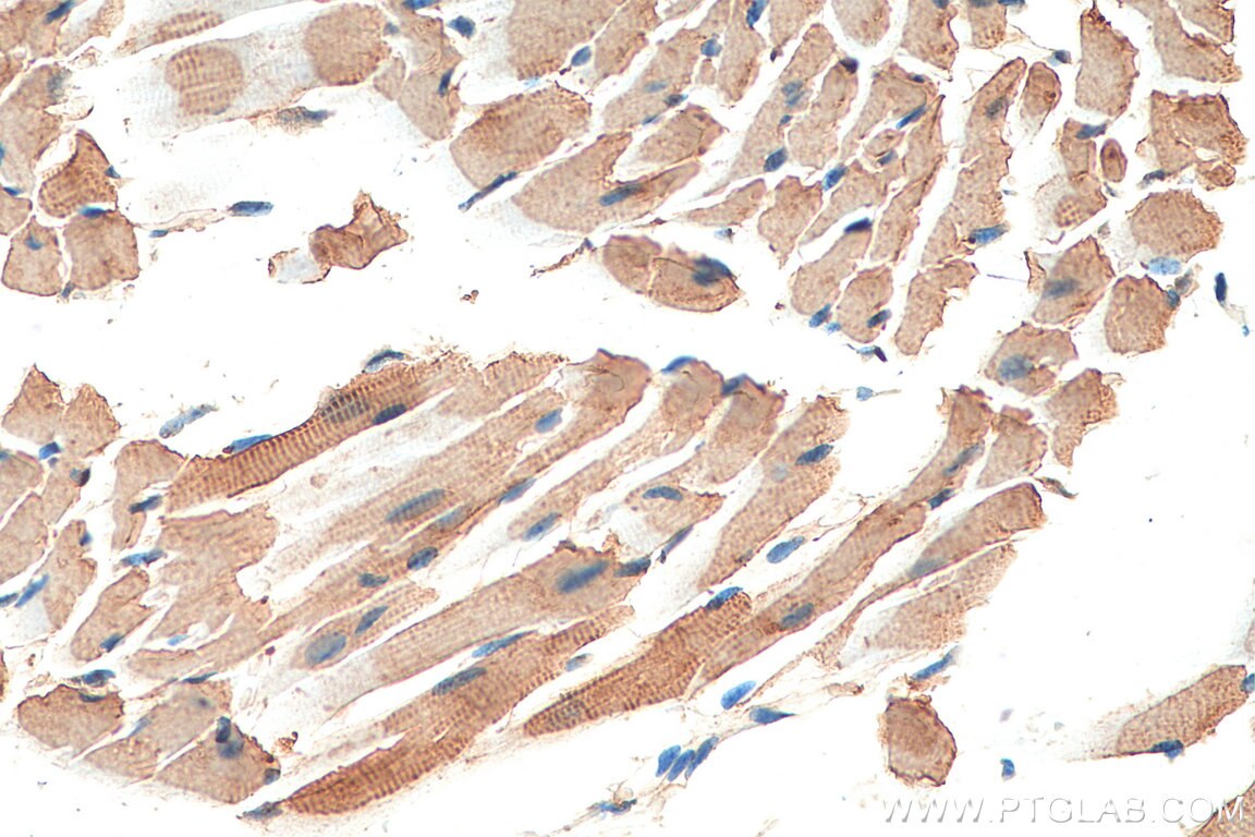
| Résultats positifs en IHC | tissu de cancer du sein humain, tissu cardiaque de souris, tissu cardiaque humain, tissu de muscle squelettique humain il est suggéré de démasquer l'antigène avec un tampon de TE buffer pH 9.0; (*) À défaut, 'le démasquage de l'antigène peut être 'effectué avec un tampon citrate pH 6,0. |
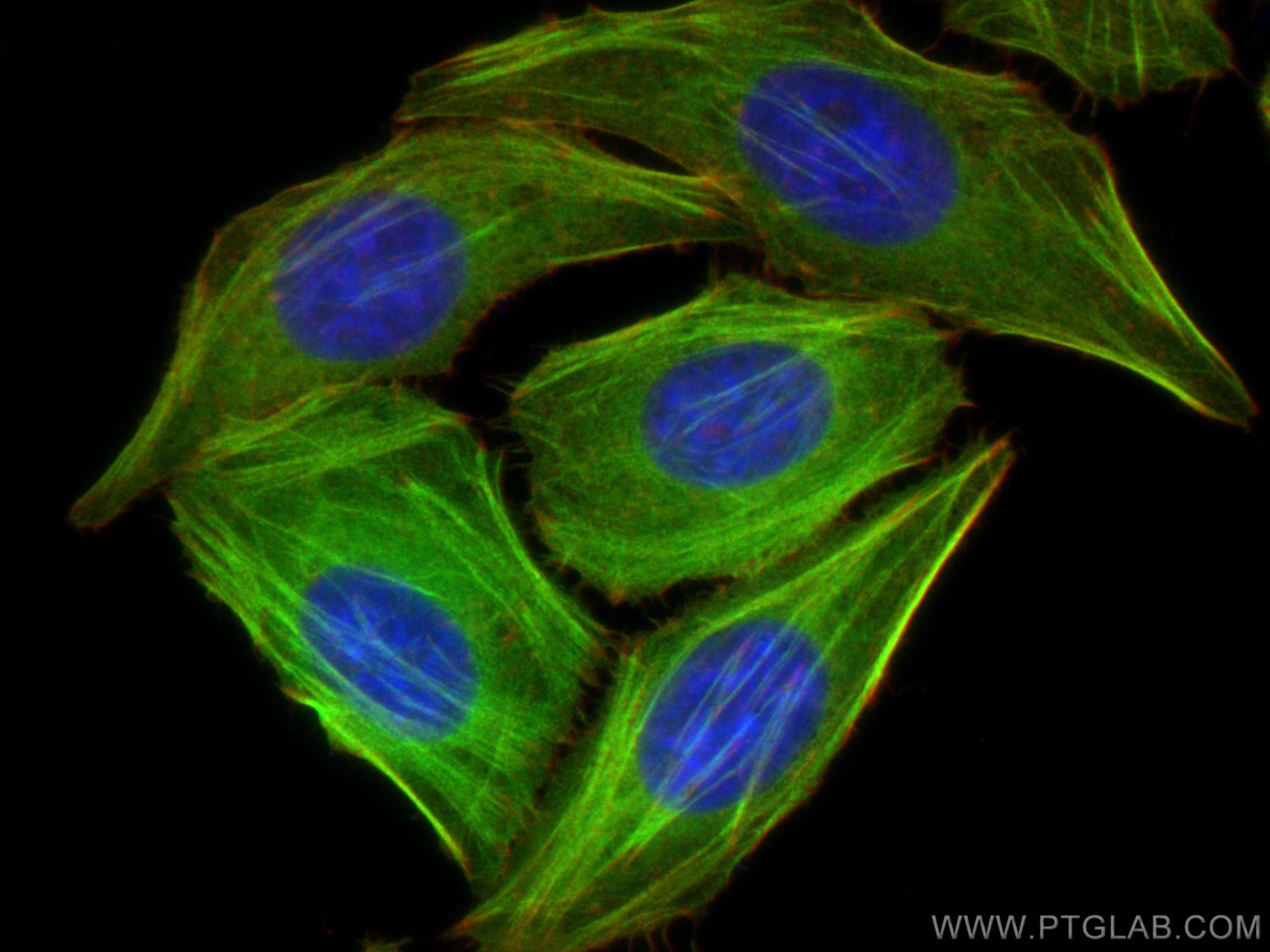
| Résultats positifs en IF/ICC | cellules HepG2, |
Dilution recommandée
| Application | Dilution |
|---|---|
| Western Blot (WB) | WB : 1:1000-1:4000 |
| Immunoprécipitation (IP) | IP : 0.5-4.0 ug for 1.0-3.0 mg of total protein lysate |
| Immunohistochimie (IHC) | IHC : 1:50-1:500 |
| Immunofluorescence (IF)/ICC | IF/ICC : 1:50-1:500 |
| It is recommended that this reagent should be titrated in each testing system to obtain optimal results. | |
| Sample-dependent, check data in validation data gallery | |
Applications publiées
| WB | See 5 publications below |
| IHC | See 2 publications below |
| IP | See 1 publications below |
Informations sur le produit
11038-1-AP cible TPM2 dans les applications de WB, IHC, IF/ICC, IP, ELISA et montre une réactivité avec des échantillons Humain, rat, souris
| Réactivité | Humain, rat, souris |
| Réactivité citée | rat, Humain, souris |
| Hôte / Isotype | Lapin / IgG |
| Clonalité | Polyclonal |
| Type | Anticorps |
| Immunogène | TPM2 Protéine recombinante Ag1508 |
| Nom complet | tropomyosin 2 (beta) |
| Masse moléculaire calculée | 33 kDa |
| Poids moléculaire observé | 29-33 kDa |
| Numéro d’acquisition GenBank | BC011776 |
| Symbole du gène | TPM2 |
| Identification du gène (NCBI) | 7169 |
| Conjugaison | Non conjugué |
| Forme | Liquide |
| Méthode de purification | Purification par affinité contre l'antigène |
| Tampon de stockage | PBS with 0.02% sodium azide and 50% glycerol |
| Conditions de stockage | Stocker à -20°C. Stable pendant un an après l'expédition. L'aliquotage n'est pas nécessaire pour le stockage à -20oC Les 20ul contiennent 0,1% de BSA. |
Informations générales
TPM2, also named as TMSB, belongs to the tropomyosin family. It binds to actin filaments in muscle and non-muscle cells. TPM2 plays a central role, in association with the troponin complex, in the calcium dependent regulation of vertebrate striated muscle contraction. In non-piamuscle cells, TPM2 is implicated in stabilizing cytoskeleton actin filaments. Defects in TPM2 are the cause of nemaline myopathy type 4 (NEM4) and distal arthrogryposis type 1 (DA1).
Protocole
| Product Specific Protocols | |
|---|---|
| WB protocol for TPM2 antibody 11038-1-AP | Download protocol |
| IHC protocol for TPM2 antibody 11038-1-AP | Download protocol |
| IF protocol for TPM2 antibody 11038-1-AP | Download protocol |
| IP protocol for TPM2 antibody 11038-1-AP | Download protocol |
| Standard Protocols | |
|---|---|
| Click here to view our Standard Protocols |
Publications
| Species | Application | Title |
|---|---|---|
Bone Res Impairment of rigidity sensing caused by mutant TP53 gain of function in osteosarcoma | ||
Cell Biosci TPM2 attenuates progression of prostate cancer by blocking PDLIM7-mediated nuclear translocation of YAP1 | ||
PLoS One Proteomic changes in the hippocampus of large mammals after total-body low dose radiation | ||
Int J Mol Sci Modulation of Titin and Contraction-Regulating Proteins in a Rat Model of Heart Failure with Preserved Ejection Fraction: Limb vs. Diaphragmatic Muscle | ||
Cancer Prev Res (Phila) Stool Protein Mass Spectrometry Identifies Biomarkers for Early Detection of Diffuse-type Gastric Cancer | ||
Leukemia Single-cell transcriptomics of pediatric Burkitt lymphoma reveals intra-tumor heterogeneity and markers of therapy resistance |
Avis
The reviews below have been submitted by verified Proteintech customers who received an incentive for providing their feedback.
FH Marco (Verified Customer) (07-03-2024) | A view unspecific bands near the region of the protein, but enough blocking (o.N.) got rid of the problem. A well visible band.
|
