- Phare
- Validé par KD/KO
Anticorps Polyclonal de lapin anti-RPS3
RPS3 Polyclonal Antibody for WB, IP, IHC, ELISA
Hôte / Isotype
Lapin / IgG
Réactivité testée
Humain, rat, souris et plus (1)
Applications
WB, IP, IF, IHC, ELISA
Conjugaison
Non conjugué
N° de cat : 11990-1-AP
Synonymes
Galerie de données de validation
Applications testées
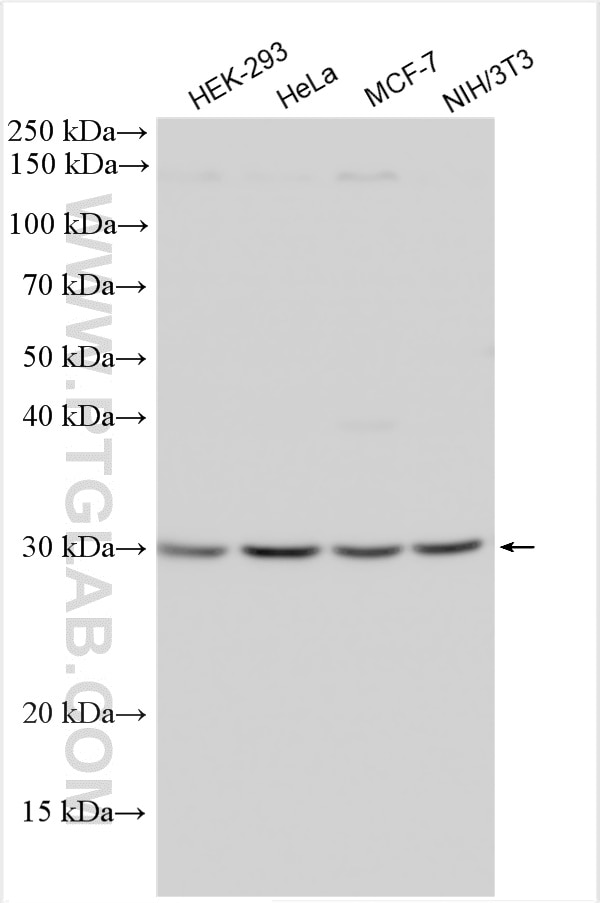
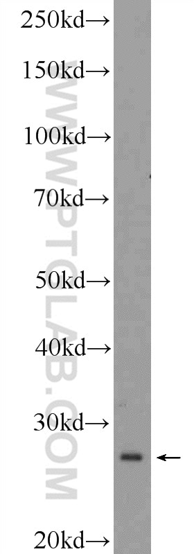
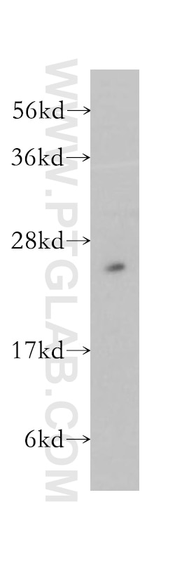
| Résultats positifs en WB | cellules HEK-293, cellules HeLa, cellules MCF-7, cellules NIH/3T3, cellules PC-12, tissu testiculaire de souris |
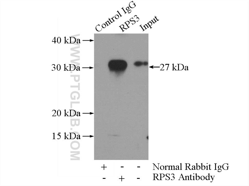
| Résultats positifs en IP | tissu testiculaire de souris |
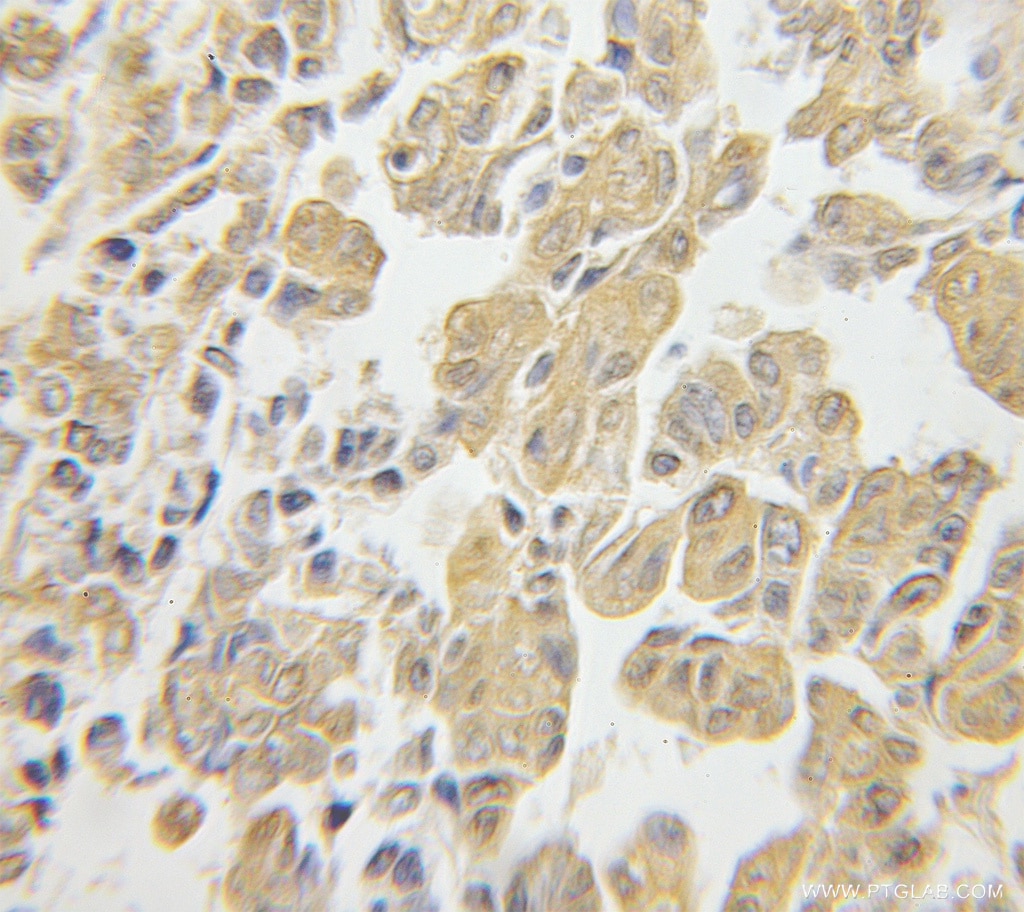
| Résultats positifs en IHC | tissu de tumeur ovarienne humain il est suggéré de démasquer l'antigène avec un tampon de TE buffer pH 9.0; (*) À défaut, 'le démasquage de l'antigène peut être 'effectué avec un tampon citrate pH 6,0. |
Dilution recommandée
| Application | Dilution |
|---|---|
| Western Blot (WB) | WB : 1:1000-1:6000 |
| Immunoprécipitation (IP) | IP : 0.5-4.0 ug for 1.0-3.0 mg of total protein lysate |
| Immunohistochimie (IHC) | IHC : 1:20-1:200 |
| It is recommended that this reagent should be titrated in each testing system to obtain optimal results. | |
| Sample-dependent, check data in validation data gallery | |
Applications publiées
| KD/KO | See 1 publications below |
| WB | See 14 publications below |
| IHC | See 2 publications below |
| IF | See 4 publications below |
| IP | See 2 publications below |
Informations sur le produit
11990-1-AP cible RPS3 dans les applications de WB, IP, IF, IHC, ELISA et montre une réactivité avec des échantillons Humain, rat, souris
| Réactivité | Humain, rat, souris |
| Réactivité citée | Humain, porc, souris |
| Hôte / Isotype | Lapin / IgG |
| Clonalité | Polyclonal |
| Type | Anticorps |
| Immunogène | RPS3 Protéine recombinante Ag3918 |
| Nom complet | ribosomal protein S3 |
| Masse moléculaire calculée | 26 aa, 7 kDa |
| Poids moléculaire observé | 26.7 kDa |
| Numéro d’acquisition GenBank | BC034149 |
| Symbole du gène | RPS3 |
| Identification du gène (NCBI) | 6188 |
| Conjugaison | Non conjugué |
| Forme | Liquide |
| Méthode de purification | Purification par affinité contre l'antigène |
| Tampon de stockage | PBS avec azoture de sodium à 0,02 % et glycérol à 50 % pH 7,3 |
| Conditions de stockage | Stocker à -20°C. Stable pendant un an après l'expédition. L'aliquotage n'est pas nécessaire pour le stockage à -20oC Les 20ul contiennent 0,1% de BSA. |
Informations générales
40S ribosomal protein S3 (RPS3), also named as SW-cl.26, is a 243 amino acid protein,which contains one KH type-2 domain and belongs to the ribosomal protein S3P family. RPS3 localizes in the cytoplasm. RPS3 is identified in a IGF2BP1-dependent mRNP granule complex, which contains untranslated mRNAs. RPS3 plays a role in repairing various DNA damage acting as a repair UV endonuclease. Nuclear accumulation of RPS3 results in an increase in DNA repair activity to some extent, thereby sustaining neuronal survival.
Protocole
| Product Specific Protocols | |
|---|---|
| WB protocol for RPS3 antibody 11990-1-AP | Download protocol |
| IHC protocol for RPS3 antibody 11990-1-AP | Download protocol |
| IP protocol for RPS3 antibody 11990-1-AP | Download protocol |
| Standard Protocols | |
|---|---|
| Click here to view our Standard Protocols |
Publications
| Species | Application | Title |
|---|---|---|
Nat Commun Kaposi's sarcoma-associated herpesvirus induces specialised ribosomes to efficiently translate viral lytic mRNAs | ||
Nat Commun Cilia locally synthesize proteins to sustain their ultrastructure and functions. | ||
Curr Biol RPL10L Is Required for Male Meiotic Division by Compensating for RPL10 during Meiotic Sex Chromosome Inactivation in Mice. | ||
J Anim Sci Biotechnol Glyphosate exposure deteriorates oocyte meiotic maturation via induction of organelle dysfunctions in pigs. | ||
Front Oncol RPS3 Promotes the Metastasis and Cisplatin Resistance of Adenoid Cystic Carcinoma. | ||
Avis
The reviews below have been submitted by verified Proteintech customers who received an incentive forproviding their feedback.
FH Boyan (Verified Customer) (12-16-2021) | This antibody recognized a clean band at 27 kd.
|
FH Huanzhou (Verified Customer) (11-26-2018) |
|
