- Phare
- Validé par KD/KO
Anticorps Polyclonal de lapin anti-KPNA2
KPNA2 Polyclonal Antibody for WB, IF, IHC, ELISA
Hôte / Isotype
Lapin / IgG
Réactivité testée
Humain, rat, souris
Applications
WB, IHC, IF/ICC, IP, CoIP, ChIP, ELISA
Conjugaison
Non conjugué
N° de cat : 10819-1-AP
Synonymes
Galerie de données de validation
Applications testées
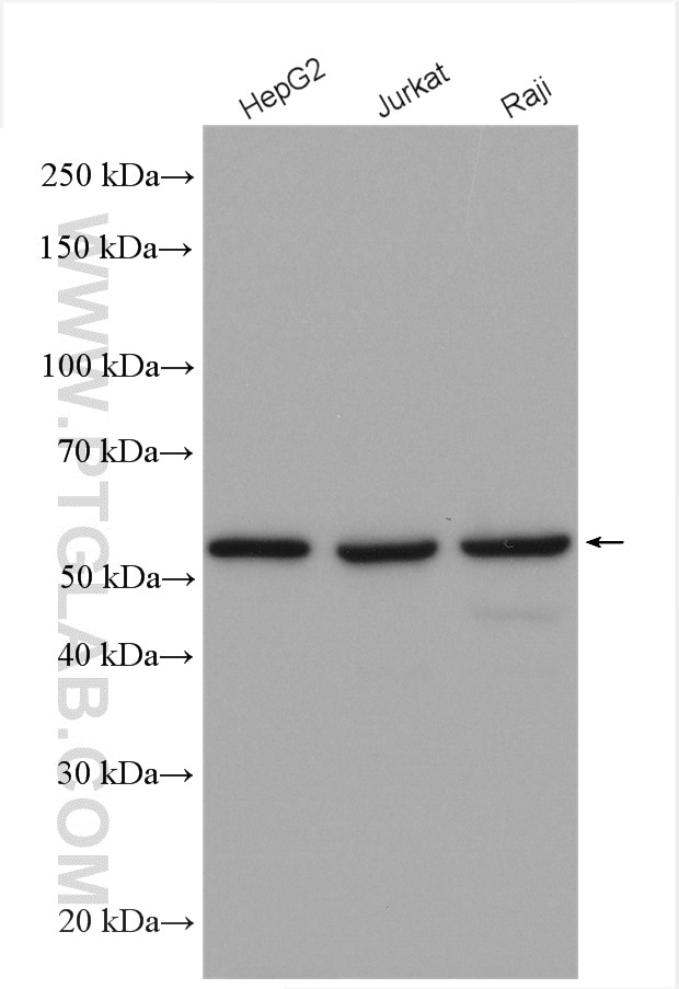
| Résultats positifs en WB | cellules HepG2, cellules Jurkat, cellules Raji |
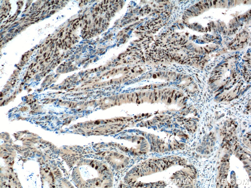
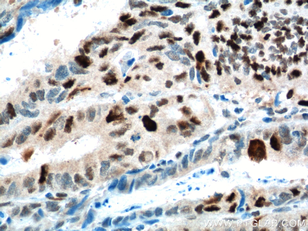
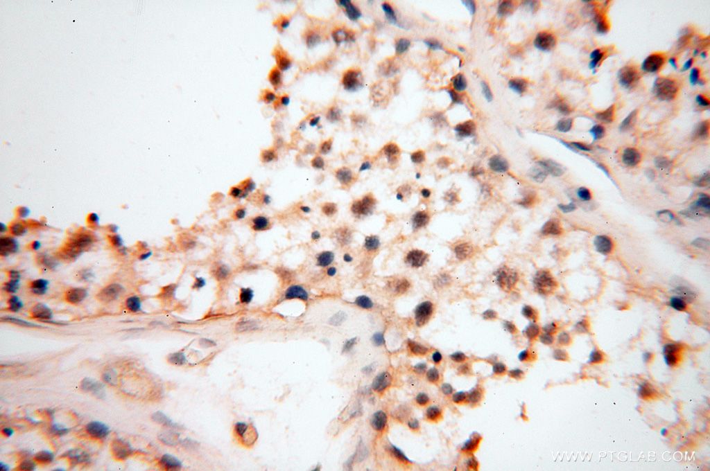
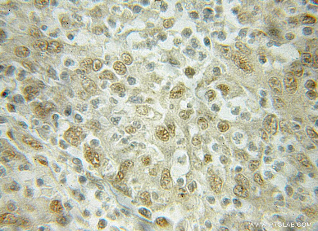
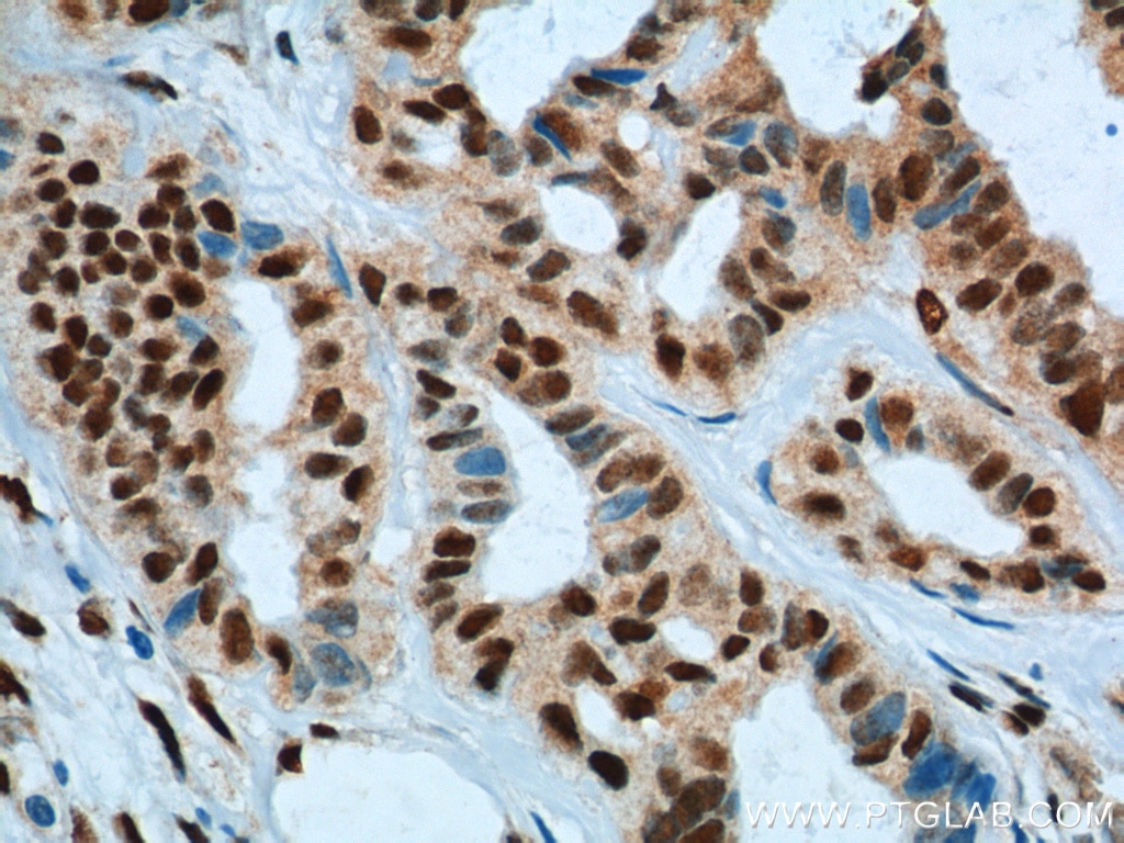
| Résultats positifs en IHC | tissu de cancer du côlon humain il est suggéré de démasquer l'antigène avec un tampon de TE buffer pH 9.0; (*) À défaut, 'le démasquage de l'antigène peut être 'effectué avec un tampon citrate pH 6,0. |
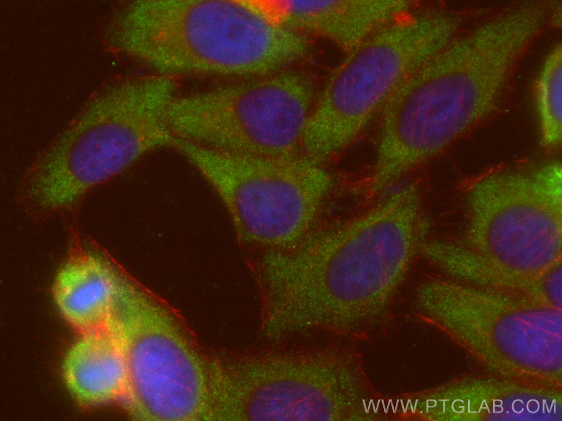
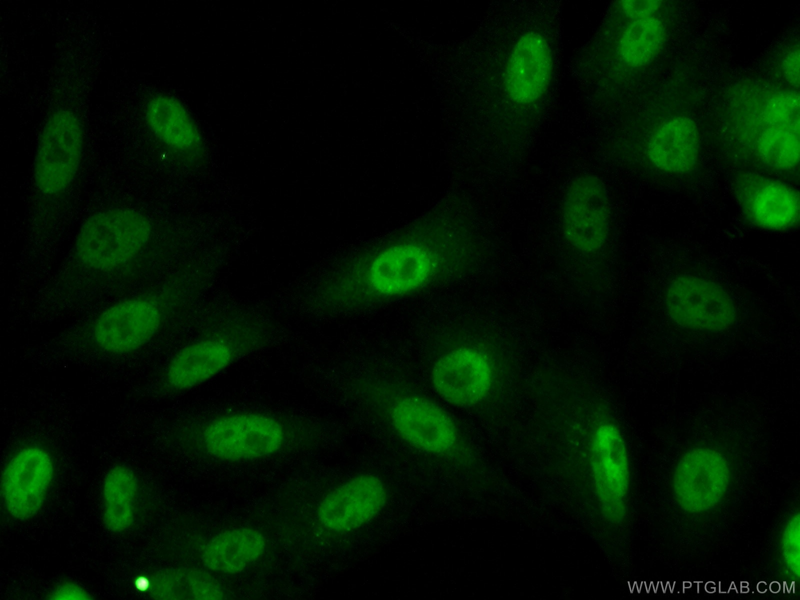
| Résultats positifs en IF/ICC | cellules HepG2, |
Dilution recommandée
| Application | Dilution |
|---|---|
| Western Blot (WB) | WB : 1:500-1:3000 |
| Immunohistochimie (IHC) | IHC : 1:20-1:400 |
| Immunofluorescence (IF)/ICC | IF/ICC : 1:50-1:500 |
| It is recommended that this reagent should be titrated in each testing system to obtain optimal results. | |
| Sample-dependent, check data in validation data gallery | |
Applications publiées
| KD/KO | See 9 publications below |
| WB | See 28 publications below |
| IHC | See 11 publications below |
| IF | See 6 publications below |
| IP | See 2 publications below |
| CoIP | See 5 publications below |
| ChIP | See 1 publications below |
Informations sur le produit
10819-1-AP cible KPNA2 dans les applications de WB, IHC, IF/ICC, IP, CoIP, ChIP, ELISA et montre une réactivité avec des échantillons Humain, rat, souris
| Réactivité | Humain, rat, souris |
| Réactivité citée | Humain, souris |
| Hôte / Isotype | Lapin / IgG |
| Clonalité | Polyclonal |
| Type | Anticorps |
| Immunogène | KPNA2 Protéine recombinante Ag1153 |
| Nom complet | karyopherin alpha 2 (RAG cohort 1, importin alpha 1) |
| Masse moléculaire calculée | 57.8 kDa |
| Poids moléculaire observé | 55-58 kDa |
| Numéro d’acquisition GenBank | BC005978 |
| Symbole du gène | KPNA2 |
| Identification du gène (NCBI) | 3838 |
| Conjugaison | Non conjugué |
| Forme | Liquide |
| Méthode de purification | Purification par affinité contre l'antigène |
| Tampon de stockage | PBS avec azoture de sodium à 0,02 % et glycérol à 50 % pH 7,3 |
| Conditions de stockage | Stocker à -20°C. Stable pendant un an après l'expédition. L'aliquotage n'est pas nécessaire pour le stockage à -20oC Les 20ul contiennent 0,1% de BSA. |
Informations générales
KPNA2 (karyopherin α2) is a nuclear import factor involved in the nucleocytoplasmic transport system. It is functionally involved in the tumor progression and is elevated in multiple cancers. KPNA2 expression may be a useful prognostic biomarker to monitor cancer prognosis.
Protocole
| Product Specific Protocols | |
|---|---|
| WB protocol for KPNA2 antibody 10819-1-AP | Download protocol |
| IHC protocol for KPNA2 antibody 10819-1-AP | Download protocol |
| IF protocol for KPNA2 antibody 10819-1-AP | Download protocol |
| Standard Protocols | |
|---|---|
| Click here to view our Standard Protocols |
Publications
| Species | Application | Title |
|---|---|---|
Cell Res Neddylation of PTEN regulates its nuclear import and promotes tumor development. | ||
J Exp Clin Cancer Res ISG15 and ISGylation modulates cancer stem cell-like characteristics in promoting tumor growth of anaplastic thyroid carcinoma
| ||
Cell Death Differ lncRNA THAP7-AS1, transcriptionally activated by SP1 and post-transcriptionally stabilized by METTL3-mediated m6A modification, exerts oncogenic properties by improving CUL4B entry into the nucleus. | ||
Mol Cell Proteomics Quantitative proteomics reveals regulation of karyopherin subunit alpha-2 (KPNA2) and its potential novel cargo proteins in nonsmall cell lung cancer.
| ||
Virol Sin African Swine Fever Virus MGF360-12L Inhibits Type I Interferon Production by Blocking the Interaction of Importin α and NF-κB Signaling Pathway. | ||
Avis
The reviews below have been submitted by verified Proteintech customers who received an incentive forproviding their feedback.
FH María (Verified Customer) (02-08-2022) | Works for WB (1:1000) and IF in permeabilized cells (1:100). Validated with siRNA
|
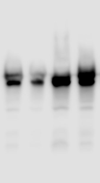
FH Chandra (Verified Customer) (09-16-2019) | Works best at a 1:1000 antibody dilution.
 |
