Anticorps Polyclonal de lapin anti-Complement factor B
Complement factor B Polyclonal Antibody for WB, IHC, ELISA
Hôte / Isotype
Lapin / IgG
Réactivité testée
Humain, rat et plus (1)
Applications
WB, IHC, ELISA, IF
Conjugaison
Non conjugué
N° de cat : 10170-1-AP
Synonymes
Galerie de données de validation
Applications testées
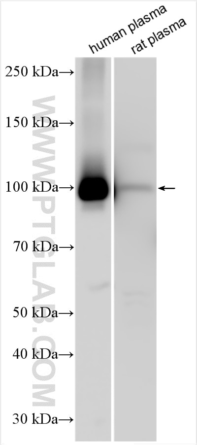
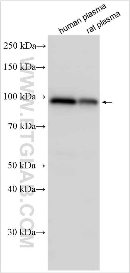
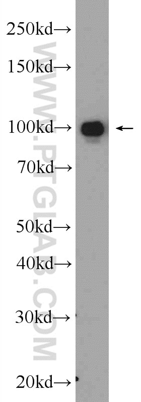
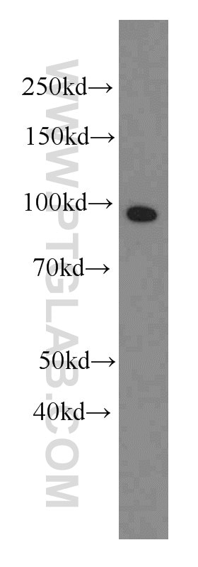
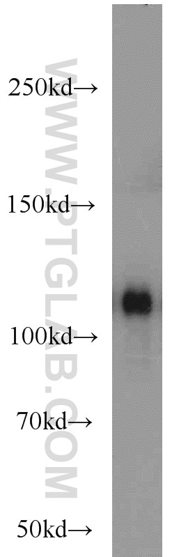
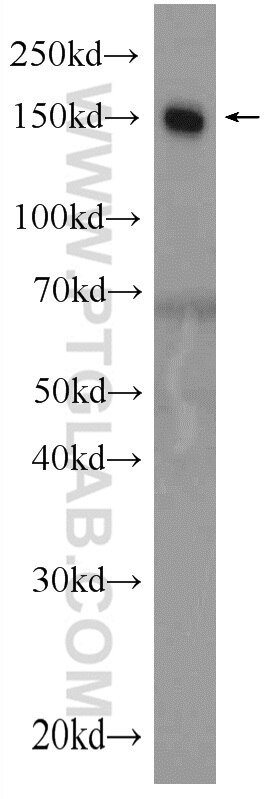
| Résultats positifs en WB | plasma humain, sang humain, Sanguin humain |
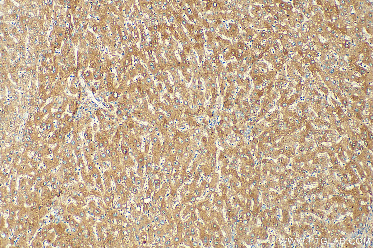
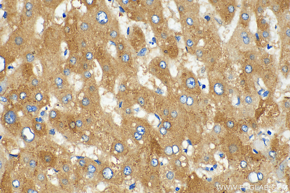
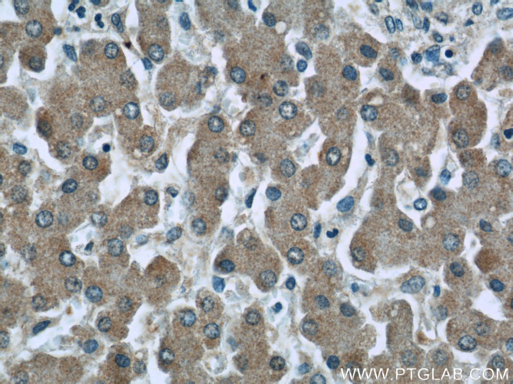
| Résultats positifs en IHC | tissu hépatique humain, il est suggéré de démasquer l'antigène avec un tampon de TE buffer pH 9.0; (*) À défaut, 'le démasquage de l'antigène peut être 'effectué avec un tampon citrate pH 6,0. |
Dilution recommandée
| Application | Dilution |
|---|---|
| Western Blot (WB) | WB : 1:1000-1:8000 |
| Immunohistochimie (IHC) | IHC : 1:200-1:800 |
| It is recommended that this reagent should be titrated in each testing system to obtain optimal results. | |
| Sample-dependent, check data in validation data gallery | |
Applications publiées
| WB | See 8 publications below |
| IHC | See 1 publications below |
| IF | See 2 publications below |
Informations sur le produit
10170-1-AP cible Complement factor B dans les applications de WB, IHC, ELISA, IF et montre une réactivité avec des échantillons Humain, rat
| Réactivité | Humain, rat |
| Réactivité citée | rat, Humain, souris |
| Hôte / Isotype | Lapin / IgG |
| Clonalité | Polyclonal |
| Type | Anticorps |
| Immunogène | Complement factor B Protéine recombinante Ag0226 |
| Nom complet | complement factor B |
| Masse moléculaire calculée | 86 kDa |
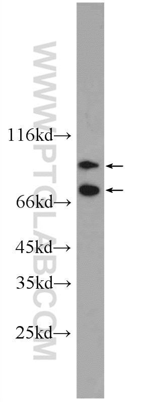
| Poids moléculaire observé | 93-100 kDa, 60-69 kDa |
| Numéro d’acquisition GenBank | BC007990 |
| Symbole du gène | Complement factor B |
| Identification du gène (NCBI) | 629 |
| Conjugaison | Non conjugué |
| Forme | Liquide |
| Méthode de purification | Purification par affinité contre l'antigène |
| Tampon de stockage | PBS avec azoture de sodium à 0,02 % et glycérol à 50 % pH 7,3 |
| Conditions de stockage | Stocker à -20°C. Stable pendant un an après l'expédition. L'aliquotage n'est pas nécessaire pour le stockage à -20oC Les 20ul contiennent 0,1% de BSA. |
Informations générales
Complement factor B (CFB) is a component of the alternative pathway of complement activation. Complement factor B is a 93-100 kDa glycoprotein and is cleaved into Ba (33 kDa) and Bb (60 kDa) by factor D in the presence of C3b (PMID: 11025450). The active subunit Bb is a serine protease which associates with C3b to form the alternative pathway C3 convertase. Bb is involved in the proliferation of preactivated B lymphocytes, while Ba inhibits their proliferation. This antibody raised against 573-761aa of human complement factor B can recognize complement factor B and Bb fragment.
Protocole
| Product Specific Protocols | |
|---|---|
| WB protocol for Complement factor B antibody 10170-1-AP | Download protocol |
| IHC protocol for Complement factor B antibody 10170-1-AP | Download protocol |
| Standard Protocols | |
|---|---|
| Click here to view our Standard Protocols |
Publications
| Species | Application | Title |
|---|---|---|
Nat Commun The classical pathway triggers pathogenic complement activation in membranous nephropathy | ||
J Cell Physiol IL-1/IL-1R signaling induced by all-trans-retinal contributes to complement alternative pathway activation in retinal pigment epithelium. | ||
Cell Oncol (Dordr) Complement factor B regulates cellular senescence and is associated with poor prognosis in pancreatic cancer. | ||
Br J Cancer Identification of GlcNAcylated alpha-1-antichymotrypsin as an early biomarker in human non-small-cell lung cancer by quantitative proteomic analysis with two lectins. | ||
Nephrol Dial Transplant Resveratrol delays polycystic kidney disease progression through attenuation of nuclear factor κB-induced inflammation. | ||
Chem Res Toxicol 4-Hydroxy-7-oxo-5-heptenoic Acid Lactone Is a Potent Inducer of the Complement Pathway in Human Retinal Pigmented Epithelial Cells. |
Avis
The reviews below have been submitted by verified Proteintech customers who received an incentive forproviding their feedback.
FH mark (Verified Customer) (03-30-2020) | worked with colourmetric immunohistochemistry
|
