mouse kidney tissue were subjected to SDS PAGE followed by western blot with 15136-1-AP (NME3 antibody) at dilution of 1:500 incubated at room temperature for 1.5 hours.
mouse kidney tissue were subjected to SDS PAGE followed by western blot with 15136-1-AP (NME3 antibody) at dilution of 1:500 incubated at room temperature for 1.5 hours.
WB analysis using 15136-1-AP
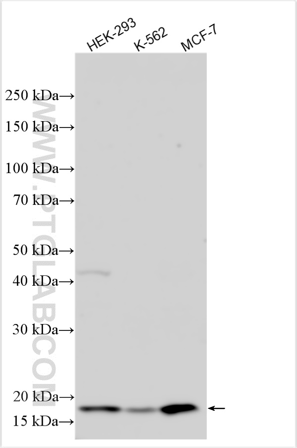
Various lysates were subjected to SDS PAGE followed by western blot with 15136-1-AP (NME3 antibody) at dilution of 1:1000 incubated at room temperature for 1.5 hours.
Various lysates were subjected to SDS PAGE followed by western blot with 15136-1-AP (NME3 antibody) at dilution of 1:1000 incubated at room temperature for 1.5 hours.
WB analysis of MCF-7 using 15136-1-AP
MCF-7 cells were subjected to SDS PAGE followed by western blot with 15136-1-AP (NME3 Antibody) at dilution of 1:300 incubated at room temperature for 1.5 hours.
MCF-7 cells were subjected to SDS PAGE followed by western blot with 15136-1-AP (NME3 Antibody) at dilution of 1:300 incubated at room temperature for 1.5 hours.
IP experiment of HEK-293 using 15136-1-AP
IP result of anti-NME3 (IP:15136-1-AP, 4ug; Detection:15136-1-AP 1:300) with HEK-293 cells lysate 2400 ug.
IP result of anti-NME3 (IP:15136-1-AP, 4ug; Detection:15136-1-AP 1:300) with HEK-293 cells lysate 2400 ug.
IHC staining of human breast cancer using 15136-1-AP
Immunohistochemical analysis of paraffin-embedded human breast cancer tissue slide using 15136-1-AP (NME3 Antibody) at dilution of 1:200 (under 10x lens). Heat mediated antigen retrieval with Tris-EDTA buffer (pH 9.0).
Immunohistochemical analysis of paraffin-embedded human breast cancer tissue slide using 15136-1-AP (NME3 Antibody) at dilution of 1:200 (under 10x lens). Heat mediated antigen retrieval with Tris-EDTA buffer (pH 9.0).
IHC staining of human breast cancer using 15136-1-AP
Immunohistochemical analysis of paraffin-embedded human breast cancer tissue slide using 15136-1-AP (NME3 Antibody) at dilution of 1:200 (under 40x lens). Heat mediated antigen retrieval with Tris-EDTA buffer (pH 9.0).
Immunohistochemical analysis of paraffin-embedded human breast cancer tissue slide using 15136-1-AP (NME3 Antibody) at dilution of 1:200 (under 40x lens). Heat mediated antigen retrieval with Tris-EDTA buffer (pH 9.0).
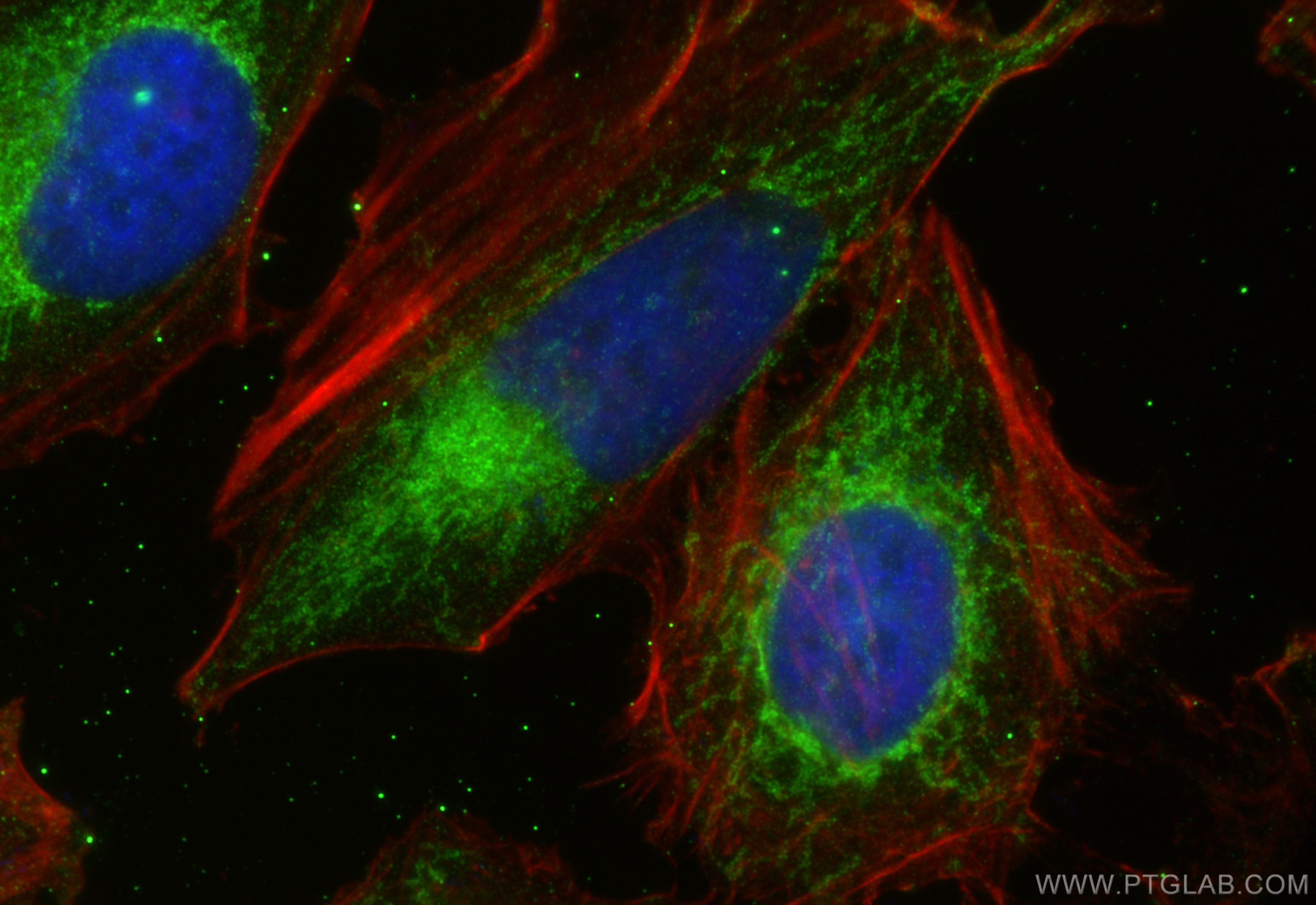
IF Staining of HeLa using 15136-1-AP
Immunofluorescent analysis of (4% PFA) fixed HeLa cells using NME3 antibody (15136-1-AP) at dilution of 1:400 and Multi-rAb CoraLite ® Plus 488-Goat Anti-Rabbit Recombinant Secondary Antibody (H+L) (RGAR002), CL594-Phalloidin (red).
Immunofluorescent analysis of (4% PFA) fixed HeLa cells using NME3 antibody (15136-1-AP) at dilution of 1:400 and Multi-rAb CoraLite ® Plus 488-Goat Anti-Rabbit Recombinant Secondary Antibody (H+L) (RGAR002), CL594-Phalloidin (red).
IF Staining of MCF-7 using 15136-1-AP
Immunofluorescent analysis of (-20℃ Ethanol) fixed MCF-7 cells using 15136-1-AP (NME3 antibody) at dilution of 1:50 and Alexa Fluor 488-conjugated AffiniPure Goat Anti-Rabbit IgG(H+L).
Immunofluorescent analysis of (-20℃ Ethanol) fixed MCF-7 cells using 15136-1-AP (NME3 antibody) at dilution of 1:50 and Alexa Fluor 488-conjugated AffiniPure Goat Anti-Rabbit IgG(H+L).
The Proteintech guarantee covers Proteintech antibodies in any species and any application, including those not listed on the datasheet. If the antibody doesn’t perform, you can receive a hassle-free refund or credit note.
human breast cancer tissue Note: suggested antigen retrieval with TE buffer pH 9.0; (*) Alternatively, antigen retrieval may be performed with citrate buffer pH 6.0
Positive IF/ICC detected in
HeLa cells, MCF-7 cells
Recommended dilution
Application
Dilution
Western Blot (WB)
WB : 1:500-1:2000
Immunoprecipitation (IP)
IP : 0.5-4.0 ug for 1.0-3.0 mg of total protein lysate
Immunohistochemistry (IHC)
IHC : 1:50-1:500
Immunofluorescence (IF)/ICC
IF/ICC : 1:200-1:800
It is recommended that this reagent should be titrated in each testing system to obtain optimal results.
Sample-dependent, Check data in validation data gallery.
PBS with 0.02% sodium azide and 50% glycerol , pH 7.3
Storage Conditions
Store at -20°C. Stable for one year after shipment. Aliquoting is unnecessary for -20oC storage. 20ul sizes contain 0.1% BSA.
Background Information
NME3 (NME/NM23 Nucleoside Diphosphate Kinase 3), also named as NM23-H3; DR-NM23, is a member of the nucleoside diphosphate kinase (NDPK) family that binds to the mitochondrial outer membrane to stimulate mitochondrial fusion. The depletion of NME3 will cause dysfunction in mitochondrial dynamics. NME3 has been found to facilitate DNA-repair mechanisms binds to several NPHPs, including NEK8, CEP164, and ankyrin repeat and sterile α motif domain-containing 6 (ANKS6).
mouse kidney tissue were subjected to SDS PAGE followed by western blot with 15136-1-AP (NME3 antibody) at dilution of 1:500 incubated at room temperature for 1.5 hours.
WB analysis using 15136-1-AP
Various lysates were subjected to SDS PAGE followed by western blot with 15136-1-AP (NME3 antibody) at dilution of 1:1000 incubated at room temperature for 1.5 hours.
WB analysis of MCF-7 using 15136-1-AP
MCF-7 cells were subjected to SDS PAGE followed by western blot with 15136-1-AP (NME3 Antibody) at dilution of 1:300 incubated at room temperature for 1.5 hours.
IHC Figures
IHC staining of human breast cancer using 15136-1-AP
Immunohistochemical analysis of paraffin-embedded human breast cancer tissue slide using 15136-1-AP (NME3 Antibody) at dilution of 1:200 (under 10x lens). Heat mediated antigen retrieval with Tris-EDTA buffer (pH 9.0).
IHC staining of human breast cancer using 15136-1-AP
Immunohistochemical analysis of paraffin-embedded human breast cancer tissue slide using 15136-1-AP (NME3 Antibody) at dilution of 1:200 (under 40x lens). Heat mediated antigen retrieval with Tris-EDTA buffer (pH 9.0).
IP Figures
IP experiment of HEK-293 using 15136-1-AP
IP result of anti-NME3 (IP:15136-1-AP, 4ug; Detection:15136-1-AP 1:300) with HEK-293 cells lysate 2400 ug.
IF/ICC Figures
IF Staining of HeLa using 15136-1-AP
Immunofluorescent analysis of (4% PFA) fixed HeLa cells using NME3 antibody (15136-1-AP) at dilution of 1:400 and Multi-rAb CoraLite ® Plus 488-Goat Anti-Rabbit Recombinant Secondary Antibody (H+L) (RGAR002), CL594-Phalloidin (red).
IF Staining of MCF-7 using 15136-1-AP
Immunofluorescent analysis of (-20℃ Ethanol) fixed MCF-7 cells using 15136-1-AP (NME3 antibody) at dilution of 1:50 and Alexa Fluor 488-conjugated AffiniPure Goat Anti-Rabbit IgG(H+L).
The species listed in Tested Reactivity are in-house verified and applicable species. For unlisted species, please refer to the homology analysis of the immunogen sequence and related species. For rabbit polyclonal antibodies, homology >70% is recommended. For mouse monoclonal antibodies and rabbit recombinant antibodies, homology >90% is recommended. Generally, the higher the homology, the greater the applicability. However, there will be certain differences in protein expression in different species, tissues or cells. Therefore, the homology analysis results are for reference only and do not serve as a guarantee.
At Proteintech, we pride ourselves on our antibody quality, customer service and transparency. As such, we are comparing our antibodies with other vendors, enabling easy identification and comparisons of key data to help you choose the suitable antibody for your needs.
We have selected the top cited antibodies from these vendors for you to compare.
Proteintech
KD/KO VALIDATED
NME3 Polyclonal antibody
Catalog Number
15136-1-AP
Citations
1
Dilutions
WB : 1:500-1:2000 IP : 0.5-4.0 ug for IP and 0.5-4.0 ug for 1.0-3.0 mg of total protein lysate for WB IHC : 1:50-1:500 IF/ICC : 1:200-1:800
Applications
WB, IHC, IF/ICC, IP, ELISA
Reactivity
human, mouse, rat, zebrafish
Product Guarantee
Covers any species including not listed on datasheet
Covers any applications including not listed on datasheet
 at dilution of 1:500 incubated at room temperature for 1.5 hours.")
 at dilution of 1:1000 incubated at room temperature for 1.5 hours.")
 at dilution of 1:300 incubated at room temperature for 1.5 hours.")
 with HEK-293 cells lysate 2400 ug.")
 at dilution of 1:200 (under 10x lens). Heat mediated antigen retrieval with Tris-EDTA buffer (pH 9.0).")
 at dilution of 1:200 (under 40x lens). Heat mediated antigen retrieval with Tris-EDTA buffer (pH 9.0).")
 fixed HeLa cells using NME3 antibody (15136-1-AP) at dilution of 1:400 and Multi-rAb CoraLite ® Plus 488-Goat Anti-Rabbit Recombinant Secondary Antibody (H+L) (RGAR002), CL594-Phalloidin (red).")
 fixed MCF-7 cells using 15136-1-AP (NME3 antibody) at dilution of 1:50 and Alexa Fluor 488-conjugated AffiniPure Goat Anti-Rabbit IgG(H+L).")