WB result of APEX1 antibody (10323-1-AP; 1:4000; incubated at room temperature for 1.5 hours) with sh-Control and sh-APEX1 transfected HeLa cells.
WB analysis of HepG2 using 10323-1-AP
HepG2 cells were subjected to SDS PAGE followed by western blot with 10323-1-AP (APEX1 antibody) at dilution of 1:500 incubated at room temperature for 1.5 hours.
HepG2 cells were subjected to SDS PAGE followed by western blot with 10323-1-AP (APEX1 antibody) at dilution of 1:500 incubated at room temperature for 1.5 hours.
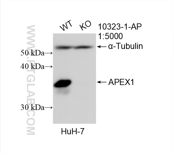
WB analysis of HuH-7 using 10323-1-AP
WB result of APEX1 antibody (10323-1-AP; 1:5000; room temperature for 1.5 hours) with wild-type and APEX1 knockout HuH-7 cells.
WB result of APEX1 antibody (10323-1-AP; 1:5000; room temperature for 1.5 hours) with wild-type and APEX1 knockout HuH-7 cells.
WB analysis of mouse liver using 10323-1-AP
mouse liver tissue were subjected to SDS PAGE followed by western blot with 10323-1-AP (APEX1 Antibody) at dilution of 1:600 incubated at room temperature for 1.5 hours.
mouse liver tissue were subjected to SDS PAGE followed by western blot with 10323-1-AP (APEX1 Antibody) at dilution of 1:600 incubated at room temperature for 1.5 hours.
WB analysis of HepG2 using 10323-1-AP
HepG2 cells were subjected to SDS PAGE followed by western blot with 10323-1-AP (APEX1 antibody) at dilution of 1:500 incubated at room temperature for 1.5 hours.
HepG2 cells were subjected to SDS PAGE followed by western blot with 10323-1-AP (APEX1 antibody) at dilution of 1:500 incubated at room temperature for 1.5 hours.
WB analysis of Raji using 10323-1-AP
Raji cells were subjected to SDS PAGE followed by western blot with 10323-1-AP (APEX1 antibody) at dilution of 1:500 incubated at room temperature for 1.5 hours.
Raji cells were subjected to SDS PAGE followed by western blot with 10323-1-AP (APEX1 antibody) at dilution of 1:500 incubated at room temperature for 1.5 hours.
WB analysis of HEK-293 using 10323-1-AP
HEK-293 cells were subjected to SDS PAGE followed by western blot with 10323-1-AP (APEX1 antibody) at dilution of 1:500 incubated at room temperature for 1.5 hours.
HEK-293 cells were subjected to SDS PAGE followed by western blot with 10323-1-AP (APEX1 antibody) at dilution of 1:500 incubated at room temperature for 1.5 hours.
WB analysis of K-562 using 10323-1-AP
K-562 cells were subjected to SDS PAGE followed by western blot with 10323-1-AP (APEX1 antibody) at dilution of 1:500 incubated at room temperature for 1.5 hours.
K-562 cells were subjected to SDS PAGE followed by western blot with 10323-1-AP (APEX1 antibody) at dilution of 1:500 incubated at room temperature for 1.5 hours.
IP experiment of HepG2 using 10323-1-AP
IP result of anti-APEX1 (IP:10323-1-AP, 4ug; Detection:10323-1-AP 1:500) with HepG2 cells lysate 2200 ug.
Immunohistochemical analysis of paraffin-embedded human cervical cancer using 10323-1-AP (APEX1 antibody) at dilution of 1:50 (under 10x lens).
IF Staining of HepG2 using 10323-1-AP
Immunofluorescent analysis of HepG2 cells using 10323-1-AP (APEX1 antibody) at dilution of 1:50 and Alexa Fluor 488-conjugated AffiniPure Goat Anti-Rabbit IgG(H+L).
Immunofluorescent analysis of HepG2 cells using 10323-1-AP (APEX1 antibody) at dilution of 1:50 and Alexa Fluor 488-conjugated AffiniPure Goat Anti-Rabbit IgG(H+L).
The Proteintech guarantee covers Proteintech antibodies in any species and any application, including those not listed on the datasheet. If the antibody doesn’t perform, you can receive a hassle-free refund or credit note.
human cervical cancer tissue Note: suggested antigen retrieval with TE buffer pH 9.0; (*) Alternatively, antigen retrieval may be performed with citrate buffer pH 6.0
Positive IF/ICC detected in
HepG2 cells
Recommended dilution
Application
Dilution
Western Blot (WB)
WB : 1:500-1:1000
Immunoprecipitation (IP)
IP : 0.5-4.0 ug for 1.0-3.0 mg of total protein lysate
Immunohistochemistry (IHC)
IHC : 1:20-1:200
Immunofluorescence (IF)/ICC
IF/ICC : 1:20-1:200
It is recommended that this reagent should be titrated in each testing system to obtain optimal results.
Sample-dependent, Check data in validation data gallery.
Product Information
10323-1-AP targets APEX1 in WB, IHC, IF/ICC, IP, ELISA applications and shows reactivity with human, mouse, rat samples.
PBS with 0.02% sodium azide and 50% glycerol pH 7.3.
Storage Conditions
Store at -20°C. Stable for one year after shipment. Aliquoting is unnecessary for -20oC storage. 20ul sizes contain 0.1% BSA.
Background Information
APEX1, also named as APE, APE1, HAP1 and REF-1, belongs to the DNA repair enzymes AP/ExoA family. It is a multifunctional protein that plays a central role in the cellular response to oxidative stress. The two major activities of APEX1 are in DNA repair and redox regulation of transcriptional factors. APEX nuclease is a DNA repair enzyme having apurinic/apyrimidinic (AP) endonuclease, 3-prime,5-prime-exonuclease, DNA 3-prime repair diesterase, and DNA 3-prime-phosphatase activities. On the other hand, APEX1 also exerts reversible nuclear redox activity to regulate DNA binding affinity and transcriptional activity of transcriptional factors by controlling the redox status of their DNA-binding domain, such as the FOS/JUN AP-1 complex after exposure to IR. APEX1 is involved in calcium-dependent down-regulation of parathyroid hormone (PTH) expression by binding to negative calcium response elements (nCaREs). When acetylated at Lys-6 and Lys-7, APEX1 stimulates the YBX1-mediated MDR1 promoter activity, leading to drug resistance. It also acts as an endoribonuclease involved in the control of single-stranded RNA metabolism. It plays a role in regulating MYC mRNA turnover by preferentially cleaving in between UA and CA dinucleotides of the MYC coding region determinant (CRD). In association with NMD1, APEX1 plays a role in the rRNA quality control process during cell cycle progression. 10323-1-AP is a rabbit polyclonal antibody raised against a fusion protein corresponding to an internal region of human APEX1.
WB result of APEX1 antibody (10323-1-AP; 1:4000; incubated at room temperature for 1.5 hours) with sh-Control and sh-APEX1 transfected HeLa cells.
WB analysis of HepG2 using 10323-1-AP
HepG2 cells were subjected to SDS PAGE followed by western blot with 10323-1-AP (APEX1 antibody) at dilution of 1:500 incubated at room temperature for 1.5 hours.
WB analysis of HuH-7 using 10323-1-AP
WB result of APEX1 antibody (10323-1-AP; 1:5000; room temperature for 1.5 hours) with wild-type and APEX1 knockout HuH-7 cells.
WB analysis of mouse liver using 10323-1-AP
mouse liver tissue were subjected to SDS PAGE followed by western blot with 10323-1-AP (APEX1 Antibody) at dilution of 1:600 incubated at room temperature for 1.5 hours.
WB analysis of HepG2 using 10323-1-AP
HepG2 cells were subjected to SDS PAGE followed by western blot with 10323-1-AP (APEX1 antibody) at dilution of 1:500 incubated at room temperature for 1.5 hours.
WB analysis of Raji using 10323-1-AP
Raji cells were subjected to SDS PAGE followed by western blot with 10323-1-AP (APEX1 antibody) at dilution of 1:500 incubated at room temperature for 1.5 hours.
WB analysis of Raji using 10323-1-AP
Raji cells were subjected to SDS PAGE followed by western blot with 10323-1-AP (APEX1 antibody) at dilution of 1:500 incubated at room temperature for 1.5 hours.
WB analysis of HEK-293 using 10323-1-AP
HEK-293 cells were subjected to SDS PAGE followed by western blot with 10323-1-AP (APEX1 antibody) at dilution of 1:500 incubated at room temperature for 1.5 hours.
WB analysis of HEK-293 using 10323-1-AP
HEK-293 cells were subjected to SDS PAGE followed by western blot with 10323-1-AP (APEX1 antibody) at dilution of 1:500 incubated at room temperature for 1.5 hours.
WB analysis of K-562 using 10323-1-AP
K-562 cells were subjected to SDS PAGE followed by western blot with 10323-1-AP (APEX1 antibody) at dilution of 1:500 incubated at room temperature for 1.5 hours.
WB analysis of K-562 using 10323-1-AP
K-562 cells were subjected to SDS PAGE followed by western blot with 10323-1-AP (APEX1 antibody) at dilution of 1:500 incubated at room temperature for 1.5 hours.
IHC Figures
IHC staining of human cervical cancer using 10323-1-AP
Immunohistochemical analysis of paraffin-embedded human cervical cancer using 10323-1-AP (APEX1 antibody) at dilution of 1:50 (under 10x lens).
IP Figures
IP experiment of HepG2 using 10323-1-AP
IP result of anti-APEX1 (IP:10323-1-AP, 4ug; Detection:10323-1-AP 1:500) with HepG2 cells lysate 2200 ug.
IF/ICC Figures
IF Staining of HepG2 using 10323-1-AP
Immunofluorescent analysis of HepG2 cells using 10323-1-AP (APEX1 antibody) at dilution of 1:50 and Alexa Fluor 488-conjugated AffiniPure Goat Anti-Rabbit IgG(H+L).
The species listed in Tested Reactivity are in-house verified and applicable species. For unlisted species, please refer to the homology analysis of the immunogen sequence and related species. For rabbit polyclonal antibodies, homology >70% is recommended. For mouse monoclonal antibodies and rabbit recombinant antibodies, homology >90% is recommended. Generally, the higher the homology, the greater the applicability. However, there will be certain differences in protein expression in different species, tissues or cells. Therefore, the homology analysis results are for reference only and do not serve as a guarantee.
At Proteintech, we pride ourselves on our antibody quality, customer service and transparency. As such, we are comparing our antibodies with other vendors, enabling easy identification and comparisons of key data to help you choose the suitable antibody for your needs.
We have selected the top cited antibodies from these vendors for you to compare.
Proteintech
KD/KO VALIDATED
APEX1 Polyclonal antibody
Catalog Number
10323-1-AP
Citations
-
Dilutions
WB : 1:500-1:1000 IP : 0.5-4.0 ug for IP and 0.5-4.0 ug for 1.0-3.0 mg of total protein lysate for WB IHC : 1:20-1:200 IF/ICC : 1:20-1:200
Applications
WB, IHC, IF/ICC, IP, ELISA
Reactivity
human, mouse, rat
Product Guarantee
Covers any species including not listed on datasheet
Covers any applications including not listed on datasheet
 with sh-Control and sh-APEX1 transfected HeLa cells.")
 at dilution of 1:500 incubated at room temperature for 1.5 hours.")
 with wild-type and APEX1 knockout HuH-7 cells.")
 at dilution of 1:600 incubated at room temperature for 1.5 hours.")
 at dilution of 1:500 incubated at room temperature for 1.5 hours.")
 at dilution of 1:500 incubated at room temperature for 1.5 hours.")
 at dilution of 1:500 incubated at room temperature for 1.5 hours.")
 at dilution of 1:500 incubated at room temperature for 1.5 hours.")
 at dilution of 1:500 incubated at room temperature for 1.5 hours.")
 at dilution of 1:500 incubated at room temperature for 1.5 hours.")
 at dilution of 1:500 incubated at room temperature for 1.5 hours.")
 with HepG2 cells lysate 2200 ug.")
 at dilution of 1:50 (under 10x lens).")
 at dilution of 1:50 and Alexa Fluor 488-conjugated AffiniPure Goat Anti-Rabbit IgG(H+L).")