
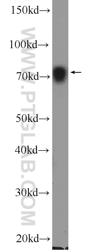
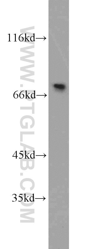
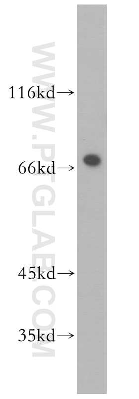
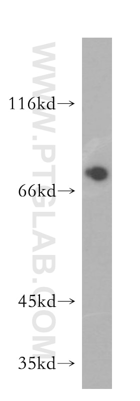

Tested Applications
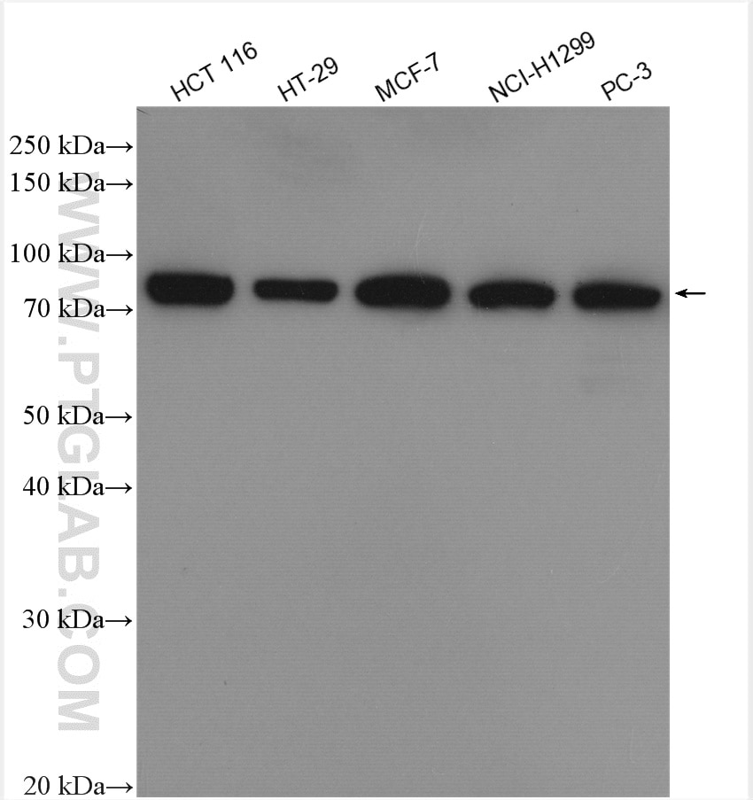
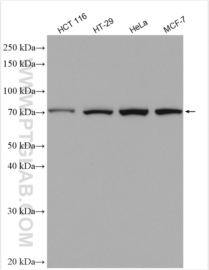
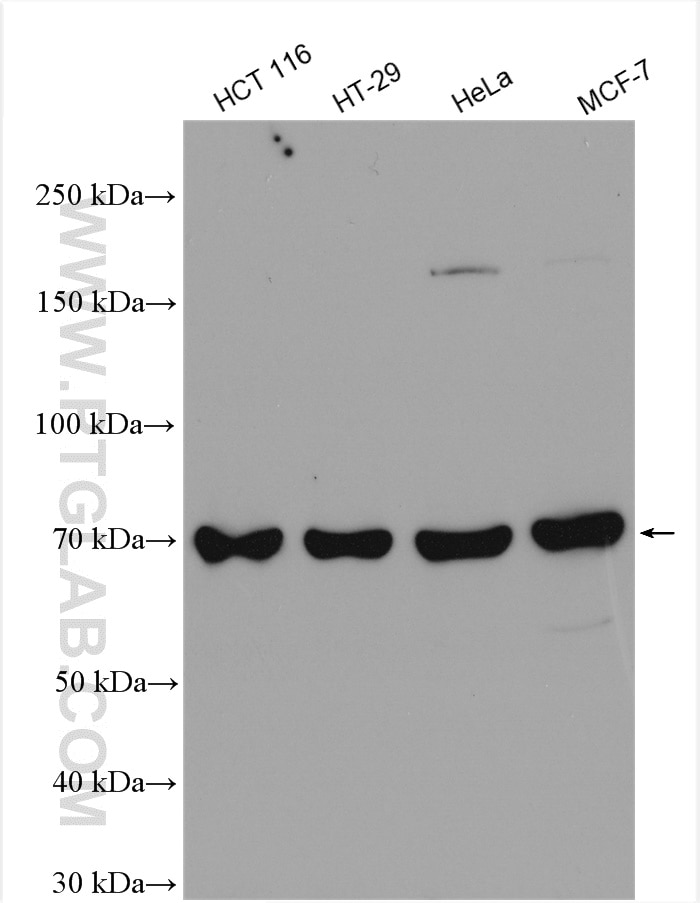
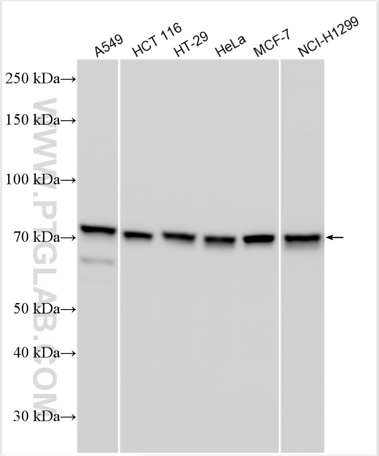
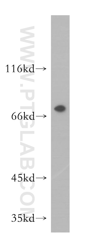
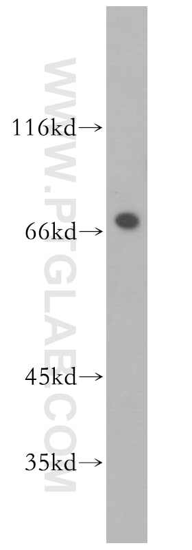
| Positive WB detected in | HCT 116 cells, mouse testis tissue, PC-3 cells, HeLa cells, A549 cells, rat testis tissue, HT-29 cells, MCF-7 cells, NCI-H1299 cells, HCT-116 cells |
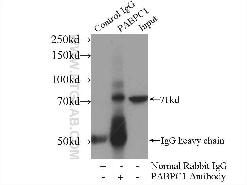
| Positive IP detected in | mouse testis tissue |
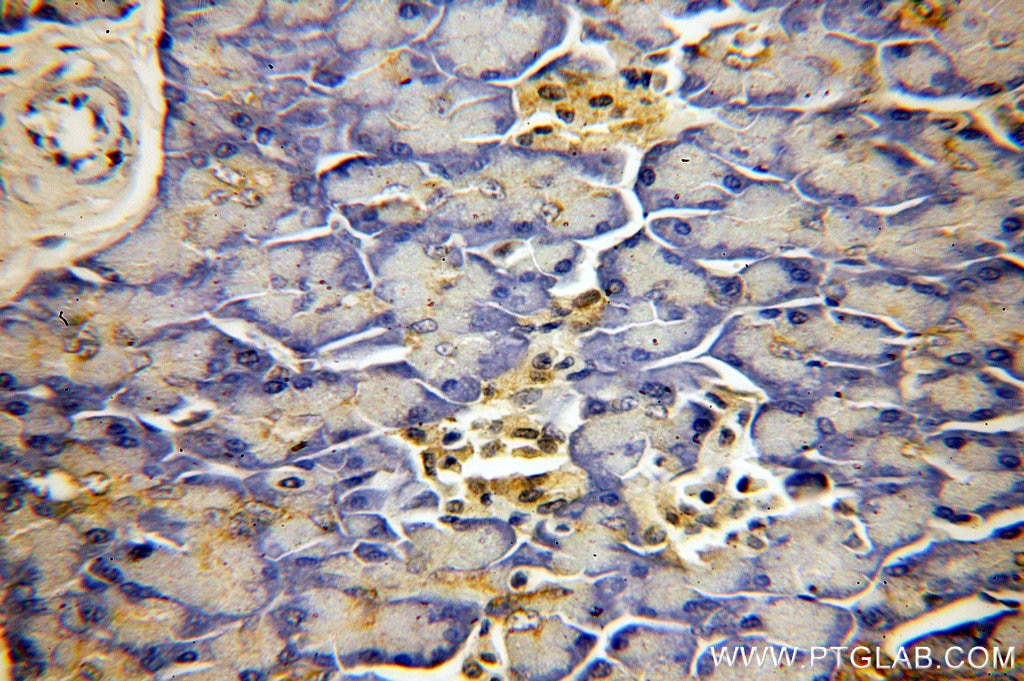
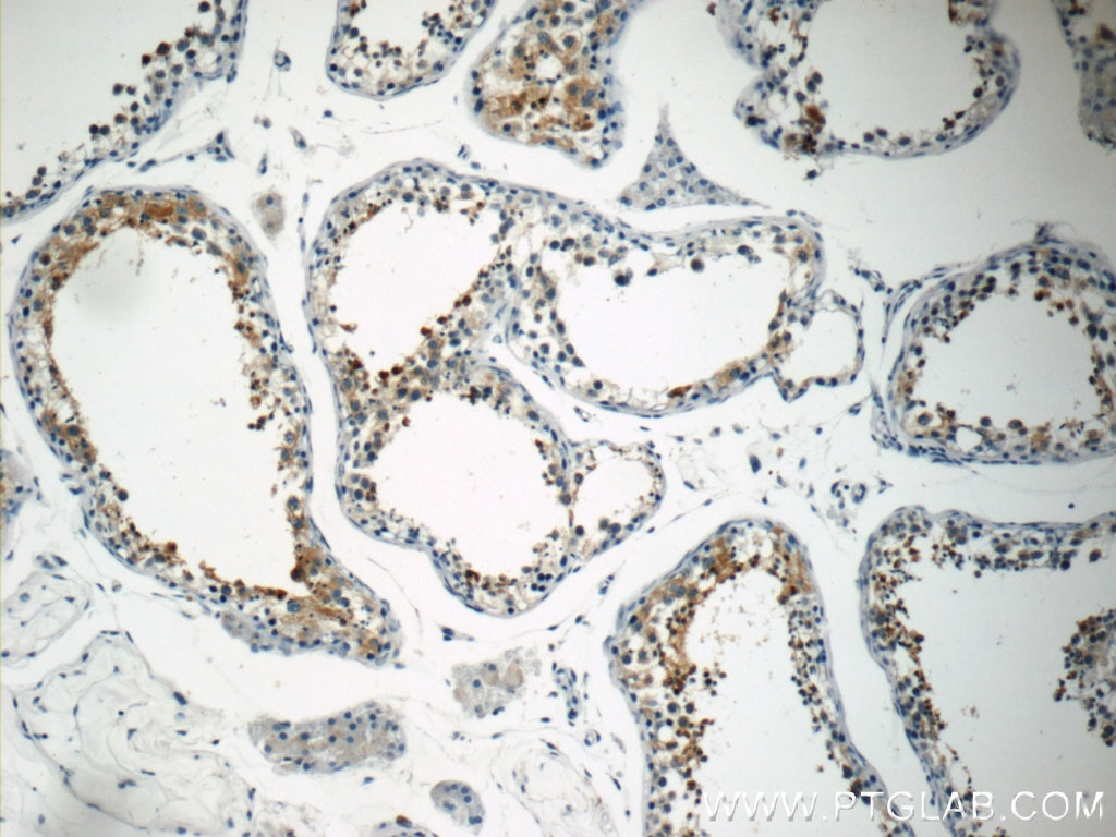
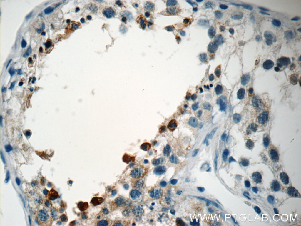
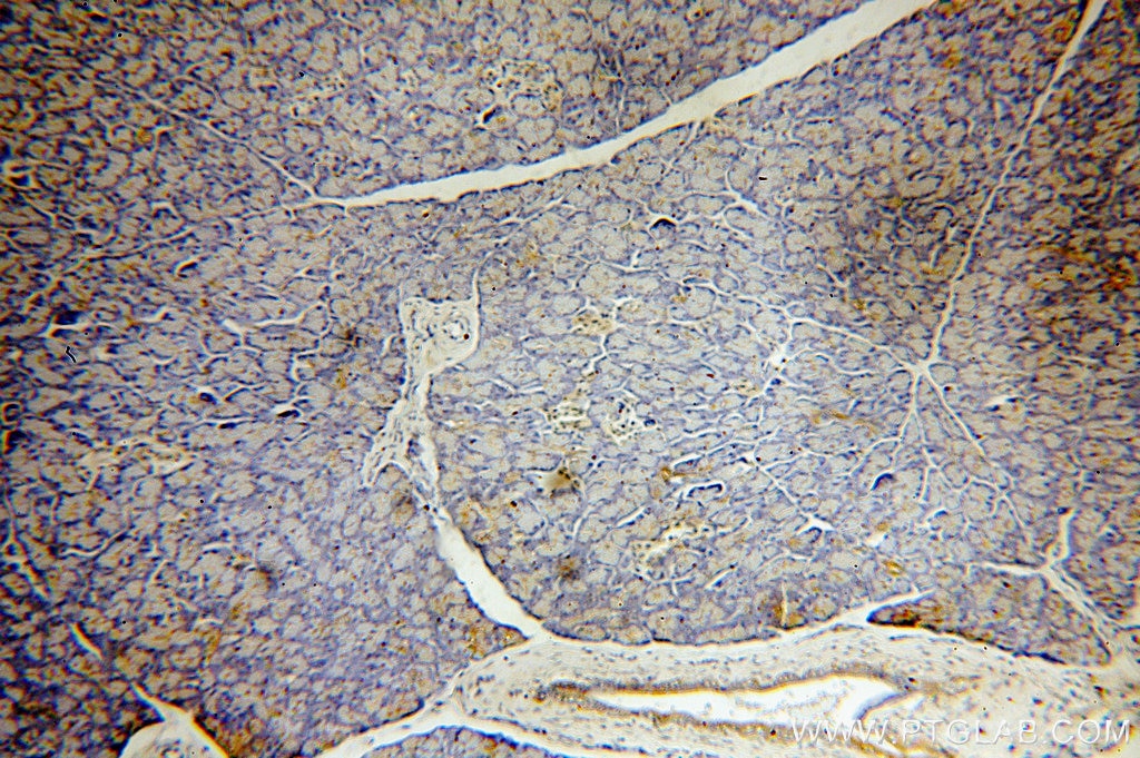
| Positive IHC detected in | human pancreas tissue, human testis tissue Note: suggested antigen retrieval with TE buffer pH 9.0; (*) Alternatively, antigen retrieval may be performed with citrate buffer pH 6.0 |
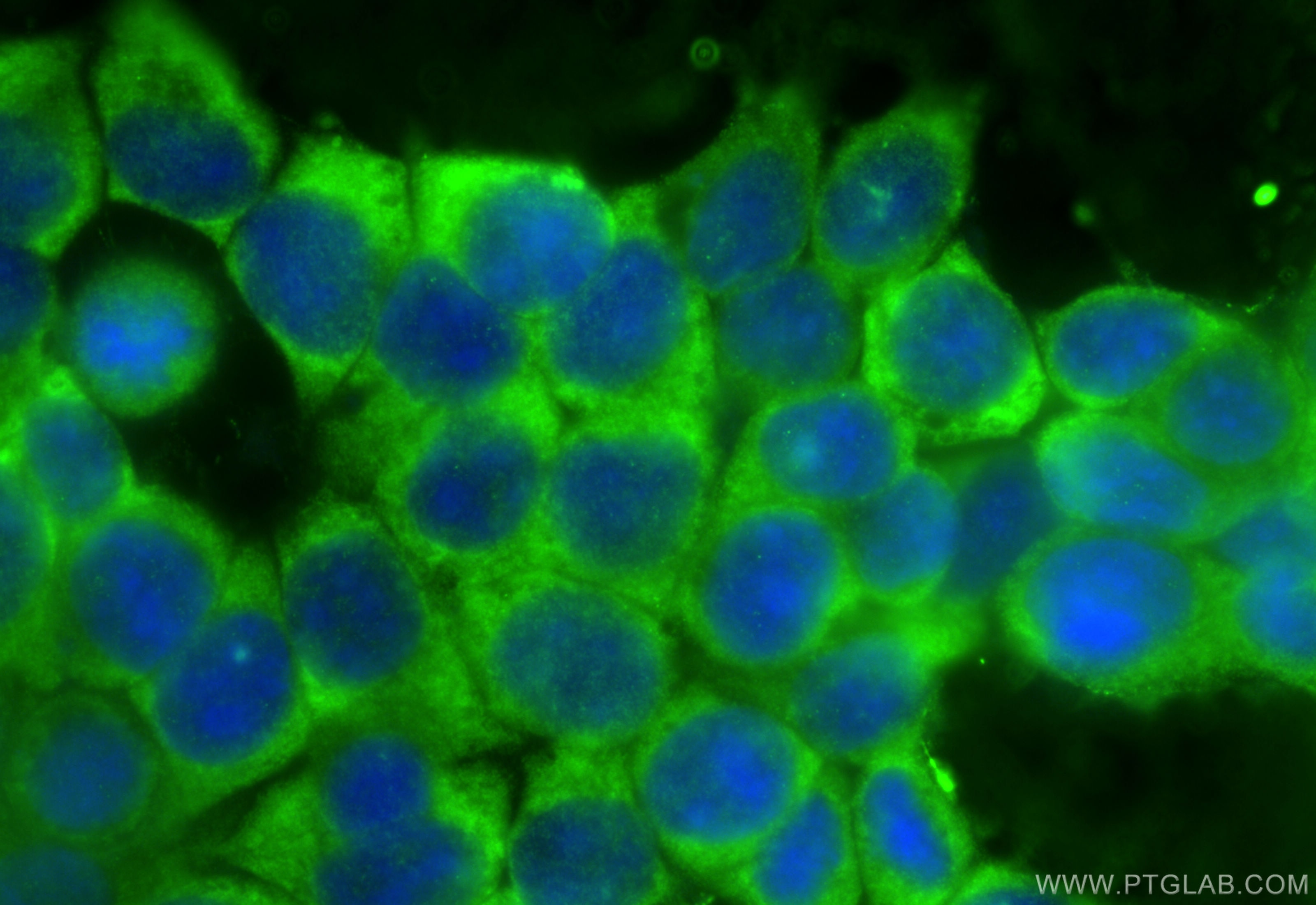
| Positive IF/ICC detected in | MCF-7 cells |
Recommended dilution
| Application | Dilution |
|---|---|
| Western Blot (WB) | WB : 1:1000-1:5000 |
| Immunoprecipitation (IP) | IP : 0.5-4.0 ug for 1.0-3.0 mg of total protein lysate |
| Immunohistochemistry (IHC) | IHC : 1:20-1:200 |
| Immunofluorescence (IF)/ICC | IF/ICC : 1:200-1:800 |
| It is recommended that this reagent should be titrated in each testing system to obtain optimal results. | |
| Sample-dependent, Check data in validation data gallery. | |
Product Information
10970-1-AP targets PABPC1,PABP in WB, IHC, IF/ICC, IP, CoIP, RIP, ELISA applications and shows reactivity with human, mouse, rat samples.
| Tested Reactivity | human, mouse, rat |
| Cited Reactivity | human, mouse, rat, pig |
| Host / Isotype | Rabbit / IgG |
| Class | Polyclonal |
| Type | Antibody |
| Immunogen |
CatNo: Ag1422 Product name: Recombinant human PABPC1,PABP protein Source: e coli.-derived, PGEX-4T Tag: GST Domain: 287-636 aa of BC015958 Sequence: RITRYQGVNLYVKNLDDGIDDERLRKEFSPFGTITSAKVMMEGGRSKGFGFVCFSSPEEATKAVTEMNGRIVATKPLYVALAQRKEERQAHLTNQYMQRMASVRAVPNPVINPYQPAPPSGYFMAAIPQTQNRAAYYPPSQIAQLRPSPRWTAQGARPHPFQNMPGAIRPAAPRPPFSTMRPASSQVPRVMSTQRVANTSTQTMGPRPAAAAAAATPAVRTVPQYKYAAGVRNPQQHLNAQPQVTMQQPAVHVQGQEPLTASMLASAPPQEQKQMLGERLFPLIQAMHPTLAGKITGMLLEIDNSELLHMLESPESLRSKVDEAVAVLQAHQAKEAAQKAVNSATGVPTV Predict reactive species |
| Full Name | poly(A) binding protein, cytoplasmic 1 |
| Calculated Molecular Weight | 71 kDa |
| Observed Molecular Weight | 71 kDa |
| GenBank Accession Number | BC015958 |
| Gene Symbol | PABPC1 |
| Gene ID (NCBI) | 26986 |
| RRID | AB_10596918 |
| Conjugate | Unconjugated |
| Form | Liquid |
| Purification Method | Antigen affinity purification |
| UNIPROT ID | P11940 |
| Storage Buffer | PBS with 0.02% sodium azide and 50% glycerol, pH 7.3. |
| Storage Conditions | Store at -20°C. Stable for one year after shipment. Aliquoting is unnecessary for -20oC storage. 20ul sizes contain 0.1% BSA. |
Background Information
The poly(A)-binding protein (PABP), which is found complexed to the 3-prime poly(A) tail of eukaryotic mRNA, is required for poly(A) shortening and translation initiation [PMID: 21989405]. Polyadenylate-binding protein 1 (PABPC1) is a cytoplasmic-nuclear shuttling protein important for protein translation initiation, and both RNA processing and stability. In the cytoplasm, PABPC1 binds to the 3' poly(A) tail of eukaryotic mRNAs through its RNA-recognition motifs (RRM) and interacts with the N-terminus of eIF4G, part of the eIF4F complex associated with the 5' cap structure [PMID:20009508, 17381337].
Protocols
| Product Specific Protocols | |
|---|---|
| IF protocol for PABPC1,PABP antibody 10970-1-AP | Download protocol |
| IHC protocol for PABPC1,PABP antibody 10970-1-AP | Download protocol |
| IP protocol for PABPC1,PABP antibody 10970-1-AP | Download protocol |
| WB protocol for PABPC1,PABP antibody 10970-1-AP | Download protocol |
| Standard Protocols | |
|---|---|
| Click here to view our Standard Protocols |
Publications
What published studies show
Anti-PABPC1 (catalog no. 10970-1-AP) has 64 citations across journals including Nature, Nature Communications, Nature Cell Biology, Cancer Research, Molecular Cell, Neuron, PNAS, Journal of Experimental Medicine, Oncogene, and Advanced Science. Research fields span post-transcriptional RNA regulation (m6A modification, lncRNA/circRNA), cancer biology (lung, breast, bladder, gastric, pancreatic, liver), stress granule biology, translation control, virology, neurodegeneration, immunology, and stem cell biology.
| Species | Application | Title |
|---|---|---|
Nat Cell Biol The atypical E3 ligase HOIL-1 safeguards the ribosome during cellular stress. | ||
Nat Cell Biol Cyclophilin A supports translation of intrinsically disordered proteins and affects haematopoietic stem cell ageing | ||
Cancer Res CRTC2 Forms Co-condensates with YTHDF2 that Enhance Translational Efficiency of m6A-Modified mRNAs to Drive Hepatocarcinogenesis and Lenvatinib Resistance | ||
Nat Commun Engineering bi-directional chemically-modulated synthetic condensates for cellular control | ||
Nat Commun Ribosome biogenesis as a potential therapeutic target in KRAS mutant colorectal cancer. |
Reviews
What customers say
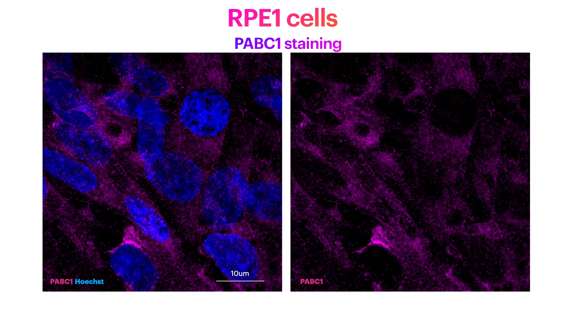
Users report consistent performance across multiple applications. For Western Blot (mouse heart tissue, 1:1000), the antibody "worked very well" with a 5-star rating. In Immunofluorescence (RPE-1 cells, 1:200), it produced clear cytoplasmic PABPC1 staining. For IHC (spinal cord/cortical slice, 1:100), reviewers noted minimal endogenous background with clear dispersed nuclear and cytoplasmic signal. Overall ratings average 4.3/5, with users highlighting low background and reliable specificity across cell lines and tissue types.
The reviews below have been submitted by verified Proteintech customers who received an incentive for providing their feedback.
Elisa (Verified Customer) (01-13-2023) | PABPC1 (Poly(A) Binding Protein Cytoplasmic 1) staining (in magenta) and Hoechst (nuclear staining) in blue. PABPC1 ab shows a clear citoplasma staining. Method: RPE1 cells were fixed in cold methanol for 10' at -20C. Cells were then rehydrated with PBS for 5'. Membrane permeabilization was then performed with 0.1% Triton + 0.1% Tween +0.01%SDS in PBS for 10'. Cells were finally incubated with blocking buffer (5% BSA+ 0.1% Tween in PBS) for 30' at RT. Primary antibody was diluted in blocking buffer 1:200 and incubated for 1h at room temperature. Alexa-488-Anti-rabbit was used as secondary antibody (1:600 dilution) (1h at room temperature).
 |
Zee (Verified Customer) (01-28-2020) | It worked very well when I performed western blot.
|
George (Verified Customer) (09-11-2019) | Rb PABP Ig shows very little endogenous background staining with clear dispersed staining seen both in the nucleus and cytoplasm. 90mins heat shock at 42oC led to granular formation in the cytoplasm
|


